Soft Tissue Biological Response to
Zirconia and Metal Implant Abutments
Compared With Natural Tooth:
Microcirculation Monitoring as
a Novel Bioindicator
Norihiro Kajiwara, DDS,* Chihiro Masaki, DDS, PhD,†Taro Mukaibo, DDS, PhD,‡Yusuke Kondo, DDS, PhD,§ Tetsuji Nakamoto, DDS, PhD,k and Ryuji Hosokawa, DDS, PhD¶
D
ue to the recent developmentof computer-assisted designing/ computer-assisted manufactur-ing, ceramic materials such as glass ceramics, poly-crystalline alumina, and zirconia-based ceramics have begun to be used as dental materials.1Inparticu-lar, zirconia is widely applied clinically as a substriate for conventional metals, and it is used in crowns and copings for natural teeth and in the framework of fixed partial dentures.2 Given its
excel-lent strength and resistance to fracture, zirconia has begun to be used widely in dentistry3–5; furthermore, in dental
implant treatment, as the price of zirco-nia is more stable than that of noble metals, it is used in abutments and implant-supported all-ceramic zirconia
frameworks.6Moreover, because
zirco-nia offers sufficient strength and esthetic properties without the risk of releasing metal ions, it is an implant fixture material expected to replace conventional titanium implants for pa-tients with metal allergies.7 Reports
indicate significantly reduced bone loss around zirconia implants compared with titanium implants8 and that zirco-nia accumulates less bacterial plaque
than titanium,9,10suggest the high
bio-compatibility of the material. Given its excellent mechanical properties and biocompatibility, zirconia has gained wide acceptance as a dental material.11
High-strength ceramics such as alu-mina and zirconia have also been devel-oped as implant abutment materials.12,13
The advantages of ceramic abutments include less mucosal discoloration com-pared with metal abutments,14 less
*Graduate Student, Department of Oral Reconstruction and Rehabilitation, Graduate School, Kyushu Dental University, Fukuoka, Japan.
†Assistant Professor, Department of Oral Reconstruction and Rehabilitation, Kyushu Dental University, Fukuoka, Japan.
‡Assistant Professor, Department of Oral Reconstruction and Rehabilitation, Kyushu Dental University, Fukuoka, Japan. §Assistant Professor, Department of Oral Reconstruction and Rehabilitation, Kyushu Dental University, Fukuoka, Japan. kAssociate Professor, Department of Oral Reconstruction and Rehabilitation, Kyushu Dental University, Fukuoka, Japan. ¶Professor, Department of Oral Reconstruction and Rehabilitation, Kyushu Dental University, Fukuoka, Japan.
Reprint requests and correspondence to: Chihiro Masaki, DDS, PhD, Department of Oral Reconstruction and Rehabilitation, Kyushu Dental University, 2-6-1 Manazuru, Kokurakita, Kitakyushu, Fukuoka 803-8580, Japan, Phone: 285-3100, Fax: +81-93-592-3230, E-mail: [email protected]
ISSN 1056-6163/15/02401-037 Implant Dentistry
Volume 24Number 1
Copyright © 2014 by Lippincott Williams & Wilkins DOI: 10.1097/ID.0000000000000167
Introduction: Zirconia is often used for implant abutments for es-thetics. The aim of this clinical study was to compare the effects of zirco-nia and metal abutments on periim-plant soft tissue.
Materials and Methods: Ten
maxillary anterior implant patients, 5 with metal abutments and 5 with zirconia abutments, were enrolled in this trial. The soft tissue around the implant abutments was evaluated by 2-dimensional laser speckle imaging and thermography. The bloodflow in soft tissue around natural teeth was also measured to correct for differ-ences among the subjects.
Results: Significantly greater blood flow was detected in the zirconia abutment group (95.64 6 5.17%) relative to the metal
abut-ment group (82.2568.92%) in free gingiva (P ¼ 0.0317). Reduced bloodflow (by almost 18%) was de-tected in the tissue surrounding metal abutments compared with the tissue surrounding natural teeth. The surface temperature showed no significant difference for all meas-urements.
Conclusions:These results sug-gest that blood flow in tissue sur-rounding zirconia abutments is similar to that in soft tissue around natural teeth. Moreover, zirconia abutments could be advantageous for the maintenance of immune func-tion by improving blood circulafunc-tion. (Implant Dent 2015;24:37–41)
Key Words: blood flow, laser
speckle imaging, thermograph, zir-conia abutment
bacterial adhesion compared with tita-nium abutments, and, in animal studies, more favorable soft tissue integration compared with titanium abutments.15
Although high-strength ceramics such as alumina and zirconia generally have high fracture resistance, zirconia, in par-ticular, has sufficient fracture resistance for use as an abutment material. Zirconia abutments supporting anterior and pre-molar single crowns have shown high survival rates in some studies,16–18and
a high 5-year survival rate was reported in a randomized-controlled clinical trial of zirconia and titanium abutments in posterior regions.19
For the long-term stability of favor-able outcomes following implant treat-ment, the health of the soft tissue around implants must be maintained (ie, plaque control and bloodflow preservation are indispensable). In bone-level implants, because implant abutments come into contact with the soft tissue around the implants, selection of the implant abut-ment material is considered very impor-tant for the health of periimplant soft tissue. However, the effects of the implant abutment material on the peri-implant mucosa, particularly on its bloodflow, have not been clarified. In this study, therefore, we selected zirco-nia, which is used widely as an implant abutment material because of its excel-lent esthetic properties and biocompat-ibility, and compared the effects of zirconia and metal abutments on peri-implant soft tissue by evaluating the microcirculatory dynamics and surface temperature.
M
ATERIALS ANDM
ETHODSSubjects
This study was performed with the approval of the Ethics Committee of Kyushu Dental College (Fukuoka, Japan; nos. 10‒25). Subjects were cho-sen from those who had undergone implant placement in the anterior max-illa at Kyushu Dental College Hospital (Fukuoka, Japan), and showed a favor-able prognosis, no signs of surrounding bone resorption on a follow-up dental x-ray, and no signs of soft tissue infl am-mation. The patients received a detailed explanation of the study, and then signed a consent form. Those who had
Fig. 1. Examples of acquired images.A, Oral photograph (#11 is a natural tooth, #12 is an implant-supported prosthesis).B, Bloodflow image: The color scale shows the bloodflow from 0 (dark blue) to 64 mL$min−1$100 g−1tissue (red).C, Thermograph: The color scale
shows the surface temperature from 32 (dark blue) to 37.5°C (red).D, Regions of interest:B, free gingiva;C, free gingiva (implant);
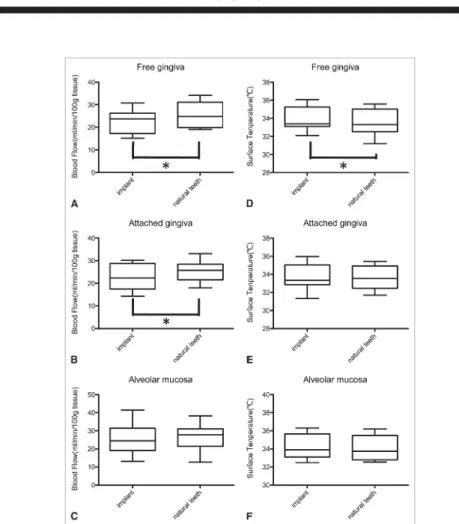
O
, attached gingiva;:, attached gingiva (implant);h, alveolar mucosa; andn, alveolar mucosa (implant).Fig. 2. Bloodflow and surface temperature around implants versus natural teeth. Bloodflow from LSI (left panels) and surface temperature from thermograph (right panels) in free gingiva (AandD), attached gingiva (BandE), and alveolar mucosa (CandF) was compared between implants and natural teeth. *P,0.05 with pairedttest. Significantly greater bloodflow was detected in natural teeth relative to implants in free gingiva and attached gingiva (free gingiva,
P¼0.0055; attached gingiva,P¼0.0052). The surface temperature was significantly higher in the implant in free gingiva (P¼0.0079).
a systemic disease such as diabetes or blood-blood vessel disease were excluded. A total of 10 subjects were included in the study (mean age, 51 years; range, 21–78 years; 5 men and 5 women).
Blood Flow and Surface Temperature Imaging
Blood flow was monitored with a 2-dimensional blood flow analysis machine using infrared laser speckle imaging (LSI; 780 nm) (OZ-1; Omega-wave, Tokyo, Japan); its effective mea-suring depth was within 1 mm of the surface. A CCD camera was set at a right angle to the buccal surface at a 25-cm distance, and 10 images were obtained from each subject. Subjects were asked to wear goggles (YL331; Yamamoto Kogaku Co., Ltd., Higashiosaka, Japan) to avoid possible eye discom-fort. The surface temperature was then monitored by thermography (Thermo GEAR; NEC Avio, Tokyo, Japan) at a 10-cm distance to obtain 4.3 cm high and 5.7 cm wide images. Additionally, a spot thermometer (Thermo-Hunter PT-7LD; Optics Co., Ltd., Otsu, Japan) was used to verify the temperature from the thermograph. Subjects were asked to remain quiet on a dental chair for at least 10 minutes before each measurement. Bloodflow imaging and temperature measurements were then performed.
Data Analysis
Ten-pixel regions of interest (ROIs) were set on the buccal free gingiva, attached gingiva, interdental papilla, implant-dental papilla, and alveolar mucosa for bloodflow imag-ing, as shown in Figure 1.
Temperature data were also ob-tained from the thermograph in refer-ence to the ROI on bloodflow images. For the comparison of blood flow between zirconia and metal abutments, our values were corrected by the value on the gingiva surrounding adjacent natural teeth. A pairedttest was used to compare the subjects’ natural teeth and implants. The Mann-Whitney test was used for comparisons between abutments. In all cases,P ,0.05 was considered to indicate a significant difference.
R
ESULTSLaser Speckle Imaging
Implants versus natural teeth (Figure 2). Significantly greater blood flow was detected in natural teeth (25.808 6 5.370 mL$min−1
$100 g−1 tissue) than
in implants (22.628 6 5.182 mL$min−1$100 g−1tissue) in the free
gin-giva (P¼0.0055) and attached gingiva
(tooth: 25.36264.685; implant: 22.682
66.084 mL$min−1
$100 g−1tissue;P¼
0.0052); however, there was no signif-icant difference in the alveolar mucosa (tooth: 25.703 6 8.985; implant: 25.36264.685 mL$min−1$100 g−1
tis-sue;P¼0.19).
Zirconia versus metal abutments (Figure 3).As a result of correction in the value of natural teeth, the bloodflow Fig. 3. Bloodflow and the surface temperature around zirconia abutments (ZA) versus metal abutments (MA). Relative bloodflow from LSI (left panels) and surface temperature from ther-mograph (right panels) in free gingiva (AandD), attached gingiva (BandE), and alveolar mucosa (CandF) was compared between zirconia and metal abutments. Relative bloodflow values were corrected by the value on the gingiva surrounding adjacent natural teeth. *P,0.05 with Mann-Whitney test. Significantly greater bloodflow was detected with a zirconia abutment (95.646 5.17%) relative to a metal abutment (82.2568.93%) in free gingiva (*P¼0.0317).
was significantly higher in zirconia abut-ments (95.64 6 5.17%) than in metal abutments (82.2568.93%) in the free gingiva (P ¼ 0.0317); however, there was no significant difference in the attached gingiva (zirconia: 88.70 6 11.51; metal: 88.01610.22%) or alve-olar mucosa (zirconia: 84.92 6 9.59; metal: 108.34626.59%).
Temperature Measurement Using Thermography
Implants versus natural teeth. The surface temperature was significantly higher in implants (33.9761.34; tooth: 33.61°C 6 1.45°C) than in natural teeth (33.61°C 6 1.45°C) in the free gingiva (P ¼ 0.0079); however, there was no significant difference in the attached gingiva (implant: 33.63 6 1.42; tooth: 33.58°C 6 1.22°C) or alveolar mucosa (implant: 34.27 6 1.36; tooth: 34.16°C61.34°C). Zirconia versus metal abutments. The surface temperatures were not signifi -cantly different among the free gingiva (zirconia: 34.4661.72; metal: 33.48°C6 0.68°C), attached gingiva (zirconia: 34.21 6 1.89; metal: 33.05°C 6 0.35°C), and alveolar mucosa (zirconia: 35.0261.42; metal: 35.51°C60.85°C).
D
ISCUSSIONRecently, blood flow has attracted attention as a method of assessing oral soft tissue, and noncontrast and noninva-sive laser Dopplerflowmetry (LDF) was developed to evaluate blood flow in marginal tissue.20,21The use of an infrared laser to irradiate red blood cells in micro-vessels causes reflection and scattering, and the Doppler shift causes a frequency change between the incidence and refl ec-tion that is proporec-tionate to the blood flow. In dentistry, much research has been conducted using this method, including analyses of the relationship between peri-odontal disease and gingival bloodflow,22
changes in gingival bloodflow after peri-odontal surgery,23,24and the relationship
between smoking and bloodflow.25,26
However, although LDF is advan-tageous for the real-time measurement of local microcirculatory dynamics, as it is a technique for point measurement with a narrow measuring range of
1 mm2, comparing the blood flow dynamics among different regions is difficult, and the technique has not been applied clinically. To solve this prob-lem, LSI was recently developed.27,28
These measurement methods are basi-cally the same, but in LSI a laser is applied to the object of measurement, and granular changes called speckles are rapidly scanned with a CCD camera and measured as an index of the blood flow dynamics. By LSI, noncontact, noninvasive measurement is possible, and the blood flow dynamics are ex-tracted as 2-dimensional images and captured as visual information, permit-ting blood flow measurement over a wide area as well as conventional point measurement. Also, as multiple ROIs can be displayed on the same image, quantitative analyses (eg, com-parison of the blood flow dynamics among sites at the same time point) have become possible. Given these advan-tages, LSI has been clinically applied to cerebrovascular surgery,29–31skin
dis-eases,29–31and blood recovery from burn
injury.32
Moreover, we found that 2-dimensional temperature measurement by thermography could be used to clarify the assessment of and diagnos-tic criteria for soft tissues. Thermogra-phy provides 2-dimensional images of temperature distribution, similar to LSI, and is used in many fields for a variety of purposes.
Using these methods, we evaluated differences in the bloodflow and sur-face temperature between tissues around implants and natural teeth. We also evaluated the effects of the abut-ment material on periimplant soft tissue. We found that the blood flow was significantly lower in periimplant soft tissue than in soft tissue around natural teeth in free and attached gin-giva. These results are in agreement with those we reported previously.33In
addition, when comparisons were made among abutment types, the bloodflow was;4% lower in the zirconia group and ;18% lower in the metal group than in the free gingiva around natural teeth, indicating that a richer bloodflow can be secured in periimplant soft tissue around zirconia than around metal abut-ments (P¼0.0317). Because the pocket
depth of the periimplant mucosa was
,2 mm in all patients, and no bleeding was noted on probing, the effect of local inflammation could be excluded.
In contrast, the proportion of leuko-cytes in the epithelium has been reported to be lower around zirconia than titanium abutments or other cast-to-abutments,34
suggesting the superiority of the mucosal seal of zirconia. Also,in vitroandin vivo data indicate that bacterial colonization on the abutment surface differs by abut-ment type, that the amount of plaque attachment is significantly smaller on zir-conia than on titanium, and that zirzir-conia may be involved in the formation of the periimplant mucosa. These characteris-tics of zirconia may have affected the bloodflow in this study. The results of this study suggest that zirconia abut-ments promote microcirculatory dynam-ics in periimplant mucosa that are closer to those around natural teeth. Moreover, securing a rich bloodflow in soft tissues around implants is considered to be advantageous for the maintenance of immune function.
It is important to consider not only the resistance and esthetic prop-erties of abutment materials, but also their effects on soft tissues for long-term preservation of the health of periimplant soft tissues. However, as various factors are involved in a com-plex manner in the selection of abut-ments for implant treatment, it is necessary to select an appropriate abutment design and material based on the state of plaque control, esthetic effect, and prosthetic design in each patient.
C
ONCLUSIONSBlood flow in soft tissue around zirconia abutments is similar to that around natural teeth, and significantly greater blood flow was maintained around zirconia abutments compared with metal abutments. Moreover, zirco-nia abutments could be advantageous for the maintenance of immune function by improving blood circulation.
D
ISCLOSUREThe authors claim to have no financial interest, either directly or
indirectly, in the products or informa-tion listed in the article.
R
EFERENCES1. Miyazaki T, Nakamura T, Matsumura H, et al. Current status of zirconia restoration. J Prosthodont Res.2013;57:236–261.
2. Beuer F, Schweiger J, Eichberger M, et al. High-strength CAD/CAM-fabricated veneering material sintered to zirconia copingsdA new fabrication mode for all-ceramic restorations.Dent Mater.2009;25: 121–128.
3. Gargari M, Gloria F, Cappello A, et al. Strength of zirconia fixed partial dentures: Review of the literature. Oral Implantol (Rome).2010;3:15–24.
4. Tinschert J, Zwez D, Marx R, et al. Structural reliability of alumina-, feldspar-, leucite-, mica- and zirconia-based ceramic. J Dent.2000;28:529–535.
5. Takaba M, Tanaka S, Ishiura Y, et al. Implant-supported fixed dental prostheses with CAD/CAM-fabricated porcelain crown and zirconia-based frame-work.J Prosthodont.2013;22:402–407.
6. Peláez J, Cogolludo PG, Serrano B, et al. A prospective evaluation of zirconia posterior fixed dental prostheses: Three-year clinical results. J Prosthet Dent. 2012;107:373–379.
7. Andreiotelli M, Wenz HJ, Kohal RJ, et al. Are ceramic implants a viable alter-native to titanium implants? A systematic literature review. Clin Oral Implants Res. 2009;20:32–47.
8. Andersson B, Schärer P, Simion M, et al. Ceramic implant abutments used for short-span fixed partial dentures: A pro-spective 2-year multicenter study. Int J Prosthodont.1999;12:318–324.
9. Rimondini L, Cerroni L, Carrassi A, et al. Bacterial colonization of zirconia ceramic surfaces: An in vitro and in vivo study. Int J Oral Maxillofac Implants. 2002;17:793–798.
10. Scarano A, Piattelli M, Caputi S, et al. Bacterial adhesion on commercially pure titanium and zirconium oxide disks: An in vivo human study. J Periodontol. 2004;75:292–296.
11. Piconi C, Maccauro G. Zirconia as a ceramic biomaterial.Biomaterials. 1999; 20:1–25.
12. Denry I, Kelly JR. State of the art of zirconia for dental applications. Dent Mater.2008;24:299–307.
13. Kim SS, Yeo IS, Lee SJ, et al. Clin-ical use of alumina-toughened zirconia abutments for implant-supported restora-tion: Prospective cohort study of survival analysis.Clin Oral Implants Res.2013;24: 517–522.
14. Jung RE, Pjetursson BE, Glauser R, et al. A systematic review of the 5-year survival and complication rates of implant-supported single crowns. Clin Oral Im-plants Res.2008;19:119–130.
15. Abrahamsson I, Berglundh T, Glantz PO, et al. The mucosal attachment at different abutments. An experimental study in dogs. J Clin Periodontol. 1998; 25:721–727.
16. Lops D, Bressan E, Chiapasco M, et al. Zirconia and titanium implant abut-ments for single-tooth implant prostheses after 5 years of function in posterior re-gions. Int J Oral Maxillofac Implants. 2013;28:281–287.
17. Glauser R, Sailer I, Wohlwend A, et al. Experimental zirconia abutments for implant-supported single-tooth restorations in esthetically demanding regions: 4-Year re-sults of a prospective clinical study. Int J Prosthodont.2004;17:285–290.
18. Ekfeldt A, Fürst B, Carlsson GE. Zirconia abutments for single-tooth implant restorations: A retrospective and clinical follow-up study.Clin Oral Implants Res.2011;22:1308–1314.
19. Zembic A, Bösch A, Jung RE, et al. Five-year results of a randomized controlled clinical trial comparing zirconia and titanium abutments supporting single-implant crowns in canine and posterior regions.Clin Oral Im-plants Res.2013;24:384–390.
20. Nilsson GE, Tenland T, Oberg PA. Evaluation of a laser Dopplerflowmeter for measurement of tissue blood flow. IEEE Trans Biomed Eng.1980;27:597–604.
21. Nilsson GE. Signal processor for laser Doppler tissueflowmeters.Med Biol Eng Comput.1984;22:343–348.
22. Gleissner C, Kempski O, Peylo S, et al. Local gingival bloodflow at healthy and inflamed sites measured by laser Doppler flowmetry. J Periodontol. 2006; 77:1762–1771.
23. Donos N, D’Aiuto F, Retzepi M, et al. Evaluation of gingival blood flow by the use of laser doppler flowmetry
following periodontal surgery. A pilot study. J Periodontal Res.2005;40:129–137.
24. Retzepi M, Tonetti M, Donos N. Comparison of gingival bloodflow during healing of simplified papilla preservation and modified widmanflap surgery: A clin-ical trial using laser doppler flowmetry. J Clin Periodontol.2007;34:903–911.
25. Ketabi M, Hirsch RS. The effects of local anesthetic containing adrenaline on gingival blood flow in smokers and non-smokers. J Clin Periodontol. 1997; 24:888–892.
26. Mavropoulos A, Brodin P, Rösing CK, et al. Gingival blood flow in perio-dontitis patients before and after peri-odontal surgery assessed in smokers and non-smokers. J Periodontol.2007; 78:1774–1782.
27. Forrester KR, Stewart C, Tulip J, et al. Comparison of laser speckle and laser doppler perfusion imaging: Measure-ment in human skin and rabbit articular tissue. Med Biol Eng Comput. 2002;40: 687–697.
28. Forrester KR, Tulip J, Leonard C, et al. A laser speckle imaging technique for measuring tissue perfusion. IEEE Trans Biomed Eng.2004;51:2074–2084.
29. Ayata C, Dunn AK, Gursoy-OZdemir Y, et al. Laser speckleflowmetry for the study of cerebrovascular physiol-ogy in nor- mal and ischemic mouse cor-tex.J Cereb Blood Flow Metab.2004;24: 744–755.
30. Dunn AK, Bolay H, Moskowitz MA, et al. Dynamic imaging of cerebral blood flow using laser speckle. J Cereb Blood Flow Metab.2001;21:195–201.
31. Zakharov P, Völker AC, Wyss MT, et al. Dynamic laser speckle imaging of cerebral blood flow. Opt Express. 2009; 17:13904–13917.
32. Stewart CJ, Frank R, Forrester KR, et al. A comparison of two laser-based methods for determination of burn scar per-fusion: Laser Doppler versus laser speckle imaging.Burns. 2005;31:744–752.
33. Nakamoto T, Kanao M, Kondo Y, et al. Two-dimensional real-time bloodflow and temperature of soft tissue around maxillary anterior implants. Implant Dent. 2012;21:522–527.
34. Welander M, Abrahamsson I, Berglundh T. The mucosal barrier at implant abutments of different materials. Clin Oral Implants Res.2008;19:635–641.