Sealants for preventing and
arresting pit-and-
fi
ssure occlusal
caries in primary and permanent molars
A systematic review of randomized controlled
trials
—
a report of the American Dental Association
and the American Academy of Pediatric Dentistry
John T. Wright, DDS, MS; Malavika P. Tampi, MPH; Laurel Graham, MLS; Cameron Estrich, MPH; James J. Crall, DDS, MS, ScD; Margherita Fontana, DDS, PhD; E. Jane Gillette, DDS; Brian B. Nový, DDS; Vineet Dhar, BDS, MDS, PhD; Kevin Donly, DDS, MS; Edmond R. Hewlett, DDS; Rocio B. Quinonez, DMD, MS, MPH; Jeffrey Chaffin, DDS, MPH, MBA, MHA; Matt Crespin, MPH, RDH; Timothy Iafolla, DMD, MPH; Mark D. Siegal, DDS, MPH; Alonso Carrasco-Labra, DDS, MSc, PhD(c)
C
aries prevalence hasdeclined in developed countries over the past several decades; however, many populations within these nations still carry a large burden of this disease.1National Health and Nutrition Examination Survey
2011-2012data indicated that, in the United States, nearly one-fourth of children and over one-half of
Copyrightª2016 American Academy of Pediatric Dentistry and American Dental Association. All rights reserved. This article is being published concurrently inPediatr Dent.
2016;38(4):282-294. The articles are identical. Either citation can be used when citing this article.
ABSTRACT
Background.National Health and Nutrition Examination Survey 2011-2012 data indicated that, in the United States, nearly one-fourth of children and over one-half of adolescents experienced dental caries in their permanent teeth. The purpose of this review was to summarize the available clinical evidence regarding the effect of dental sealants for the prevention and management of pit-and-fissure occlusal carious lesions in primary and permanent molars, compared with a control without sealants, withfluoride varnishes, or with other head-to head comparisons. Type of Studies Reviewed.The authors included parallel and split-mouth randomized controlled trials that included at least 2 years of follow-up, which they identified using MEDLINE (via PubMed), Embase, LILACS, the Cochrane Central Register of Controlled Trials, and registers of ongoing trials. Pairs of reviewers independently conducted the selection of studies, data extraction, risk of bias as-sessments, and quality of the evidence assessments by using the Grading of Rec-ommendations Assessment, Development and Evaluation approach.
Results.Of 2,869 records screened, the authors determined that 24 articles (representing 23 studies) proved eligible. Moderate-quality evidence suggested that participants who received sealants had a reduced risk of developing carious lesions in occlusal surfaces of permanent molars compared with those who did not receive sealants (odds ratio [OR], 0.15; 95% confidence interval [CI], 0.08-0.27) after 7 or more years of follow-up. When the authors compared studies whose investigators had compared sealants withfluoride varnishes, they found that sealants reduced the incidence of carious lesions after 7 or more years of follow-up (OR, 0.19; 95% CI, 0.07-0.51); however, thisfinding was supported by low-quality evidence. On the basis of the evidence, the authors could not provide a hierarchy of effectiveness among the studies whose investigators had conducted head-to-head comparisons. The investigators of 2 trials provided information about adverse events, but they did not report any adverse events.
Conclusions and Practical Implications.Available evidence suggests that sealants are effective and safe to prevent or arrest the progression of noncavitated carious lesions compared with a control without sealants orfluoride varnishes. Further research is needed to provide information about the relative merits of the different types of sealant materials.
Key Words.Glass ionomer sealants; resin-based sealants; caries prevention; caries arrest; pit-and-fissure sealants; systematic review.
JADA 2016:147(8):631-645
adolescents experienced dental carious lesions in their permanent teeth.2Occlusal surfaces, especially those on permanent molars, contain grooves called pits andfi s-sures that can trap debris and microorganisms, thereby increasing the risk of developing dental carious lesions. Indeed, the caries that are found in the adolescent pop-ulation are represented disproportionately in the pits and
fissures of teeth compared with the smooth surfaces.3 Fluorides and other caries preventive approaches (for example, mechanical plaque control) seem to be less effective for preventing carious lesions in pit-and-fissure surfaces compared with smooth surfaces.3Pit-and-fissure sealants, or simply sealants, were developed to help manage these sites of dental stagnation that are resistant to other therapeutic approaches and contribute to a significant portion of caries disease burden in the pop-ulation. Sealants are an underused therapy; only30% of children 6to8years old have at least 1dental sealant.4
Sealants are dental materials that dentists apply to the pit-and-fissure surfaces of teeth. The sealant material penetrates pits andfissures and then hardens, acting as a physical barrier that stops or inhibits the ingress of bacteria and nutrients. Researchers conducted thefirst clinical trials in the late 1960s and early1970s using a variety of materials. Today there are multiple commer-cially available sealant materials, including resin-based sealants such as urethane dimethacrylate or bisphenol A-glycidyl methacrylate monomers that are polymerized by means of either a chemical activation-initiation or a light activation system. Glass ionomer (GI) cements are another type of sealant material that have been widely recognized and used for theirfluoride-release properties, which stem from the acid-base reaction between afl uo-roaluminosilicate glass powder and an aqueous-based polyacrylic acid solution. Polyacid-modified resin seal-ants, also referred to as compomers, combine resin-based material found in traditional resin-based sealants with thefluoride-release and adhesive properties of GI seal-ants. Resin-modified GI sealants are essentially GI sealants with resin components that allow for light polymerization.5These dental materials differ in many of their physical properties, including hydrophobicity, fracture resistance, thermal expansion, and bond strength. Also, investigators have found that topical
fluoride varnishes (sodiumfluoride) substantially prevent dental caries in children and adolescents by decreasing demineralization, promoting remineraliza-tion, and possibly inhibiting the effects of bacterial biofilm.6
Investigators have conducted a number of systematic reviews to determine the clinical effectiveness, cost-effectiveness, and safety of pit-and-fissure sealants compared with another type of sealant material, a control without sealants, andfluoride varnishes. The authors of1 review reported that sealants were effective in preventing occlusal and proximal carious lesions in the molars of
children when compared with controls without sealants.7 The authors of this review also reported inconclusive and inconsistent results related to the potential superiority of any of the sealant materials in head-to-head compari-sons.7The authors of another systematic review sug-gested that sealants may be more effective thanfluoride varnishes in preventing occlusal carious lesions in molars in children, but the quality of the evidence was low.6 The investigators of both of these systematic reviews6,7 reported that the authors of most of the included studies did not mention adverse events, and even when authors did mention adverse events, they did not report any adverse events that had occurred in their studies.6,7
The purpose of this review was to summarize the available evidence regarding the effect of dental sealants for the prevention of pit-and-fissure occlusal caries in primary and permanent molars on children, adolescents, and adults compared with a control without sealants, withfluoride varnishes, or with another head-to-head comparison to inform the development of a joint evidence-based clinical practice guideline by the American Dental Association and the American Academy of Pediatric Dentistry.8
METHODS
This report follows the guidance of the Preferred Reporting Items for Systematic Reviews and Meta-Analyses statement.9
Selection criteria for the studies in this review.
Type of studies.We included parallel and split-mouth
randomized controlled trials (RCTs) with at least2years of follow-up. We excluded quasirandomized trials, nonrandomized trials, and observational studies.
Type of participants.We included studies that
involved children, adolescents, and adults from the general population who did or did not have a history of carious lesions and who had either a sound occlusal surface or a noncavitated carious lesion in primary and permanent molars.
Type of interventions.For this systematic review,
we defined4categories of sealant materials: resin-based sealants, GI cements or GI sealants, resin-modified GI sealants, and polyacid-modified resins. We classified resin-modified GI sealants as a subcategory of the GI sealants category and polyacid-modified resins as a subcategory of the resin-based sealants category.5 We defined“intervention”as any of the4types of sealant materials described previously, irrespective of the application technique. We excluded studies whose investigators used sealant materials that were not
ABBREVIATION KEY.GI:Glass ionomer.GRADE:
commercially available at the time of this review. We defined“comparison”as any type of sealant material irrespective of the application technique, the nonplace-ment of sealants, or the use of fluoride varnishes.
Type of outcome measures.We defined“caries
inci-dence”as the identification of a new carious lesion on the occlusal surface of a primary or permanent molar that compromised dentin tissue. We defined“lack of retention”as the complete detachment or retention loss of the sealant material from the grooves and pits in the occlusal surface of a tooth with no macroscopically visible sealant material. We defined“adverse effects”as any potential adverse effect defined by the authors of the primary studies. For all outcomes, we grouped the studies into3categories according to the length of follow-up:2to3years,4to7years, and7or more years.
Search methods for the identification of studies.
Electronic databases.We searched MEDLINE (via
PubMed), Embase, LILACS, and the Cochrane Central Register of Controlled Trials (CENTRAL) from January
1971to May2013. We searched MEDLINE (via PubMed) and the Cochrane Central Register of Controlled Trials (CENTRAL) from June 2013to May2016. We used a combination of key words and controlled vocabulary that we adapted for each electronic database. We usedfilters, such as the Cochrane Highly Sensitive Search Strategy, for identifying randomized trials (Appendix, available online at the end of this article).10
Other type of resources.We searchedClinicalTrials.
govto identify completed or ongoing RCTs that were not yet published and indexed in the regular electronic indices. We also screened the reference lists of included studies from previous systematic reviews to ensure that we had not omitted relevant studies. We did not exclude any studies on the basis of the status or language of publication.
Data collection and analysis. Selection of studies.In thefirst stage,2reviewers (M.T., L.G.) independently screened the titles and abstracts of all retrieved references by using a standardized form. Because they used an inclusive criterion, when the reviewers disagreed on the eligibility status for a particular reference, they included the citation in question at this stage and resolved the disagreement at the full-text screening stage. In the second stage,2reviewers independently screened the full text of all potentially eligible studies. They resolved any disagreement by means of discussion. When consensus was elusive, a third reviewer (C.E.), acting as an arbiter, decidedfinal eligibility.
Data extraction and management.Using a
stan-dardized form, 2reviewers (M.T., L.G.) independently extracted data from all the included studies. The form included instructions to extract the main characteristics of the studies, including the type of study design (par-allel, split-mouth), population (age, sex, selection criteria, caries history, clinical diagnosis of the occlusal surface to be sealed), type of sealant material and the comparison (nonuse of sealant or an active comparator), and the
outcomes (specific definition from the primary study and results). When these reviewers identified discrepancies that they were unable to clarify, a third reviewer (C.E.) acted as arbiter.
Assessment of the risk of bias of included studies.Two
reviewers (M.T., A.C.L.) independently conducted an assessment of the risk of bias for each included study by using the Cochrane risk of bias tool.11We assessed the following types of bias in each study: selection bias (Was allocation randomized and concealed to ensure compara-bility between groups?), detection bias (Were the patients and outcome assessors unaware of which treatment was applied?), attrition bias (Were dropout rates sufficiently low to ensure that groups were still comparable at follow-up?), reporting bias (Did investigators selectively report outcomes?), and other sources of bias. For each domain, we determined whether a study had a high, low, or unclear risk of bias. We considered randomization sequence generation and allocation concealment to be the most important domains for the overall assessment of risk of bias. We resolved any disagreements by means of discussion until we reached consensus.
Measures of treatment effect and missing data.We
analyzed caries incidence, lack of retention, and adverse events as dichotomous outcomes. For studies in which the investigators reported sealants as being fully retained, partially retained, and not retained, we grouped the fully and partially retained events and compared them with the sealants that were not retained to create the estimate. We calculated odds ratios (OR) and95% confidence intervals (CI) for both outcomes. For each study, we calculated the proportion of missing participant data, and we determined to what extent the amount of missing data was substantial enough to change the magnitude and direction of the estimates to the point of dramati-cally changing the conclusions, as suggested by Akl and colleagues.12Otherwise, we used complete case analysis.
Assessment of heterogeneity.We conducted the
assessment of heterogeneity by following the guidance of theCochrane Handbook for Systematic Reviews of Intervention.13We used the
c
2test to determine the presence of statistical heterogeneity, and we set the level of significance at .1. In addition, we quantified the amount of heterogeneity among studies using theI2statistic, in which we considered a value ofI240% or less to be unimportant heterogeneity, a value ofI2from
30% through 60% to be moderate heterogeneity, a value ofI2from50% through 90% to be substantial heterogeneity, and a value ofI2from70% through100% to be considerable heterogeneity.
Assessment of publication bias.We conducted the
Data synthesis.Investigators of RCTs who measured the effectiveness of interventions to prevent carious le-sions typically used1of2designs: split-mouth or parallel. In RCTs whose investigators used a parallel design, the investigators allocated study participants to receive either the experimental treatment or a control. In split-mouth trials, the investigators randomly assigned 1of2 treat-ments (for example, sealant versus no sealant) to the same type of tooth on the right and left sides of the participant’s mouth. One advantage of conducting split-mouth trials is that these types of RCTs minimize vari-ability among study participants, as the intervention and control teeth are in the same person’s mouth. One po-tential issue, however, is that the preventive benefits of the intervention may carry over to the control teeth. We judged these carryover effects to be minimal for sealants, and therefore, we pooled thefindings from studies whose investigators had used each of these designs to create a single effect estimate by using the methodology proposed by Lesaffre and colleagues15and Elbourne and col-leagues.16We used Review Manager (RevMan), Version
5.3(Cochrane Collaboration) to conduct the analysis. To obtain the pooled estimate, we used the generic inverse-variance method with a random-effects model. When we included fewer than4studies in the meta-analysis, we used afixed-effects model.
Subgroup analysis.We conducted subgroup analysis
to determine whether the studies whose investigators had enrolled participants with noncavitated pit-and-fissure occlusal carious lesions, sound occlusal surfaces, and those who had both (that is, a population who had a mix of both sound occlusal surfaces and noncavitated carious lesions) had different treatment effects. For the interac-tion test, we used a level of significance of .05.
Assessment of the quality of the evidence.We
determined the quality of the evidence (certainty in the estimates of effect) for each outcome by using the
Grading of Recommendations Assessment, Development and Evaluation (GRADE) approach.17With the GRADE approach, RCTs start as high-quality evidence; however, the quality or certainty in the body of evidence decreases to moderate-, low-, or very low–quality evidence if serious or very serious issues related to risk of bias, imprecision, inconsistency, indirectness, and publication bias are present (Table1).18Two reviewers (M.T., A.C.L.) independently conducted these evaluations.
RESULTS
Results of the search.The search process resulted in
2,869references, which we screened to assess their titles and abstracts; we excluded2,419references at that stage of the search process. Next, we excluded426articles, which we had assessed by means of full-text screenings, and we included24 articles,1,19-41which represented23 studies, in this review (Figure1).
Characteristics of included studies.We included24
articles (representing 23studies) published from1976 through2016,1,19-41whose investigators had reported data related to the effectiveness of sealants compared with a control without sealants,1,19-26fluoride varnishes,20,22,27 or other head-to-head comparisons.28-40Nine studies’ investigators used a parallel design,20,22,24,26,28,31,33,38,39,41 whereas14studies’investigators used a split-mouth design.1,19,21,23,25,27,29,30,32,34-37,40Table2summarizes the characteristics of the included populations, which in-vestigators described as including children and adoles-cents aged3to16years who were living in settings with and without waterfluoridation. We did not identify any studies that met the selection criteria whose investigators had provided information about the effect of sealants in an adult population.
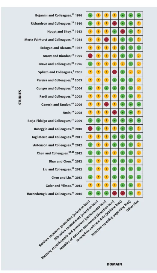
Risk of bias of included studies.Poor quality of
reporting of the included studies prevented us from conducting a complete assessment of the risk of bias. For most of the studies, we assessed the key3domains of random sequence generation, allocation concealment, and masking of participants and personnel as having an unclear risk of bias. Of these3domains, we determined that allocation concealment was the most serious and underreported methodological issue (Figure2).
Effects of the interventions. Comparison1. Sealants
versus nonuse of sealants. Caries incidence.The results
of9studies1,19-26(3,542participants) informed the com-parison and outcome for the2- to3-year follow-up category. In relative terms, participants who received sealants reduced their risk of developing new carious lesions by76% (odds ratio [OR],0.24;95% confidence interval [CI],0.19-0.30;P<.00001) compared with participants who did not receive sealants. The hetero-geneity was moderate (
c
2P¼.09;I2¼41%); however, the investigators of all of the individual studies reported the same direction of effect with an overlap of CIs (eFigure1, available online at the end of this article). In a TABLE 1Levels of quality of evidence (certainty
in the evidence).
*
QUALITY LEVEL
DEFINITION
High We are very confident that the true effect lies close to that of the estimate of the effect
Moderate We are moderately confident in the effect estimate; the true effect is likely to be close to the estimate of the effect, but there is a possibility that it is substantially different
Low Our confidence in the effect estimate is limited; the true effect may be substantially different from the estimate of the effect
Very Low We have very little confidence in the effect estimate; the true effect is likely to be substantially different from the estimate of effect
subgroup analysis conducted to determine whether the treatment effect differed among studies with patients who had noncavitated occlusal carious lesions, sound occlusal surfaces, and a population with mixed features, we did notfind statistically significant results (interaction testP¼.58). We assessed the quality of the evidence for this outcome as moderate, owing to serious issues related to risk of bias (Table3).
The results of3studies20,21,23(752participants) informed the comparison and outcome for the4- to
7-year follow-up category. In relative terms, participants who received sealants had a reduction in the risk of developing new carious lesions by79% (OR,0.21;95% CI,
0.10-0.44;P <.0001) compared with participants who did not receive sealants (eFigure2, available online at the end of this article). Because the investigators of all 3of these studies included only participants with sound occlusal surfaces, we did not perform a subgroup anal-ysis. Serious issues of inconsistency (
c
2P¼.01; I2¼77%) and risk of bias warranted us to determine that low-quality evidence informed this outcome (Table3).
The results of2studies20,23 (446participants) informed the comparison and outcome for the7or more years of follow-up category. In relative terms, partici-pants who received sealants had a reduction in the risk of developing new carious lesions by85% (OR,0.15;95% CI,
0.08-0.27;P<.00001) compared with participants who did not receive sealants (eFigure3, available online at the end of this article). The heterogeneity was moderate to high (
c
2P¼.16;I2¼50%); however, the investigators of all of the individual studies found the same direction of effect with an overlap of CIs. Because the investigators of the 2studies included only participants with sound occlusal surfaces, we did not perform a subgroup anal-ysis. We assessed the quality of the evidence for this outcome as moderate, owing to serious issues related to risk of bias (Table3).Lack of retention.The nature of the comparison did not allow us to obtain information to compare the use versus the nonuse of sealants.
Comparison2. Sealants versusfluoride varnishes.
Caries incidence.The results of3studies20,22,27(1,715 participants) informed the comparison and outcome for the2- to 3-year follow-up category. In relative terms, participants who received sealants had a 73% reduction in the risk of developing new carious lesions (OR,0.27;
95% CI,0.11-0.69;P¼.006) compared with participants who received fluoride varnishes (eFigure 4, available online at the end of this article). In a subgroup analysis conducted to determine whether the treatment effect differed among studies with patients having noncavitated occlusal carious lesions, sound occlusal surfaces, and a population with mixed features, we found statistically significant results (interaction testP¼.04); however, this subgroup analysis did not explain the heterogeneity of the results. The investigators of both subgroups of studies
with sound occlusal surfaces (OR,0.19;95% CI,0.07-0.47;
P¼.0004) and with a mixed population of participants with and without noncavitated carious lesions (OR,0.66;
95% CI,0.30-1.44;P¼.3) found that there was a
bene-ficial effect when using sealants; however, this difference was not statistically significant in the latter study.22We assessed the quality of the evidence for this outcome as low, owing to serious issues related to inconsistency (
c
2P¼.002;I2¼88%) and risk of bias (eTable1, available online at the end of this article).
The results of2studies20,27(472participants) informed the comparison and outcome for the4- to7-year follow-up category. In relative terms, participants who received sealants had an81% reduction in the risk of developing new carious lesions (OR,0.19;95% CI,0.07-0.51;P¼
.0008) compared with participants who receivedfluoride varnishes (eFigure5, available online at the end of this article). Because the investigators of the2studies included only participants with sound occlusal surfaces, we did not perform a subgroup analysis. We assessed the quality of the evidence for this outcome as low, owing to serious issues of inconsistency (
c
2P¼.03;I2¼80%) and risk of bias (eTable1, available online at the end of this article).Records identified through database searching (n = 4,483)
Additional records identified through other sources (n = 487)
Records screened (n = 2,869)
Duplicates and non-English publications excluded
(n = 2,101)
Records excluded (n = 2,419)
Full-text articles assessed for
eligibility (n = 450)
Full-text articles excluded (n = 426)
Studies included in the quantitative synthesis
(meta-analysis) (n = 22 [23 articles])
Studies included in the qualitative synthesis (n = 1)
TABLE 2
Characteristics of the included studies.
STUDY COUNTRY DESIGN PARTICIPANTS AGE RANGE,
Y (MEAN) Bojanini and Colleagues,19
1976
Colombia Split-mouth Children with erupted, sound PM†; setting was not clearly defined 6-8
Richardson and Colleagues,25 1980
Canada Split-mouth Children with erupted, sound or carious PFM‡; setting was an elementary school clinic
7-8
Houpt and Shey,271983 United
States Split-mouth (mobile unit)Children with erupted, sound PFM; setting was a dental van 6-10
Mertz-Fairhurst and Colleagues,23 1984
United States
Split-mouth Children with erupted, sound PFM; setting was a dental school clinic
6-8
Erdogan and Alacam,211987 Turkey Split-mouth Children with erupted, sound PFM; setting was not described 8-10
Arrow and Riordan,301995 Australia Split-mouth Children with sound PFM; setting was a school clinic 7 (0.72)
Bravo and Colleagues,201996 Spain Parallel Children with erupted, sound PM; setting was a school clinic 6-8
Splieth and Colleagues,12001 Germany Split-mouth Children with erupted, sound or carious PFM; setting was a private practice office
5-8
Pereira and Colleagues,242003 Brazil Parallel Children with erupted, sound PFM; setting was a dental school clinic
6-8
Gungor and Colleagues,372004 Turkey Split-mouth Children with erupted PFM; setting was a dental school clinic 7-10
Pardi and Colleagues,382005 Brazil Parallel Children with erupted PFM; setting was a school clinic 7-8
Ganesh and Tandon,402006 India Split-mouth Children with erupted, sound primary molars (Group 1) and erupted, sound permanent molars (Group 2)
Group 1: 3-5 Group 2: 6-7
Amin,282008 Egypt Parallel Children with sound PFM; setting was a dental school clinic 7-10
Barja-Fidalgo and Colleagues,31 2009
Brazil Parallel Children with erupted PFM; setting was a university dental clinic 6-8
Baseggio and Colleagues,322010 Brazil Split-mouth Adolescents with erupted second PM; setting was a public health service center
12-16
Tagliaferro and Colleagues,26 2011
Brazil Parallel Children with erupted, sound PFM; setting was a private practice 6-8
Antonson and Colleagues,292012 United States
Split-mouth Children with partially erupted PFM; setting not clearly defined, seems to be a university dental clinic
5-9
Chen and Colleagues,33,412012
(2 reports)
China Parallel Children with erupted, carious PFM; setting was at 5 public schools
7-9.1
Dhar and Chen,352012 India Split-mouth Children with erupted PFM; setting was a school clinic 6-10
Liu and Colleagues,222012 China Parallel Children with erupted, sound or carious PFM; setting was a school clinic
Mean¼9.1
Chen and Liu,342013 China Split-mouth Children with erupted, sound PFM; setting was a pediatric department of a university hospital
6.1-8.9
Guler and Yilmaz,362013 Turkey Split-mouth Children with erupted PFM; setting was a dental school clinic 7-13
Haznedaroglu and Colleagues,39 2016
Turkey Parallel Children with fully erupted, sound PFMs; setting was a university pediatric clinic
7-10
* Information provided corresponds with thefirst follow-up period of the study.
†PM: Permanent molar.
The results of1study20(242participants) informed the comparison and outcome for the7or more years of follow-up category. In relative terms, participants who received sealants had a71% reduction in the risk of developing new carious lesions (OR,0.29;95% CI,0.17-0.49;P<.00001) compared with participants who received fluoride
varnishes (eFigure 6, available online at the end of this article). Because the results of only1study informed this outcome, we did not perform a subgroup analysis. We assessed the quality of the evidence for this outcome as low, owing to very serious issues related to risk of bias (eTable 1, available online at the end of this article). TABLE 2 (CONTINUED)
FLUORIDE EXPOSURE INTERVENTION COMPARISON SEALANT
(N)*
COMPARISON (N)*
Community waterfluoridation Resin-based sealant (Delton, Dentsply)
No sealant 42 42
Nonfluoridated community Self-curing bisphenol A-glycidyl methacrylate sealant (3M)
No sealant 337 337
Community waterfluoridation Sealant (Delton, Dentsply) Fluoride varnish (no further description)
250 250
Community waterfluoridation Resin-based sealant (Delton, Dentsply)
No sealant 201 201
None Resin-based sealant (Delton, Dentsply)
No sealant 96 96
None GI§sealant (Ketac-fil, 3M) Resin-based sealant (Delton, Dentsply)
412 412
Community waterfluoridation at 0.07 ppm¶ offluoride
Resin-based sealant (Delton, Dentsply)
No sealant;fluoride varnish (Duraphat, Colgate-Palmolive)
238 272
Community waterfluoridation at 0.1 ppm. Fluoride tablets used forfirst year of their life only (48%), and some children took tablets during study (5%). Duraphatfluoride varnish was applied in both groups.
Resin-based sealant No sealant 176 176
Community waterfluoridation Sealant
GI sealant (Ketac bond, 3M)
No sealant;
resin-modified GI sealant (Vitremer, 3M)
342 240
Nonfluoridated water; encouraged use of fluoridated toothpaste
Poly-acid modified resin (Dyract Seal, Dentsply)
Resin-based sealant (Delton FSþ, Dentsply)
70 70
Community waterfluoridation Resin-modified GI sealant (Vitremer, 3M)
Resin-based sealant (Revolution, Kerr);
poly-acid modified resin sealant (Dyract Flow, Dentsply)
97 182
None GI sealant (Fuji VII, GC) Resin-based sealant (Concise, 3M) 100 100
Fluoridated toothpaste Resin-modified GI sealant (Fuji II LC, GC)
Resin-based sealant (Tetric Flow and Helioseal F, Ivoclor Vivadent)
24 54
Fluoridated toothpaste GI sealant (Fuji IX, GC) Resin-based sealant (Delton, Dentsply)
21 28
None Resin-modified GI sealant (Vitremer, 3M)
Resin-based sealant (Fluoroshield, Dentsply)
628 628
Community waterfluoridation at 0.7 ppm, and 93% of participants reported usingfluoridated toothpaste
Resin-modified GI sealant (Vitremer, 3M)
No sealant 91 86
None GI sealant (Fuji Triage, GC) Resin-based sealant (Delton FSþ, Dentsply)
27 27
None GI sealant (Ketac Molar Easymix, 3M)
Resin-based sealant (Clinpro, 3M)
1,282 452
None GI sealant (Fuji VII, GC) Resin-based sealant (Clinpro, 3M) 50 50 No community waterfluoridation, but 90% of
toothpastes sold in area containfluoride
Resin-based sealant (Clinpro, 3M)
No sealant;fluoride varnish (5% sodiumfluoride Duraphat, Colgate-Palmolive)
367 379
Use of 600 ppmfluoridated toothpaste. 6,000 ppm foam applied at every recall visit
GI sealant (Fuji VII, GC) Resin-based sealant (Concise, 3M)
75 75
Fluoride varnish applied after sealant placement GI sealant (Fuji VII, GC) Resin-based sealant (Admira Seal, Voco)
68 66
“Lowfluoride”in drinking water GI sealant (Fuji Triage, GC) Resin-based sealant (Ultraseal XT, Ultradent)
Lack of retention.The nature of the comparison did not allow us to obtain information to compare the use versus the nonuse of sealants.
Comparison3.
Glass ionomer sealants versus resin-based
seal-ants. Caries incidence.
The results of10 studies28-30,32-36,38,39(4,741 participants) informed the comparison and outcome for the2- to3 -year follow-up category. In relative terms, partic-ipants who received GI sealants had a29% reduction in the risk of developing new carious lesions compared with participants who received resin-based sealants (OR,0.71;95% CI,0.32-1.57); however, this difference was not statistically significant (P ¼.39) (eFigure7, available online at the end of this article). Owing to limitations in1 study’s40data presenta-tion, we did not include that study (200 partici-pants) in the meta-analysis. For that study,40 the investigators failed tofind a clinically or statistically significant difference in caries inci-dence when they applied GI sealants and resin-based sealants in the occlusal surfaces of pri-mary and permanent molars. In a subgroup analysis conducted to determine whether the treatment effect differed among studies with patients having non-cavitated occlusal carious lesions, sound occlusal surfaces, and a popula-tion with mixed clinical Random sequence generation (selection bias)
Allocation concealment (selection bias)
Masking of outcome assessment (det ection bias)
Masking of par
ticipants and personnel (per formance b
ias)
Incomplet
e outcome data (attrition bias) Selective repor
ting (r epor
ting bias)Other bias Amin,28 2008 ? ? ? – + + ?
Antonson and Colleagues,29 2012 + ? ? + + + ? Arrow and Riordan,30 1995 – ? ? ? + + ?
Barja-Fidalgo and Colleagues,31 2009 + + ? + + + +
Baseggio and Colleagues,32 2010 – + ? ? + + ? Bojanini and Colleagues,19 1976 + ? ? ? + + +
Bravo and Colleagues,20 1996 + ? ? ? ? + ?
Chen and Colleagues,33,41 2012 + + ? ? + + ?
Chen and Liu,34 2013 + ? ? + + + + Dhar and Chen,35 2012 + ? ? + + + ? Erdogan and Alacam,21 1987 ? ? ? ? + + +
Ganesh and Tandon,40 2006 ? ? – ? + + +
Guler and Yilmaz,36 2013 ? ? ? ? + + ? Gungor and Colleagues,37 2004 + ? + + + + ?
Haznedaroglu and Colleagues,39 2016 + + + – – + ? Houpt and Shey,27 1983 ? ? ? + – + ?
Liu and Colleagues,22 2012 + ? ? + + + + Mertz-Fairhurst and Colleagues,23 1984 ? ? – ? + + ?
Pardi and Colleagues,38 2005 ? ? ? ? + + ? Pereira and Colleagues,24 2003 ? ? ? + + + ? Richardson and Colleagues,25 1980 + ? ? – + + ?
Splieth and Colleagues,1 2001 ? ? ? – + ? ?
Tagliaferro and Colleagues,26 2011 ? ? ? + + + +
S
T
UD
IE
S
DOMAIN
features, we did notfind statistically significant results (interaction testP¼.19). We assessed the quality of the evidence for this outcome as very low, owing to serious issues related to risk of bias, inconsistency (
c
2P>.00001; I2¼81%), and imprecision (Table4).
The results of2studies31,39(145participants) informed the comparison and outcome for the4- to7-year follow-up category. In relative terms, participants who received GI sealants had a63% reduction in the risk of developing new carious lesions compared with participants who received resin-based sealants (OR,0.37;95% CI,0.14-1.00;
P ¼.05) (eFigure8, available online at the end of this article). Because we found only2 studies to inform this outcome, we did not perform a subgroup analysis. We assessed the quality of the evidence for this outcome as very low, owing to serious issues related to risk of bias and very serious issues related to imprecision (Table4).
We did notfind any studies whose investigators had reported data on the incidence of caries for7or more years of follow-up for this comparison.
Lack of retention.The results of10studies28-30,32-36,38,39 (4,741participants) informed the comparison and outcome for the2- to 3-year follow-up category. In relative terms, participants who received GI sealants had
5times greater chance (406% increased chance) of experiencing sealant retention loss compared with par-ticipants who received resin-based sealants (OR, 5.06;
95% CI,1.81-14.13;P¼.002) (eFigure9, available online at the end of this article). In a subgroup analysis conducted to determine whether the treatment effect differed among studies with patients who had noncavitated occlusal carious lesions, sound occlusal surfaces, and a population with mixed clinical features, we did notfind statistically significant results (interaction testP¼.29). We assessed the quality of the evidence for this outcome as low, owing to serious issues related to risk of bias and inconsistency (
c
2P<.00001;I2¼96%) (Table4).The results of2studies31,39(145participants) informed the comparison and outcome for the4- to7-year follow-up category. In relative terms, participants who received GI sealants had a108% increase in the risk of experiencing a retention loss compared with the participants who received resin-based sealants (OR,2.08;95% CI,0.15-27.95); however, this difference was not statistically significant (P¼.58) (eFigure10, available online at the end of this article). Because only2studies informed this outcome, we did not perform a subgroup analysis. We assessed the quality of the evidence for this outcome as low, owing to serious issues related to risk of bias and imprecision (Table4).
We did notfind any studies whose investigators had reported data on the incidence of lack of sealant reten-tion for 7or more years of follow-up.
Comparison4. Glass ionomer sealants versus
resin-modified glass ionomer sealants. Caries incidence.The
results of 1study24(344participants) informed the comparison and outcome for the2- to 3-year follow-up
category. In relative terms, participants who received GI sealants had a41% increased risk of developing new carious lesions compared with participants who received resin-modified GI sealants (OR,1.41; 95% CI,0.65-3.07) (eFigure11, available online at the end of this article); however, this difference was not statistically significant (P¼.38). Because only1study informed this outcome, we did not perform a subgroup analysis. We assessed the quality of the evidence for this outcome as very low, owing to serious issues related to risk of bias and very serious issues related to imprecision (eTable2, available online at the end of this article).
We did notfind any studies whose investigators had reported data on caries incidence for the4- to7-year follow-up category and the more than7years of follow-up category.
Lack of retention.The results of1study24(344 partici-pants) informed this comparison and outcome for the2- to
3-year follow-up category. In relative terms, participants who received GI sealants had3times greater chance (221% increased chance) to experience sealant retention loss compared with the participants who received resin-modified GI sealants (OR,3.21;95% CI,1.87-5.51;P<.0001) (eFigure12, available online at the end of this article). Because only1study informed this outcome, we did not perform a subgroup analysis. We assessed the quality of the evidence as moderate, owing to serious issues related to risk of bias (eTable2, available online at the end of this article). We did notfind any studies whose investigators had reported data on caries incidence for the4- to7-year follow-up category and the more than7years of follow-up category for this comparison and outcome.
Comparison5. Resin-modified glass ionomer sealants
versus polyacid-modified resin sealants. Caries
inci-dence.The results of1study38(186participants) informed the comparison and outcome for the2- to3-year follow-up category. In relative terms, participants who received resin-modified GI sealants had a56% reduction in the risk of developing new carious lesions compared with partic-ipants who received polyacid-modified resin sealants (OR,
0.44;95% CI,0.11-1.82); however, this difference was not statistically significant (P¼.26) (eFigure13, available on-line at the end of this article). Because only1study informed this outcome, we did not perform a subgroup analysis. We assessed the quality of the evidence for this outcome as very low, owing to serious issues related to risk of bias and very serious issues related to imprecision (eTable3, available online at the end of this article).
We did notfind any studies whose investigators had reported data on caries incidence for the4- to7-year follow-up category and the more than7years of follow-up category for this comparison and outcome.
GI sealants had a17% increased risk of experiencing sealant retention loss compared with the participants who received polyacid-modified resin sealants (OR,1.17;
95% CI,0.52-2.66); however, this difference was not statistically significant (P¼.70) (eFigure14, available online at the end of this article). Because only1study informed this outcome, we did not perform subgroup analysis. We assessed the quality of the evidence as very low, owing to serious issues related to risk of bias and very serious issues related to imprecision (eTable3, available online at the end of this article).
We did notfind any studies whose investigators had reported data for this comparison with regard to the outcome of lack of sealant retention for the 4- to7-year follow-up category and the more than7years of follow-up category.
Comparison6. Polyacid-modified resin sealants
versus resin-based sealants. Caries incidence.The results
of2studies37,38(322participants) informed the comparison and outcome for the2- to3-year follow-up category. In
relative terms, participants who received polyacid-modified resin sealants had a1% increased risk of devel-oping new carious lesions compared with participants who received resin-based sealants (OR,1.01;95% CI,0.48-2.14); however, this difference was not statistically significant (P¼.97) (eFigure15, available online at the end of this article). We were unable tofind evidence of heterogeneity (
c
2P¼.39;I2¼0%). Because the investigators of the2 studies included only participants with sound occlusal surfaces, we did not perform a subgroup analysis. We assessed the quality of the evidence for this outcome as very low, owing to serious issues related to risk of bias and very serious issues related to imprecision (eTable4, available online at the end of this article).We did notfind any studies whose investigators had reported data on caries incidence for the4- to7-year follow-up category and the more than7years of follow-up category for this comparison and outcome.
Lack of retention.The results of2studies37,38(322 participants) informed the comparison and outcome for
TABLE 3
Evidence pro
fi
le: sealants compared with nonuse of sealants in pit-and-
fi
ssure
occlusal surfaces in children and adolescents.
*
QUALITY ASSESSMENT
No. of Studies Study Design Risk of Bias Inconsistency Indirectness Imprecision Other Considerations
Caries incidence (follow-up: range 2-3 y)‡
9 Randomized trials Serious§ Not serious Not serious Not serious None
Caries incidence (follow-up: range 4-7 y)#
3 Randomized trials Serious§ Serious** Not serious Not serious None
Caries incidence (follow-up: range 7 y or more)#
2 Randomized trials Serious§ Not serious Not serious Not serious None
Lack of retention (follow-up: range 2-3 y)
9 Randomized trials Serious§ Not serious Not serious Not serious None
* Sources: Splieth and colleagues,1Bojanini and colleagues,19Bravo and colleagues,20Erdogan and colleagues,21Liu and colleagues,22 Mertz-Fairhurst and colleagues,23Pereira and colleagues,24Richardson and colleagues,25Tagliaferro and colleagues.26
†The percentages (30% and 70%) indicate the control group baseline risk (caries prevalence).
‡A subgroup analysis conducted to determine whether there was a difference in the caries incidence depending on whether the sealant was placed in patients with noncavitated carious lesions or deepfissures and pits, no caries in the occlusal surface, and a mix of caries free and noncavitated carious lesions, showed no statistically significant differences (P¼.58). Studies including a mixed population (recruiting both patients with noncavitated initial occlusal caries and caries-free occlusal surfaces) showed a 76% reduction in caries incidence after 2- to 3-y follow-up (odds ratio, 0.24; 95% confidence interval, 0.19-0.30).
§ Most studies were classified as unclear for the“allocation concealment”and“masking”domains. ¶ 4 of 9 studies reported being conducted in water-fluoridated communities.
# Studies only reported data for this outcome in patients who were caries-free. Patients with noncavitated carious lesions or deep pits andfissures were not included in the studies.
** Unexplained heterogeneity (P<.0001,I2¼77%).
††2 of 3 studies reported being conducted in water-fluoridated communities.
the2- to 3-year follow-up category. In relative terms, participants who received polyacid-modified resin seal-ants had a 23% reduction in the risk of experiencing sealant retention loss compared with participants who received resin-based sealants (OR,0.87;95% CI,
0.12-6.21); however, this difference was not statistically significant (P¼.89) (eFigure16, available online at the end of this article). Because the investigators of the2 studies included only participants with sound occlusal surfaces, we did not perform a subgroup analysis. We assessed the quality of the evidence for this outcome as very low, owing to serious issues related to risk of bias, inconsistency (
c
2P¼.02; I2¼81%), and imprecision (eTable 4, available online at the end of this article).We did notfind any studies whose investigators had reported data for this comparison with regard to the outcome of lack of sealant retention for the 4- to7-year follow-up category and the more than7years of follow-up category.
Safety of sealants.The investigators of2 studies22,42 sought to measure adverse events associated with the use of sealants. The investigators of these RCTs were unable to identify any adverse events among the participants.
DISCUSSION
Summary of the results.The results of this systematic review suggest that children and adolescents who receive sealants in sound occlusal surfaces or noncavitated pit-and-fissure carious lesions in their primary or permanent molars (compared with a control without sealants) experienced a76% reduction in the risk of developing
new carious lesions after2years of follow-up. Even after
7or more years of follow-up, children and adolescents with sealants had a caries incidence of29%, whereas those without sealants had a caries incidence of74%. We assessed the quality of the evidence as being mod-erate, owing to serious issues related to the risk of bias. Furthermore, low-quality evidence (owing to serious issues related to the risk of bias and inconsistency) suggested that sealants applied to the pits andfissures of primary and permanent molars may be more benefi -cial compared with the application offluoride varnishes after7or more years of follow-up (that is, 290fewer carious lesions over1,000; ranging from176fewer carious lesions over1,000, to381fewer carious lesions over
1,000). We did not identify any studies whose in-vestigators provided information about the effect of sealants in adults.
The head-to-head analysis of the effect of sealant materials on caries incidence and retention loss did not provide enough evidence for us to reliably offer a description of the relative merits of each sealant material. When making clinical decisions, we suggest that clini-cians take into account the likelihood that their patients will experience a lack of retention inherent to the sealant material as well as their ability to isolate and maintain a dryfield during placement.
Quality of the evidence.We found moderate-quality evidence for the outcome of caries incidence in the comparison of sealants versus the control without seal-ants. When we tried to make more specific comparisons, we found that the quality of the evidence decreased to
TABLE 3 (CONTINUED)
PATIENTS (N) EFFECT QUALITY IMPORTANCE
Sealants Nonuse of
Sealants† Relative Odds Ratio(95% Confidence
Interval)
Absolute (95% Confidence Interval)
194/1,799 (10.8%) 584/1,743 (33.5%)¶ 0.24 (0.19-0.30) 248 fewer per 1,000 (221-271 fewer) Moderate Critical 30.0% 207 fewer per 1,000 (186-225 fewer)
70.0% 341 fewer per 1,000 (288-393 fewer)
74/368 (20.1%) 206/384 (53.6%)†† 0.21 (0.10-0.44) 341 fewer per 1,000 (199-433 fewer) Low Critical 30.0% 217 fewer per 1,000 (141-259 fewer)
70.0% 371 fewer per 1,000 (193-511 fewer)
62/215 (28.8%) 170/231 (73.6%)‡‡ 0.15 (0.08-0.27) 441 fewer per 1,000 (307-554 fewer) Moderate Critical 30.0% 240 fewer per 1,000 (196-267 fewer)
70.0% 441 fewer per 1,000 (313-543 fewer)
Including all sealant material types and tooth preparation techniques, 55.6% of sealants were fully retained at 2 y, and 59.3% were fully or partially retained at 2 y; at 3 y, 56.4% of all sealants were fully retained, and 58.8% were fully or partially retained after 3.6 y
low or very low for most of the outcomes measured related to the head-to-head sealant comparisons. The main issues we identified among the comparisons related to risk of bias, inconsistency, and imprecision.
Comparison with previous reviews.The authors of
1Cochrane review published in20137summarized the effect of sealants compared with a control without sealants and multiple head-to-head comparisons. Although for our study, we differed in the inclusion and exclusion of some of the studies they included, their results also suggested that sealants prevent carious le-sions in children and adolescents. Their assessment of the quality of the evidence at different end points also decreased from the shortest to the longest follow-up, in agreement with the results of our evaluation. The au-thors of another Cochrane review conducted in 201643
summarized the evidence on the effect of sealants versus
fluoride varnishes in children aged5to10 years. Again, although we differed in the inclusion and exclusion of some studies, their conclusions in relation to the effect of sealants and the assessment of the quality of the evidence coincide with ours.43The authors of yet another systematic review published in201644aimed to determine the effec-tiveness of high-viscosity GI sealants compared with resin-based sealants. Finally, the authors of a systematic review published in2016on the use of adhesive systems under
fissure sealants45concluded that bonding agents could increase the retention of sealants. These authors did not include dental caries as an outcome, and they further concluded that there was insufficient evidence to make comparisons among different generations of adhesive systems.45
TABLE 4
Evidence pro
fi
le: glass ionomer sealants compared with resin-based sealants in
pit-and-
fi
ssure occlusal surfaces in children and adolescents.
*
QUALITY ASSESSMENT
No. of Studies Study Design Risk of Bias Inconsistency Indirectness Imprecision Other Considerations
Caries incidence (follow-up: range 2-3 y)‡,§
10 Randomized trials Serious¶ Serious# Not serious Serious** None
Caries incidence (follow-up: range 4-7 y)‡‡
2 Randomized trials Serious§§ Not serious Not serious Very serious¶¶ None
Caries incidence (follow-up: range 7 y or more)—not reported
—## — — — — — —
Lack of retention (follow-up: range 2-3 y)
10 Randomized trials Serious¶ Serious*** Not serious Not serious None Lack of retention (follow-up: range 4-7 y)
2 Randomized trials Serious§§ Not serious Not serious Serious††† None Lack of retention—not reported
— — — — — — —
* Sources: Amin,28Antonson and colleagues,29Arrow and Riordan,30Baseggio and colleagues,32Chen and colleagues,33,41Chen and Liu,34Dhar and Chen,35Guler and Yilmaz,36Pardi and colleagues,38and Haznedaroglu and Guner.39
†The percentages (30% and 70%) indicate the control group baseline risk (caries prevalence).
‡A subgroup analysis conducted to determine whether there was a difference in the caries incidence depending on whether the sealant was placed in noncavitated carious lesions or deepfissures and pits, no caries in the occlusal surface, and a mix of caries free and noncavitated carious lesions, showed no statistically significant differences (odds ratio, 1.53; 95% confidence interval, 0.58-4.07;P¼.19).
§ One additional study including 200 participants that was not included in the meta-analysis due to the data presentation failure to show a clinically or statistically significant difference in caries incidence when glass ionomer sealants and resin-based sealants were placed in the occlusal surfaces of primary and permanent teeth.
¶ Most studies were classified as unclear for the“allocation concealment”and“masking”domains. # Unexplained heterogeneity (P<.00001,I2
¼81%).
** 95% confidence interval suggests large benefit and a large harm (95% confidence interval, 68% reduction-57% increase).
††1 of 10 studies reported being conducted in water-fluoridated communities.
‡‡Only 2 studies reported this outcome. No subgroup analysis was conducted.
§§ The“randomization”and“allocation concealment”domains were classified as“unclear”risk of bias for most studies. ¶¶ 95% confidence interval suggests a large benefit and a large harm (95% confidence interval, 96% reduction-0% increase). ## Dashes indicate data not available.
*** Unexplained heterogeneity (P#.00001,I2¼97%).
Strength and limitations of this review.The strength of this systematic review lies in the rigor of its method-ology, which follows the recommendations in the
Cochrane Handbook for Systematic Reviews of Interven-tion.46For example, we conducted screening and data extraction in duplicate, pooled the results of split-mouth and parallel design trials, adjusting for the dependence of the observations, and we assessed the quality of the evidence using the GRADE approach.17Limitations included our inability to contact primary authors of the studies to clarify issues related to risk of bias or specific study features owing to the fact that most of the included trials were published more than20years ago, and the inability to assess publication bias by means of using a funnel plot owing to the limited number of included studies per outcome.
CONCLUSIONS
In summary, we found moderate-quality evidence to suggest that the use of sealants when compared with control groups that did not have sealants reduces the incidence of carious lesions in the occlusal surfaces of permanent molars by approximately80% in children and adolescents. When comparing thisfinding with the results associated withfluoride varnishes, we found that sealants still were associated with a reduction in the incidence of carious lesions in the occlusal surfaces of permanent molars of approximately70%, which, in this case, was supported by low-quality evidence. Also, we found that
none of the investigators of the studies reported adverse outcomes. Finally, although in our analysis we failed to
find a hierarchy of effectiveness, which prevented us from making strong statements about the relative merits of each sealant material, we didfind that sealants compared with no sealants orfluoride varnishes prove superior in pre-venting carious lesions and arresting the progression of noncavitated carious lesions.n
SUPPLEMENTAL DATA
Supplemental data related to this article can be found at http://dx.doi.org/10.1016/j.adaj.2016.06.003.
Dr. Wright is a Dr. James W. Bawden Distinguished Professor of Pediatric Dentistry and the director of strategic initiatives, Department of Pediatric Dentistry, School of Dentistry, University of North Carolina at Chapel Hill, Chapel Hill, NC.
Ms. Tampi is a research assistant, Center for Evidence-Based Dentistry, Science Institute, American Dental Association,211E. Chicago Ave., Chicago, IL60611, [email protected]. Address correspondence to Ms. Tampi.
Ms. Graham is an evidence-based dentistry manager, American Academy of Pediatric Dentistry, Chicago, IL.
Ms. Estrich is a health science research analyst, Scientific Information, Science Institute, American Dental Association, Chicago, IL.
Dr. Crall is a professor and the chair, Division of Public Health and Community Dentistry, School of Dentistry, University of California, Los Angeles, Los Angeles, CA.
Dr. Fontana is a professor, Department of Cariology, Restorative Sciences, and Endodontics, School of Dentistry, University of Michigan, Ann Arbor, MI.
Dr. Gillette is an affiliate faculty member, School of Dentistry, University of Washington, Seattle, WA, and a private practitioner, Bozeman, MT. TABLE 4 (CONTINUED)
PATIENTS (N) EFFECT QUALITY IMPORTANCE
Glass Ionomer Sealants
Resin-Based
Sealants† Relative Odds Ratio(95% Confidence
Interval)
Absolute (95% Confidence Interval)
179/2,727 (6.6%) 141/2,014 (7.0%)†† 0.71 (0.32-1.57) 19 fewer per 1,000 (36 more-46 fewer) Very low Critical 30.0% 67 fewer per 1,000 (102 more-179 fewer)
70.0% 76 fewer per 1,000 (86 more-273 fewer)
6/61 (9.8%) 19/84 (22.6%) 0.37 (0.14-1.00) 154 fewer per 1,000 (0-228 fewer) Very low Critical 30.0% 163 fewer per 1,000 (0-243 fewer)
70.0% 237 fewer per 1,000 (0-454 fewer)
— — — — — Critical
1,875/2,727 (68.8%) 596/2,014 (29.6%) 5.06 (1.81-14.13) 384 more per 1,000 (136-560 more) Low Important
46/61 (75.4%) 50/84 (59.5%) 2.08 (0.15-27.95) 158 more per 1,000 (381 more-415 fewer) Low Important
Dr. Nový is the director of practice improvement, DentaQuest Institute, Westborough, MA.
Dr. Dhar is an associate professor and the chief, Division of Pediatric Dentistry, School of Dentistry, University of Maryland, Baltimore, MD. Dr. Donly is a professor and the chair, Department of Developmental Dentistry, School of Dentistry, University of Texas Health Science Center, San Antonio, TX.
Dr. Hewlett is a professor, Section of Restorative Dentistry, School of Dentistry, University of California, Los Angeles, Los Angeles, CA.
Dr. Quinonez is an associate professor, Department of Pediatric Dentistry and Pediatrics, School of Dentistry, University of North Carolina at Chapel Hill, Chapel Hill, NC.
Dr. Chaffin is the vice president and the dental director, Delta Dental of Iowa, Des Moines, IA, and an assistant professor, College of Graduate Health Studies, A.T. Still University, Mesa, AZ, and the representative for the Association of State and Territorial Dental Directors, Reno, NV.
Mr. Crespin is the associate director, Children’s Health Alliance of Wisconsin/Children’s Hospital of Wisconsin, Milwaukee, WI.
Dr. Iafolla is the chief, Program Analysis and Reports Branch, National Institute of Dental and Craniofacial Research, National Institutes of Health, Bethesda, MD.
Dr. Siegal is an adjunct faculty member, College of Dentistry, The Ohio State University, Columbus, OH. He represented the American Association of Public Health Dentistry, Springfield, IL, on the panel.
Dr. Carrasco-Labra is the director, Center for Evidence-Based Dentistry, American Dental Association, Chicago, IL; an instructor, Evidence-Based Dentistry Unit and Department of Oral and Maxillofacial Surgery, Faculty of Dentistry, University of Chile, Santiago, Chile; and a doctoral candidate, Department of Clinical Epidemiology and Biostatistics, McMaster Univer-sity, Hamilton, Ontario, Canada.
Disclosure.Dr. Fontana is a consultant for the American Dental Asso-ciation Council on Scientific Affairs. In the past, she has received funds from the National Institute of Dental and Craniofacial Research, Delta Dental, and Ivoclar Vivadent to conduct research focused on dental sealants. These grants ended before her engagement with the work involved in this manuscript. Dr. Nový’s previous continuing education lecture honoraria were provided by the following manufacturers of sealant materials: GC America, SDI, and Shofu, and his previous continuing education lecture honoraria were provided by the following dental manufacturers: Air Techniques, CariFree, GlaxoSmithKline, Ivoclar, Phillips, Solutionreach, Triodent, and Xlear. Mr. Crespin is the chair of the Children’s Dental Health Project’s sealant work group and has received funding from Children’s Dental Health Project, Delta Dental of Wisconsin, Washington Dental Services Foundation, DentaQuest Foundation, Health Resource and Services Administration Maternal and Child Health Bureau, and the Healthier Wisconsin Partnership Program. Mr. Crespin serves on the board of trustees of the American Dental Hygienists’Association. None of the other authors reported any disclosures.
The American Dental Association’s Council on Scientific Affairs commissioned this work, and the American Academy of Pediatric Dentistry partly funded this project.
The authors would like to acknowledge the contributions of their col-leagues in the expert panel: Susan Griffin, PhD, Centers for Disease Control and Prevention, Atlanta, GA; Rita Cammarata, DDS, American Dental Association Council on Dental Practice, Chicago, IL; Daniel Krantz, DDS, American Dental Association Council on Dental Benefit Programs, Chicago, IL; Brian Leroux, PhD, School of Dentistry, University of Washington, Seattle, WA; Richard Simonsen, DDS, dean, College of Dental Medicine, University of Sharjah, United Arab Emirates; Cheryl Watson-Lowry, DDS, American Dental Association Council on Access, Prevention, and Inter-professional Relations, Chicago, IL.
The panel would also like to acknowledge the following people and organizations for their valuable support and input during this project: Robert Weyant, DrPH, School of Dental Medicine, University of Pittsburgh, Pittsburgh, PA; Elliot Abt, DDS, MS, MSc, chair, Council on Scientific Affairs, American Dental Association, Chicago, IL, and Advocate Illinois Masonic Medical Center, Chicago, IL; Norman Tinanoff, DDS, MS, School of Dentistry, University of Maryland,
Baltimore, MD; Steven Offenbacher, DDS, MMSc, PhD, School of Dentistry, University of North Carolina at Chapel Hill, Chapel Hill, NC; William B. Parker, DDS, Nova Southeastern University, Davie, FL; Sharon Tracy, PhD, previously affiliated with the American Dental Association, Chicago, IL; Julie Frantsve-Hawley, PhD, RDH, previously affiliated with the American Dental Association, Chicago IL; Ruth Lipman, PhD, American Dental Association, Chicago, IL; Spiro Megremis, PhD, American Dental Association, Chicago, IL; Steve Gruninger, MS, previously affiliated with the American Dental Asso-ciation, Chicago, IL; Eugenio D. Beltrán-Aguilar, DMD, DrPH, MPH, MS, previously affiliated with the American Dental Association, Chi-cago, IL; Marcelo Araujo DDS, MS, PhD, American Dental Association, Chicago, IL; Jim Lyzniki, MS, MPH, American Dental Association, Chicago, IL; Olivia Panepinto, MPH, American Dental Association, Chicago, IL; the American Academy of Pediatric Dentistry; the American Dental Hygienists’Association; the National Institute of Dental and Craniofacial Research; the Centers for Disease Control and Prevention; the Association of State and Territorial Dental Directors; and the American Association of Public Health Dentistry.
1.Splieth C, Förster M, Meyer G. Additional caries protection by sealing permanentfirst molars compared tofluoride varnish applications in children with low caries prevalence:2-year results.Eur J Paediatr Dent. 2001;2(3):133-138.
2.Dye BA, Li X, Thornton-Evans G. Oral health disparities as determined by selected Healthy People2020oral health objectives for the United States, 2009-2010.NCHS Data Brief.2012;(104):1-8.
3.Beauchamp J, Caufield PW, Crall JJ, et al. Evidence-based clinical recommendations for the use of pit-and-fissure sealants: a report of the American Dental Association Council on Scientific Affairs.JADA.2008; 139(3):257-268.
4.Dye BA, Thornton-Evans G, Li X, Iafolla TJ. Dental caries and sealant prevalence in children and adolescents in the United States,2011-2012. NCHS Data Brief.2015;(191):1-8.
5.Anusavice KJ, Shen C, Rawls HR, Phillips RW.Phillips’Science of Dental Materials. St. Louis, MO: Elsevier;2013.
6.Ahovuo-Saloranta A, Forss H, Hiiri A, Nordblad A, Makela M. Pit and fissure sealants versusfluoride varnishes for preventing dental decay in the permanent teeth of children and adolescents.Cochrane Database Syst Rev. 2016;(1):CD003067.
7.Ahovuo-Saloranta A, Forss H, Walsh T, et al. Sealants for preventing dental decay in the permanent teeth.Cochrane Database Syst Rev.2013;(3): CD001830.
8.Wright JT, Crall JJ, Fontana M, et al. Evidence-based clinical practice guideline for the use of pit-and-fissure sealants: a report of the American Dental Association and the American Academy of Pediatric Dentistry. JADA.2016;147(8):672-682.
9.Moher D, Liberati A, Tetzlaff J, Altman DG; PRISMA Group. Preferred reporting items for systematic reviews and meta-analyses: the PRISMA statement.J Clin Epidemiol.2009;62(10):1006-1012.
10. Lefebvre C, Manheimer E, Glanville J. Chapter6: searching for studies. In: Higgins JPT, Green S, eds.Cochrane Handbook for Systematic Reviews of Interventions. Version5.1.0(updated March2011). The Cochrane Collaboration;2011. Available at:http://handbook.cochrane.org. Accessed June11,2016.
11. Higgins JPT, Altman DG, Sterne JAC. Chapter8: assessing risk of bias in included studies. In: Higgins JPT, Green S, eds.Cochrane Handbook for Systematic Reviews of Interventions. Version5.1.0(updated March2011). The Cochrane Collaboration;2011. Available at:http://handbook.cochrane. org. Accessed June11,2016.
12.Akl EA, Johnston BC, Alonso-Coello P, et al. Addressing dichoto-mous data for participants excluded from trial analysis: a guide for sys-tematic reviewers.PloS One.2013;8(2):e57132.
13. Deeks JJ, Higgins JPT, Altman DG. Chapter9: analysing data and undertaking meta-analyses. In: Higgins JPT, Green S, eds.Cochrane Handbook for Systematic Reviews of Interventions. Version5.1.0(updated March2011). The Cochrane Collaboration;2011. Available at:http:// handbook.cochrane.org. Accessed June11,2016.