FUNCTIONAL CHARACTERIZATION OF DJ-1: AN OXIDATIVE RESPONSE PROTEIN
By
Casey M. Clements
A dissertation submitted to the faculty of the University of North Carolina at Chapel Hill in partial fulfillment of the requirements for the degree of Doctor of Philosophy in the
Department of Microbiology and Immunology.
Chapel Hill 2007
Approved by:
Advisor: Dr. Jenny P.-Y. Ting, Ph.D. Dr. Blossom Damania, Ph.D. Dr. Nancy Raab-Traub, Ph.D.
ABSTRACT
FUNCTIONAL CHARACTERIZATION OF DJ-1: AN OXIDATIVE RESPONSE PROTEIN
Casey M. Clements
(Under the direction of Jenny P.-Y. Ting)
The cancer and Parkinson’s disease associated protein DJ-1 functions to protect cells from toxins. Presented here is a mechanistic analysis of DJ-1 cytoprotection. We show that DJ-1 is required for cellular responses to oxidant exposures leading to the protection and survival of cells in adverse conditions. We find that DJ-1 is required for the activity of Nrf2, the master regulator of antioxidant transcription. Furthermore, we show that DJ-1 provides this function by causing Nrf2 to dissociate from its cytsolic inhibitor protein, Keap1. This stabilizes Nrf2, preventing its ubiquitination and
degradation in the absence of oxidative stress. DJ-1, therefore, maintains an active-ready pool of Nrf2 protein in cells to respond to oxidative stress.
characterize the interaction of DJ-1 with Cezanne, a deubiquitinating enzyme and negative regulator of NF-B. We show that DJ-1 is able to inhibit deubiquitinating enzymes in vitro, including Cezanne and that DJ-1 negates Cezanne mediated inhibition of NF-B. Finally, we implicate a broader role of deubiquitinating enzymes in
antioxidant responses showing that the ubiquitin editing protein, A20, inhibits the antioxidant transcription factor Nrf2.
The results presented herein provide evidence for a mechanism of DJ-1 function as a positive regulator of gene transcription during periods of oxidative stress. DJ-1 functions in this role to protect cells from cytotoxic exposures leading to increased cell survival. Loss of DJ-1 is realized in the death of oxidative sensitive cells, such as
DEDICATION
ACKNOWLEDGEMENTS
We gratefully acknowledge Dr. Yue Xiong and Dr. Tak W. Mak for their valuable guidance and generous gift of several reagents, Dr. Yacov Hod for the gift of the flag-DJ-1 construct, Dr. Anil K. Jaiswal for generously providing the pGL2-ARE (NQOflag-DJ-1) expression plasmid, as well as Dr. Daniel T. Bergstralh and Dr. Willie June Brickey for their assistance with microarray data analysis. We are grateful to Dr. Averil Ma for
providing all of the A20 reagents. Thanks are also in order for Sean McNally and Monika Schneider for their help, and for Dr. Beckley Davis, Dr. Brian Conti, and Dr. Willie J. Brickey for their willingness to share their expertise, answers to countless questions, and where appropriate their patience with stubborn coworkers.
TABLE OF CONTENTS
Page
List of Tables………..ix
List of Figures………..…x
List of Abbreviations……….xii
Chapter I I. INTRODUCTION………1
1.1 DJ-1 – a multifunctional protein regulating cell survival………..2
1.2 DJ-1 and cancer………..3
1.3 DJ-1 and Parkinson’s disease……….4
1.4 DJ-1’s role in other neurodegenerative diseases………8
1.5 Structural biology of DJ-1 protein………...10
1.6 Animal models of DJ-1 function………..13
1.7 Drosophila models of DJ-1 function………14
1.8 Mouse models of DJ-1 function………...20
1.9 Detoxification reactions………...27
1.10 Nrf2: a master regulator of antioxidant transcription………29
1.11 Regulation of Nrf2 activity………30
II. DJ-1/PARK 7 STABILIZES THE ANTIOXIDANT TRANSCRIPTIONAL MASTER REGULATOR, Nrf2: IMPLICATIONS IN CANCER AND PARKINSON’S
DISEASE………...40
2.1 Abstract………41
2.2 Introduction………..42
2.3 Results………..44
2.4 Discussion………50
2.5 Materials and Methods……….52
III. PROTEIN INTERACTION ANALYSIS AND CHARACTERIZATION OF THE CANCER AND PARKINSON’S DISEASE ASSOCIATED PROTEIN, DJ-1……...79
3.1 Abstract………80
3.2 Introduction………..81
3.3 Results………..84
3.4 Discussion………94
3.5 Materials and Methods………97
IV. CONCLUSIONS………...118
LIST OF TABLES
LIST OF FIGURES
Page
Figure 1.1 Schematic of neuronal connections affected in Parkinson’s disease ……35
Figure 1.3 Comparison of Nrf2 regulation with HIF1………..39
Figure 2.1 siRNA mediated knockdown of DJ-1 and Affymetrix analysis…………58
Figure 2.2 Summary of Affymetrix GeneChip analysis……….60
Figure 2.3 DJ-1 is required for Nrf2 mediated transcription………...62
Figure 2.4 DJ-1 is required for Nrf2 protein stability……….64
Figure 2.5 DJ-1 is required for Nrf2 function in mouse embryonic fibroblasts…….66
Figure 2.6 Chromatin immunoprecipitation (ChIP) of the NQO1 promoter………..68
Figure 2.7 DJ-1 does not alter Keap1 mRNA expression………...70
Figure 2.8 DJ-1 is required for Nrf2 protein stability……….72
Figure 2.9 Nrf2 pathway proteins did not co-immunoprecipitate with DJ-1……….74
Figure 2.10 DJ-1 is required for Nrf2 function in mouse embryonic fibroblasts…….76
Figure 2.11 High dose tBHQ induction of mouse NQO1 and GCLM genes are less dependent on DJ-1……….78
Figure 3.1 Mass spectrometric identification of DJ-1 interacting proteins………...103
Figure 3.2 Gel filtration chromatography of DJ-1 protein complexes………..105
Figure 3.3 Cellular localization of putative DJ-1 interacting proteins………..107
Figure 3.4 DJ-1 inhibits Cezanne mediated anti-NF-B………..109
Figure 3.5 DJ-1 counteracts the function of deubiquitinating enzymes………111
Figure 3.7 A20 reduces Nrf2 protein expression independent of Nrf2
LIST OF ABBREVIATIONS (in alphabetical order)
INTRODUCTION
Mechanisms governing the balance between cell survival and cell death have remained major focuses of molecular and cellular biological research. These mechanisms play critical roles in the development of many important clinical problems. Cancer, heart disease, aging, and neurodegenerative diseases are all caused by disruption in the balance between cell survival and cell death. Major scientific advances in recent times have highlighted the importance of cell signaling cascades and apoptosis, an active mechanism governing cell death, in the cause of human diseases.
1.1 DJ-1 – a multifunctional protein regulating cell survival
toxic, leading to prolonged mutagenic conditions eventually culminating in the development of cancers.
1.2 DJ-1 and cancer
Initially described for its ability to transform mouse fibroblasts, DJ-1 is a proto-oncogene of previously unclear function that is weakly carcinogenic in cell culture on its own, but cooperates in transformation with the oncogene, H-Ras (Nagakubo et al. 1997). The combination of DJ-1 and H-Ras was three fold more efficient at transforming 3T3 cells as was the Ras/Myc combination, although DJ-1 alone is able to transform cells with similar potency as the c-Myc oncogene. While this transforming capability is striking, the clinical significance of DJ-1 with respect to cancer remains unclear. However, several lines of evidence point to DJ-1 as a potentially important biomarker. First of all, DJ-1 has been shown, by our group and others, to be expressed at higher levels in tumor tissue than in normal tissue of the same type (MacKeigan et al. 2003; Hod 2004; Kim et al. 2005). Secondly, high expression of DJ-1 in non-small cell lung
1.3 DJ-1 and Parkinson’s disease
Parkinson’s disease is a neurodegenerative disorder affecting motor neuron control of movement. Parkinson’s disease is very common, with an incidence and
prevalence that increases proportionally with aging, affecting roughly 1% of persons over 65 years of age worldwide (Dawson et al. 2003). The overwhelming majority of
Parkinson’s disease cases are idiopathic, without an identifiable cause and despite intense research, the etiology of Parkinson’s disease remains unclear. The diagnosis of
Parkinson’s disease is a clinical diagnosis based on a set of symptoms including, resting tremor, ‘ratcheting’ rigidity, bradykinesia, and postural instability (Tuite et al. 2007). Experienced clinicians can diagnose Parkinson’s disease in seconds by observing these symptoms, having a patient walk a few steps, and by conducting a neurological physical examination. While the primary deficit in Parkinson’s disease patients is their motor control, Parkinson’s disease can also affect other neurological pathways as well. These effects are not present uniformly in Parkinson’s disease patient populations, and their association with Parkinson’s disease pathology is unclear. Such symptoms include cognitive, behavioral, and autonomic neurological dysfunction. (reviewed in(Zesiewicz et al. 2006)).
There are two histological hallmarks of Parkinson’s disease. The first hallmark of Parkinson’s disease is the progressive loss of neurons that make dopamine (dopaminergic neurons), specifically those found within the substantia nigra pars compacta of the human brain (Hodaie et al. 2007). The substantia nigra lies within the basal ganglia of the brain, and dopaminergic neurons act as an inhibitory signal extending to the caudate and
schematic of the neuronal connections controlling movement as it relates to Parkinson’s disease. Loss of dopaminergic neurons within the substantia nigra, as in Parkinson’s disease, leads to hyper excitation and over activity of the cortex motor neurons producing the ridigity, tremor, and other physical symptoms and signs of Parkinson’s disease
(Hodaie et al. 2007). The second histological hallmark of Parkinson’s disease are Lewy bodies, which are cytoplasmic inclusions within neurons. Lewy bodies are large
insoluble aggregates of lipid and protein(Pollanen et al. 1993), largely comprised of -synuclein(Lippa et al. 1998), Lewy bodies also containing ubiquitin(Lowe et al. 1988) and highly oxidized proteins (Good et al. 1998; Giasson et al. 2000). While
dopaminergic neuronal loss is characteristic of Parkinson’s disease, Lewy bodies, most commonly found post-mortem in Parkinson’s disease patients, are also associated with related neurodegenerative disorders, most notably Dementia with Lewy Bodies, a syndrome resembling Alzheimer’s disease (Gibb et al. 1987).
While there is no cure for Parkinson’s disease, several treatments are used to alleviate the motor symptoms of the disease. Currently, the overwhelming majority of medical and surgical treatment modalities for Parkinson’s disease are aimed at
potentiating dopamine signals. Pharmacologic preparations of L-Dopa known as levodopa, are orally available and can cross the blood-brain barrier where they are metabolized to dopamine, globally elevating dopamine levels within the brain.
Monoamine oxidase-B (MAO-B). (reviewed in(Napolitano et al. 1995)) Inhibitors of these enzymes prolong dopamine half-life allowing dopamine to remain active within synapses for longer periods of time producing a more robust, potentiated dopamine signal. (reviewed in(Siderowf et al. 1999)) Surgical techniques are also available to help treat Parkinson’s disease symptoms, and are used in severe cases. These include deep brain stimulation, in which an electronic device is implanted into subthalamic nuclei or the globus pallidus, where the device delivers high frequency electric pulses to the surrounding tissue. It remains unclear how deep brain stimulation specifically inhibits surrounding neural pathways, but the procedure often improves patients symptoms, and it is being used with increased frequency (Wichmann et al. 2006). More drastically,
surgery can remove or destroy either the subthalamic nucleus or the globus pallidus sections of the brain. These structures contain neurons that inhibit the dopaminergic neurons of the substantia nigra. Therefore, ablation of these inhibitory neurons leads to increased dopamine neurotransmission, and decreased motor neuron excitation – reducing the symptoms of Parkinson’s disease.
wild type, to a proline in the mutant. Subsequent molecular analysis of this mutant has shown that the mutation leads to misfolding of the DJ-1 protein destabilizing it and leading to the degradation of the mutant protein product (Miller et al. 2003). Therefore the point mutation, like the genomic deletion, also results in a DJ-1 null phenotype amounting to the same effect, early onset Parkinson’s disease. Following Bonifati et. al., several other groups have identified mutations in DJ-1 within early onset Parkinson’s disease patients, these are reviewed in Lev et. al., 2006 (Lev et al. 2006).
DJ-1 deficient Parkinsonism has since been clinically characterized in several small scale clinical studies (Abou-Sleiman et al. 2003; Dekker et al. 2003; Ibanez et al. 2003; Healy et al. 2004; Hedrich et al. 2004; Tan et al. 2004; Klein et al. 2005). True DJ-1 deficient Parkinson’s disease is very rare, accounting for DJ-1-2% of early onset
Parkinson’s disease patients, a very small subset of idiopathic Parkinson’s disease, therefore making up only a minute portion of Parkinson’s disease cases as a whole. However, other studies have identified genetic polymorphisms in the park7/dj-1 gene in normal incident Parkinson’s disease that may play a role in Parkinson’s disease
While Parkinson’s disease caused by DJ-1 loss may in fact be a rare occurrence, the causative association of DJ-1 with Parkinson’s disease is of particular interest since the underlying etiology of Parkinson’s disease has remained elusive despite intense research spanning the last several decades. In this vein, the intent of research relating to DJ-1 function, is that in understanding how DJ-1 functions at a cellular or molecular level will allow an understanding of how the loss of those functions could lead to Parkinson’s disease. Subsequently this research aims to define how dysfunction of these and related mechanisms may relate to the causes of Parkinson’s disease as a whole, with the eventual goal of identifying targets of therapy treating the root causes of Parkinsonism to prevent or eliminate Parkinson’s disease as a whole. Rare genetic causes of Parkinson’s disease or Amyotrophic Lateral Sclerosis, which is also associated with monogenetic causes, arguably present the best opportunities to study the causes of neurodegenerative diseases, none of which have ever been cured.
1.4 DJ-1’s role in other neurodegenerative diseases
While the strongest disease association with DJ-1 remains Parkinson’s disease, alterations in normal DJ-1 sequence or expression have been observed in several other neurodegenerative diseases. While the clinical or biological significance of these
2004) within the brain that are associated with dementia syndromes, most notably Alzheimer's disease and Pick’s disease. Similarly, DJ-1 along with -synuclein have been identified within astrocyte cytoplasmic inclusion bodies found in patients suffering from multisystem atrophy (as opposed to -synuclein inclusion bodies in neurons, namely Lewy bodies) (Neumann et al. 2004).
Both the Dutch and Italian families initially used to associate DJ-1 with
Parkinson’s disease also suffer psychiatric symptoms of unknown significance. These families carry very different mutations, both leading to a DJ-1 null phenotype; yet both families have a history of behavioral and dystonic disturbances along with severe anxiety and some psychotic episodes (Bonifati et al. 2003). These two families provide a case study on DJ-1 and neural dysfunction, but are too few in number to adequately associate DJ-1 with psychiatric disorders at large. No further studies have addressed this question to date, but the finding is intriguing. In 2005, a different Italian family was identified carrying a DJ-1 mutation, suffering from multiple neurodegenerative disorders including Parkinson’s disease, amyotrophic lateral sclerosis (ALS), and dementia (Annesi et al. 2005). Recent research has found many overlapping mechanisms between
neurodegenerative diseases. A good example includes the role of oxidative stress in Alzheimer's disease, Parkinson’s disease, and amyotrophic lateral sclerosis. The
1.5 Structural biology of DJ-1 protein
Following the clinical correlation of DJ-1 with Parkinson’s disease, several groups rushed to crystallize DJ-1 protein, hoping to determine the indispensable role that DJ-1 plays. In the summer of 2003, five independent groups published reports of DJ-1 crystal structures (Honbou et al. 2003; Huai et al. 2003; Lee et al. 2003; Tao et al. 2003; Wilson et al. 2003). However, these reports varied significantly in their interpretation of the resolved structure.
DJ-1 belongs to an expanding protein family known as the DJ-1/PfpI/ThiJ protein family with representative orthologs spanning across evolution from archaebacteria to eukarotic species (Tao et al. 2003). This conservation of protein sequence and structure suggests that DJ-1 family proteins provide a vital function to living cells. The functions of the DJ-1 family member proteins however, are diverse and various. Proteins in this family include: PfpI and PH1704 – bacterial cysteine proteases (Halio et al. 1996; Du et al. 2000), ThiJ – a glutamine aminotransferase and kinase (Mizote et al. 1999), HPII – a bacterial catalase (Loewen et al. 1993), and Hsp31 – a protein chaperone and heatshock protein in E. Coli (Sastry et al. 2002). Obviously, unifying the common structure of these proteins with the disparate molecular functions is a daunting task.
The resolved structure of DJ-1 reveals a helix-strand-helix sandwich
helix at the carboxy terminus of the protein. This extra helix is suggested to have important consequences on any enzymatic function DJ-1 might possess since it blocks a deep groove in the DJ-1 protein that is analogous to the active site in other protein family members. One report suggested that this extra helix may regulate potential enzymatic activity of DJ-1 possibly in response to oxidation, but no experimental evidence has been shown to support this hypothesis to date (Honbou et al. 2003). This extra helix is
additionally involved in an interface between two DJ-1 molecules, forming a homodimer. All five reports found that DJ-1 crystallized as a homodimer, and Wilson et al.verified that DJ-1 also forms dimers in solution using native gel electrophoresis and gel filtration chromatography (Wilson et al. 2003). Subsequent studies have shown that DJ-1 does self-associate in cells (Gorner et al. 2007; Hulleman et al. 2007). Therefore it is likely that DJ-1 dimerization is important for DJ-1 function.
Along with wild type DJ-1, several DJ-1 mutants were crystallized over the course of these studies as well. Mutation of the site of DJ-1 sumoylation, a post-translational modification that regulates the cellular localization and activity of many proteins, Lys130, to an arginine did not affect DJ-1 structure significantly (Tao et al. 2003). On the other hand, mutation of Leu166 to a proline, one of the causative
mutations in DJ-1 linked Parkinson’s disease, severely disrupted one of the alpha helices in DJ-1, preventing DJ-1 dimerization (Tao et al. 2003). This leads to protein
destabilization and rapid degradation in the cell, a phenomenon that has been shown by several groups (Miller et al. 2003; Olzmann et al. 2004). Also of note, Lee et. al.
106, a fact that was earlier suggested given that Cys106 was sensitive to radiation induced damage during X-ray diffraction (Wilson et al. 2003). Measurements of the oxidized protein suggest that the cysteine at 106 is likely oxidized to a sulfinic acid to fit the crystal measurements (Lee et al. 2003).
While the various activities of DJ-1 family members are diverse, many of these proteins are in fact enzymes. DJ-1 protein family members containing enzymatic activity catalyze reactions by using a reactive cysteine residue to facilitate proton transfer
His126 residues are in the correct orientation to act as a catalytic diad, which are found in other important protein families including caspases.
Despite the lack of a classical catalytic triad in DJ-1, several of these studies have tested for DJ-1 enzymatic activity (Huai et al. 2003; Lee et al. 2003; Tao et al. 2003; Wilson et al. 2003). Unlike similar protein family members, DJ-1 has no protease activity against a slew of potential substrates, including unbiased short peptides. Neither was it able to act as an aminotransferase or a kinase targeting thiamine or pyridoxine derivatives, as some DJ-1 family members can. The lack of a proposed enzymatic function of DJ-1 lead Wilson et. al. to suggest that DJ-1 is likely to play a role in gene expression or as a molecular chaperone (Wilson et al. 2003). Indeed, Lee et. al. tested DJ-1, activity in preventing protein aggregation of traditional chaperone substrates, Luciferase and the protein ‘CS’. DJ-1 was able to act as a chaperone for both of these proteins in vitro (Lee et al. 2003).
1.6 Animal models of DJ-1 function
Several orthologs of DJ-1 have been cloned. Soon after human DJ-1 was identified, the rat DJ-1 ortholog was cloned as well. Designated ‘Contraceptive
Associated Protein-1’ (CAP-1), the rat DJ-1 ortholog was published to be indispensable for male fertility in rats (Wagenfeld et al. 1998). While this result has not been
in which the sequences of all DJ-1 family proteins from mammals ranging back to prokaryotes were compared using traditional methods of sequence analysis. The
researchers came to the conclusion that DJ-1 family members were very closely related, and that the researchers could not adequately be distinguish between eukaryotic species by protein sequence alone (Wei et al. 2007). This suggests that DJ-1 function is
important and has been maintained throughout recent biological history. However, one exception to this rule is drosophila DJ-1.
1.7 Drosophila models of DJ-1 function
In Drosophila Melanogaster, two separate DJ-1 genes encoding separate protein products have arisen over the course of evolution. Named DJ-1 and DJ-1, the
drosophila orthologs have different expression patterns, yet by protein sequence remain very similar to one another and to DJ-1 in higher order species (Menzies et al. 2005). DJ-1 has a similar expression pattern as human DJ-1, expressed globally throughout the fly, at varying levels based on cell type. DJ-1 on the other hand has limited expressed, most notably in the testes of the fly, and at very low levels in other tissues including the brain. Several molecular models have been used to ascertain the function of drosophila DJ-1. These models all use techniques that reduce or eliminate DJ-1 expression in the fly, either globally or in a tissue specific manner. The authors of these studies then observed the flies for resulting gross phenotypes, and changes in cellular biology.
2005; Park et al. 2005). Two of these groups targeted the DJ-1 locus, justifying their choice due to DJ-1’s similarity in expression to human DJ-1, and the apparent lack of substantial levels of DJ-1 in the drosophila brain. In both cases the resulting DJ-1 deficient flies did not display the characteristic histological hallmark of Parkinson’s disease, loss of dopamine expressing neurons (Menzies et al. 2005; Park et al. 2005). Neither did the flies have a frank Parkinson’s disease like phenotype of motor deficits, though one of the groups did observe a decreased climbing ability of the flies that progressed with age. However, this phenotype was apparent even in one-day-old flies, and no transgenic experiment was performed adding DJ-1 expression back to these flies to determine the specificity of the observed phenotype (Park et al. 2005). This draws into question both the role that DJ-1 plays in this phenotype as opposed to the role of the genetic background of the DJ-1 versus control fly strains, as well as the significance of the perceived motor deficit in these flies as it could relate to human Parkinsonian
degeneration.
DJ-1 expression is coordinately upregulated in the brains of these flies by a factor of two fold, perhaps leading to cytoprotection. This could imply that the DJ-1 protein plays a substantial role in specific dopaminergic neuronal protection in drosophila, since serotonergic neurons in the same areas of the brain were unaffected by DJ-1 alterations in this model.
Meulener et. al. used P-transposition genetic deletion to generate double knockout flies deficient in both DJ-1 and DJ-1 gene loci (Meulener et al. 2005). This strain of drosophila did not show any motor deficits, and numbers of dopaminergic neurons in the brains of these flies were unaffected by DJ-1 ablation. However, the timing of these experiments may have missed important differences since the researchers chose to study the flies at thirty days of age at the oldest. The authors report instead another important finding using this model. DJ-1 deficient flies were selectively sensitive to treatment with oxidant chemicals including rotenone, paraquat, or hydrogen peroxide. DJ-1 double knockout flies were killed at only one-tenth the dose of paraquat needed to kill wild type drosophila, and one-fifteenth the dose of hydrogen peroxide (Meulener et al. 2005). Strikingly, the dopaminergic neurons in the brains of these flies were unaffected in these oxidant treatments, indicating that these cells were not selectively sensitive in this model. This could indicate that DJ-1 may play a role more broadly cell survival throughout the body, a finding that is clinically superceded by the Parkinson’s disease association with DJ-1 loss, but perhaps still a significant physiological role of DJ-1.
mRNA level reducing DJ-1 protein expression (Yang et al. 2005). This group chose to target DJ-1 specifically. They justify this choice since DJ-1 contains the same three amino acid residues that have been proposed by structural analysis to form an enzymatic catalytic triad, although no functional data has shown enzymatic activity of DJ-1 protein to date. DJ-1 on the other hand has one of those three amino acids altered compared to human DJ-1 protein. RNAi knockdown of DJ-1 did not affect DJ-1 protein levels even though the coding sequence of both genes is very similar. When DJ-1 RNAi was expressed throughout the developing fly body, the resulting strain was lethal at the larval stage. By combining placement of the DJ-1 RNAi under control of a GAL4 responsive promoter sequence, and tissue specific expression of GAL4, the group was able to
DJ-1 expression in neurons cultured from drosophila brains were markedly sensitive to oxidative cell killing. This was true when the cultures were treated with either exogenous hydrogen peroxide or inhibitors of catalase that mimic endogenous oxidative stress. While DJ-1 was shown to be able to scavenge hydrogen peroxide to a small extent itself, it is two orders of magnitude less efficient than catalase, suggesting that its role as a scavenger is unable to account for the observed robust cytoprotective phenotype. It should be noted here that previous publications on drosophila DJ-1 were unable to show that DJ-1 overexpression was protective of oxidant cell killing (Menzies et al. 2005). These findings may not be at odds with one another if DJ-1 is not the limiting factor in these cytoprotective pathways; in which case, DJ-1 loss may evidence an important role, while over-expression may have little effect.
Expression of DJ-1 RNAi using neuronal specific GAL4 expression led to an age dependent loss of dopaminergic neurons in the dorsomedial cluster, an area of the
drosophila brain often studied as a model of Parkinson’s disease. Additionally, the overall content of dopamine in the brain was decreased in the absence of DJ-1
necessary to show the specificity of DJ-1 in dopaminergic neuronal cytoprotection in the drosophila brain, and how this may act as a model for DJ-1 deficient Parkinson’s disease.
The transgenic RNAi mediated method of DJ-1 knockdown was used to determine possible genetic interactions of DJ-1 with signaling pathways in drosophila (Yang et al. 2005). Using the dose dependence of the DJ-1 protein knockdown with the eye degeneration phenotype, they examined signaling pathway mutants that could
1.8 Mouse models of DJ-1 function
Similar to drosophila, researchers have used mice as genetic models of human gene function for many years. While technically more difficult, genetic engineering of mice provides a mammalian model that more closely relates to human beings, and therefore is usually more physiologically relevant. Following the disease association of DJ-1 malfunction with Parkinson’s disease, several research groups generated mouse models of DJ-1 function. Researchers from diverse scientific backgrounds have used mouse models to provided divergent opinions on DJ-1 function with data generated from different systems.
describe the sensitivity of DJ-1 null ES cells by inducing oxidative stress, and measuring the oxidation state inside of the ES cells. They measured the amount of reactive oxygen species directly, and as a marker of oxidative cell damage they measured the
carbonylation of cellular proteins. Cells lacking DJ-1 expression exhibited the same levels of reactive oxygen within the cells compared to wild type ES cells, and similarly, protein carbonylation one hour following oxidative treatment was indistinguishable. However, six hours after oxidative burst, the protein carbonylation measurement of oxidative cell damage was much higher in the DJ-1 null cells than in wild type ES cells. This evidence indicates that DJ-1 is involved in the response to oxidative stress.
Using established methods, Martinat et. al. differentiated their ES cells into dopaminergic neurons in culture (Martinat et al. 2004). ES cells lacking DJ-1 produced far fewer dopaminergic neurons when differentiated than wild type expressing ES cell clones. This effect of DJ-1 seems to be specific since other neuronal cell types were unaffected when generated from DJ-1 null cells. Therefore, to further test the specificity of DJ-1 function in dopaminergic neurons, differentiated cultures were treated with 6-hydroxydopamine, an oxidant compound which can serve as a substrate for the dopamine transporter, and is therefore specifically concentrated inside of dopaminergic neurons. DJ-1 null neuronal cultures were more sensitive than wild type cultures to
6-hydroxydopamine cell killing. As a correlate to these findings, Martinat et. al. used lentiviral delivery of RNAi to knockdown DJ-1 expression in the developing midbrain of normal wild type mice. Dopaminergic neurons in slices from these brains were
unaffected. The authors suggest that DJ-1 has a specific effect on dopaminergic neuron survival in response to oxidative stress. It remains unclear however, what role the general senstivity of dopaminergic neurons to oxidative cell killing (vs. other neuronal types) contributes to this effect as opposed to their model of DJ-1 specificity of function within said neurons.
Goldberg et al. generated the first live mouse lacking DJ-1 expression (Goldberg et al. 2005). This DJ-1 knockout strain was generated by homologous recombination to remove the second exon of the DJ-1 gene. This removes the translation start codon within the open reading frame, while not affecting transcription of the mutant DJ-1 transcript. The authors acknowledge that in frame start codons in the third, fifth, sixth, and seventh exons could lead to the translation of truncated forms of DJ-1, but argue that since polyclonal antibodies raised against full length DJ-1 do not detect smaller forms of mutant DJ-1 protein, this risk is minimal.
brain slices from these knockout mice. This method uses electrophysiology to measure the effect of dopamine on a tissue sample when a dopaminergic neuron is stimulated to release its stored neurotransmitter. Evoked dopamine overflow from knockout mouse brain slices was only one sixth of that from wild type controls. Dopamine overflow can be altered due to differences in dopamine release from neurons, or reuptake removing dopamine from the extracellular milieu. Treatment with the dopamine transporter
inhibitor nomifensine eliminated the difference in dopamine overflow, indicating that the difference in this case is due to reuptake by the dopamine transporter. Dopamine
transporter activity was increased in DJ-1 knockout brains independent of transporter expression, which is unaltered as measured by quantitative PCR and radioligand binding.
Treatment of brain slices with high dose dopamine causes dopaminergic neurons to hyperpolarize, preventing firing. This membrane hyperpolarization fades over time, and firing can resume (Goldberg et al. 2005). Given the effect of DJ-1 on dopamine overflow, Goldberg et. al., tested the effect of DJ-1 on this dopamine specific function. Brain slices from DJ-1 knockout and wild type control mice were treated with dopamine, and mice lacking DJ-1 interrupted firing following dopamine treatment for a much shorter period of time than wild type animals. This presynaptic inhibitory effect of dopamine is a known effect of the dopamine ‘D2’ receptor (Lacey et al. 1987; Mercuri et al. 1997). In agreement with that model, treatment of the brain slices with the D2
substantia nigra pars compacta extend to the innervate medium spiny neurons in the striatum. Dopamine mediates the effects of long term potentiation (LTP), and long term depression (LTD), in these synapses to regulate synaptic plasticity thought to be involved in motor learning (Calabresi et al. 2000). Both LTP and LTD are effects of a high
frequency of firing in the presynaptic neuron, modifying subsequent signals either strengthening them or weakening them respectively. These effects are receptor specific. While both the dopamine D1 and D2 receptors are responsible for LTP, it is specifically the dopamine D2 receptor responsible for LTD (Thomas et al. 2000). While DJ-1 knockout mice had normal presynaptic neuron firing and synaptic transmission, mice lacking DJ-1were unable to evoke an LTD while their responses to LTP remained intact (Goldberg et al. 2005). Exogenous addition of the D2 specific receptor agonist restored LTD, while the D1 agonist, SKF38393, did not.
In agreement with these findings of motor neuron dysfunction in the absence of frank dopaminergic neuronal cell death, these DJ-1 knockout mice do not develop a Parkinson’s disease phenotype, but instead are hypokinetic. When measured in an open field, DJ-1 knockout mice move less than their wild type counterparts, and spend less time “reared” up on their hind limbs. However, unlike these voluntary movements, reflexes, which do not depend on the activity of the motor cortex, were unaffected in DJ-1 knockout mice.
oxygen and glucose deprivation. Unlike with dopamine treatment, they find that DJ-1 knockout mice are more sensitive to hyperpolarization triggered by deprivation. On the contrary, these neurons are more sensitive to hyperpolarization induced by rotenone treatment, but following hyperpolarization knockout neurons are irreversibly depolarized. This led the authors to hypothesize a metabolic roll for DJ-1, possibly with respect to ion gradient maintenance. The membrane potential of neurons is maintained in an ATP dependent manner by the sodium-potassium pump (Na+/K+ pump). When they treated with Oubain, an inihibitor of the Na+/K+ pump, neurons from the knockout mice were hypersensitive to the loss of membrane potential. While they authors propose a
metabolic role for DJ-1, they fail to show a mechanism for that role, or physiological effects of DJ-1 loss on any metabolic pathways. It is important to note here that rotenone in addition to causing mitochondrial dysfunction, subsequently induces oxidative stress. The role of oxidative stress in these processes remains unclear.
A wholly separate mouse model was derived by Kim et al. (Kim et al. 2005). Similar to the previous model, these mice were generated using homologous
normal numbers of dopaminergic neurons under basal conditions. Kim et al. however went on to examine the effects of oxidative toxin exposures in these mice. Treatment of primary cortical neurons from these mice with hydrogen peroxide showed a 20%
increased cell death in the knockout mice compared to their wild type counterparts. Mice heterozygous for the mutant DJ-1 allele showed an intermediate sensitivity to hydrogen peroxide induced cell death, indicating a gene dosage effect. Indeed, this sensitivity is specific to DJ-1 expression and not a strain difference or off target effect, since
restoration of DJ-1 expression via infection with a DJ-1 encoding adenovirus protected the neurons from hydrogen peroxide. Furthermore, the authors show that infection with the L166P mutant DJ-1 actually slightly increased neuronal cell death. They suggest that this mutant may have a dominant negative effect on DJ-1. In contrast to the previous knockout mouse study, rotenone treatment in this model did reveal increased cell death of mesencephalic dopaminergic neurons in the absence of DJ-1 expression. DJ-1 knockout neurons were not sensitive to cell killing by non-oxidative treatments including the Topoisomerase I inhibitor camptothecin, or the global kinase inhibitor, staurosporine.
Measuring the movement of wild type and knockout mice in a home cage
respect to DJ-1 expression. Conversely, treatment with amphetamine, which, among other things, reverses the dopamine transporter leading to dopamine overflow, and increased movement in mice, induced a smaller increase in the movement of knockout mice than in wild type. This would suggest that, like the previous knockout mouse studies, DJ-1 indeed might have dopaminergic effects beyond cytoprotection. Injection of MPTP into knockout mice caused a greater loss of TH+ dopaminergic neurons than in wild type, and adenoviral add back of DJ-1 reversed this sensitivity compared with contralateral injection of a LacZ encoding adenovirus. These findings suggest, that loss of DJ-1 protein may not have a striking effect on cell survival under normal, controlled, laboratory conditions, but may be essential for cellular protection following exposure to oxidative environmental toxins.
1.9 Detoxification reactions
When challenged with toxic insults, cells respond by activating pathways that lead to protection of the organism. This includes inducing the activity and expression of a gamut of detoxification enzymes (Reviewed in (Wilkinson et al. 1997; Owuor et al. 2002; Hayes et al. 2005)). Systemic detoxification, in general, follows a two step series of detoxification reactions. Phase I detoxification functions to directly neutralize chemical insults to a non-toxic state, or to form activated intermediates that can be further
systemic damage to tissues most notably: mutagenesis, transformation, and cancer (Rooney et al. 2004). The most common and well studied phase I enzymes are the cytochrome P450 family. This large enzyme family has the unique ability to catalyze the oxidation of carbon in un-activated carbon-hydrogen bonds. Such oxidation alters the biological activity of most bioactive organic small molecules including pharmaceuticals and toxins alike. The enzymatic activity of cytochrome P450 enzymes produces oxygen containing free radicals as a byproduct (Yardley-Jones et al. 1991; Rashba-Step et al. 1994). These free radicals are highly toxic to cells, are an important cause of oxidative stress, and can damage DNA leading to cancer (Reviewed in (Karihtala et al. 2007)). These free radicals must therefore be removed as a component of detoxification in addition to the initial toxic compounds. Phase II detoxification is traditionally thought of as a series of conjugation reactions leading to compound excretion. Two examples include glutathione conjugation, which produces water soluble compounds that are excreted by the kidneys into the urine; and glucuronidation, in which glucuronic acid is conjugated with compounds signaling for their excretion into bile and therefore into feces. Another important component of phase II detoxification are the induction of antioxidant responses which remove oxidative species generated by phase I
detoxification, and regenerate antioxidant compounds that are depleted during
1.10 Nrf2: a master regulator of antioxidant transcription
As a component of phase II detoxification responses, Nuclear factor erythroid 2 – related factor 2 (Nrf2) is activated following oxidative or xenobiotic stresses, as well as by several non-toxic natural mimetics (Reviewed in (Kang et al. 2005)). Nrf2 activates the expression of genes containing an antioxidant response cis-element (ARE) in their promoter (Moi et al. 1994). These genes encode a wide variety of detoxification enzymes such as NAD(P)H quinone oxidoreductase I (NQO1), Heme oxygenase-1 (Hmox1), and Epoxide hydrolase (Ephx). These enzymes function to detoxify oxidative compounds, remove reactive oxygen, and to regenerate antioxidant compounds present in cells such as tocopherols like vitamin E (Nioi et al. 2004). Enzymes governing the generation and recycling of glutathione are also prototypic gene targets of Nrf2 regulated transcription. These include glutathione cysteine ligases, reductases and glutathione-S-transferases, which are key pathway components responsible for maintaining adequate levels of reduced glutathione in cells thereby maintaining normal oxidative conditions. For a list of Nrf2 regulated genes see Table 1.2.
caused by cigarette smoking is so prevalent, and because Ephx is only one of several genes controlled by Nrf2 that can act to breakdown cyclic oxidative compounds such as epoxides.
NAD(P)H quinone oxidoreductase 1 (NQO1) is a prototypic Nrf2 regulated gene and provides a good model to consider Nrf2 activity. This is due to the fact that NQO1 is promiscuous in its ability to reduce cyclic substrates (Faig et al. 2000), and because NQO1 activity is largely regulated by its expression, which is controlled by Nrf2 (Jaiswal 2000).
NQO1 catalyzes an obligate two-electron reduction of cyclic compounds. Similar to oxidation by cytochrome P450 enzymes, this obligate reduction can detoxify cyclic compounds or activate them increasing reactivity and/or toxicity (Winski et al. 2001). The clearest example of the importance of NQO1 activity is cancer prevention and its central role in benzene detoxification. Polymorphisms in the nqo1 gene render
individuals’ bone marrow susceptible to benzene toxicity (Moran et al. 1999). Likewise, decreased NQO1 activity and/or expression are associated with myelogenous hyperplasia and leukemias (Long et al. 2002; Iskander et al. 2005; Das et al. 2006). The role of NQO1 in neurodegenerative diseases has been a major focus of recent research, and NQO1 associations with Parkinson’s disease have been proposed (Harada et al. 2001; van Muiswinkel et al. 2004).
1.11 Regulation of Nrf2 activity
compounds (Kang et al. 2005). Nrf2 function is tightly regulated so as to allow for fast and robust gene induction in response to toxic stresses. Prior to toxic insult, Nrf2 is maintained in a basal state bound to its cytosolic inhibitor protein, Kelch-like ECH-associated protein 1 (Keap1) (Itoh et al. 1999). Keap1 protein binds Nrf2 via its Kelch domain, and at the opposite end of the protein, Keap1 binds Cullin-3 (Cul3) via a BTB/POZ domain. The Keap1(BTB)-Cul3-Roc1 protein complex promotes
ubiquitination of Nrf2, which is then degraded by the 26S proteosome (Kobayashi et al. 2004; Zhang et al. 2004; Furukawa et al. 2005). This constitutive degradation of Nrf2 in the absence of toxic stress, represses Nrf2 driven gene transcription while maintaining an active-ready pool of Nrf2 protein, that is constantly being translated, prepared for
activation in the event of toxic or oxidative stresses.
When cells are exposed to oxidative stress, xenobiotics, or oxidant mimetics, cellular sensors respond by causing Nrf2 to dissociate from Keap1, and Nrf2’s nuclear localization signal (NLS) is unmasked. Nrf2, now free from ubiquitination and
degradation, is stabilized and can translocate to the nucleus strongly inducing the
expression of genes containing antioxidant response elements with their promoters. This mechanism of activation is parallel to, and can be compared with, the activation of the hypoxia inducible factor 1 alpha and 2 alpha (HIF1 /HIF2). These factors are
maintained under normoxic conditions bound in the cytosol to their inhibitor protein, the Von Hippel-Lindau tumor suppressor (VHL). VHL, like Keap1, is part of a ubiquitin ligase complex composed Cullin-2 and Ring-box 1 (Rbx1) instead of Cul3-Roc1
maintaining an active-ready pool of HIF protein in the event of hypoxia. (See Figure 1.3 for an overview) It is perhaps noteworthy that the activation signals of both of these two important yet distinct systems rely on oxygenation for activation. In the case of Keap1-Nrf2, it is the excess of oxygen – in the form of free radicals, or hyperoxia, that activates the stabilization and induction of Nrf2; while conversely it is the absence of oxygen – or hypoxia, which activates the stabilization and induction of HIF regulated transcription. This is an example of convergent evolution, in which similar molecular mechanisms govern the function of these two distinct, and unrelated transcription factors.
The cellular sensors that recognize oxidative stress and induce Nrf2 stabilization remain unclear and controversial. It has been proposed that Keap1 itself is a cellular sensor of oxidative stress (Dinkova-Kostova et al. 2002). Indeed, there are thiol rich regions of the linker domain joining the BTB and Kelch domains of Keap1, that can be oxidized and physiologically attainable redox potentials (Dinkova-Kostova et al. 2002). However, to date, Keap1 has not been shown to be oxidized in vivo. And, while
1.12 Conclusions
Functional studies of DJ-1 in cell culture, drosophila, mouse, and human models have shown that DJ-1 is a pro-survival protein, that functions to protect cells from
cytotoxic insults. However, the mechanism by which DJ-1 performs this function in cells has remained elusive. DJ-1 is strongly linked to cancer, and the loss of DJ-1 causes Parkinson’s disease. Restoring DJ-1 expression to these Parkinson’s disease patients lacking DJ-1 would likely prevent their disease. Unfortunately, gene therapy is
problematic, especially gene transfer to neurons in the brain. Likewise, the reduction of DJ-1 expression in cancer cells renders them susceptible to cell killing, and DJ-1
Figure 1.1: Schematic of the neuronal connections in the brain affected during
Motor Cortex
Thalamus
C
auda
te &
Putamen G
lobus
Pallidus
Substantia Nigra
Nrf2 regulated genes:
Gene: Common name: Function:
Nqo1 NAD(P)H Quinone Oxidoreductase-1 2 election reduction of cyclic molecules
Hmox-1 Heme Oxygenase-1 detoxification of oxidative species
Gcl (m&c) Glutathione cysteine ligase (both subunits) Glutathione metabolism GST (several) Glutathione-S-transferase Glutathione metabolism Gpx (several) Glutatione peroxidase Glutathione metabolism Sod3 Superoxide dismutase-3 Detoxification of superoxide
Txn Thioredoxin disulfide reductase
CHAPTER II:
2.1 ABSTRACT:
2.2 INTRODUCTION:
Oxidative stress has been implicated as a major contributing factor to a wide variety of ailments. Cancer, cardiovascular disease, neurodegenerative disorders, and aging are all associated with increased oxidative stress in tissues. Such stress results from the accumulation of oxidative species due to their metabolic generation and environmental exposures. These oxidative species are detoxified by a gamut of antioxidant enzymes and molecules. The balance between oxidative species generation and removal determines the oxidative stress on a given tissue. Not surprisingly therefore, cellular responses to oxidative stress are major determinants of disease susceptibility. This is particularly true in tissues that are sensitive to oxidative stress such as the central nervous system. Genetic defects in oxidative responses lead to neurodegenerative diseases. Examples include mutations in SOD that lead to amyotrophic lateral sclerosis (ALS) (Beckman et al. 1993), and more recently, loss of DJ-1, which leads to early onset Parkinson ’s disease with high
penetrance (Bonifati et al. 2003).
mechanism by which DJ-1 imparts this protection remains unknown. We report here that DJ-1 is required for the activity of Nrf2, a master regulator of response to oxidative stress.
Nrf2 is a member of the Cap ‘n’ Collar (CNC) family of b-Zip transcription factors that regulate the expression of many antioxidant pathway genes (reviewed in (Cho et al. 2006)). Nrf2 is maintained at basal levels in cells by binding to its inhibitor protein, Keap1 (Itoh et al. 1999; Dhakshinamoorthy et al. 2001). Keap1 is a BTB domain containing protein that targets Nrf2 for ubiquitination by Cul3/Roc-1 leading to its constitutive degradation (Cullinan et al. 2004;
Kobayashi et al. 2004; Zhang et al. 2004; Furukawa et al. 2005). Upon exposure to oxidative stress, xenobiotics, or electrophilic compounds, Nrf2 protein is stabilized and translocates to the nucleus (Chen et al. 2004). There it forms heterodimers with other transcription regulators, such as small Maf proteins, and induces the expression of antioxidant genes (Itoh et al. 1997; Wild et al. 1999). Nrf2 drives the expression of detoxification enzymes such as NQO1, Hmox-1, and enzymes that generate antioxidant molecules such as glutathione (Venugopal et al. 1996; Alam et al. 1999). Nrf2 function and the expression of its regulated genes, including NQO1, have been implicated in the risk and/or prevention of both cancer and Parkinson’s disease (Ramos-Gomez et al. 2001; Fahey et al. 2002; Kwak et al. 2003; Cao et al. 2005; Nakaso et al. 2006; Satoh et al. 2006).
2.3 RESULTS:
siRNA mediated knockdown of DJ-1 and Affymetrix GeneChip® analysis
To explore DJ-1’s function, we examined its effect on global gene expression. DJ-1
expression was reduced by small interfering RNA (siRNA) in H157 non-small cell lung carcinoma cells (Figure 2.1 A, B and C). The characterization of the antibody used to verify DJ-1 expression is shown in Figure 2.2. The first 1 siRNA (referred to as si1#1) caused modest decrease of DJ-1 while siDJ-DJ-1#2 caused profound decrease. RNA samples from cells with siDJ-DJ-1#DJ-1, siDJ-DJ-1#2, two control scrambled oligomers (siCTL) and one mock transfected sample were subjected to
Affymetrix GeneChip profiling. To ensure that changes warranted further study, we stringently filtered expression to exclude differences less than three fold, and any genes having spots with a raw signal intensity of less than 500 units in the samples where a gene was determined to be
present. This stringent filtering produced a list of three genes that were increased and fourteen genes that were decreased in cells with siDJ-1 (Figure 2.3). As expected, siDJ-1 reduced DJ-1 expression.
Among the genes whose expression decreased in the absence of DJ-1 one of particular interest was NAD(P)H Quinone Oxidoreductase 1 (NQO1). NQO1 is a well described
upstream of the transcriptional start site of the genes identified in Figure 2.3. Seven out of seventeen genes that were changed by more than 3 fold by siDJ-1 contained an ARE-like sequence
(TMAnnRTGAYnnnGCRwwww) in their promoters (Figure 2.3, last column). We then
re-analyzed our microarray data with respect to Nrf2 and found that several Nrf2 regulated genes were altered in the absence of DJ-1 (Figure 2.4). All array data is deposited in the GEO online
repository, series number GSE5519.
DJ-1 is required for Nrf2 mediated transcription
To verify the microarray data, we used NQO1 as a prototypic target gene of DJ-1. Real-time PCR analysis shows that siDJ-1#2 reduced DJ-1 and NQO1 by over 80%. However Nrf2 mRNA expression was not changed (Figure 2.5A). This indicates that NQO1 expression differences are not due to a reduction of Nrf2 mRNA. To determine if DJ-1 affects NQO1 gene transcription via Nrf2 function, we utilized a reporter construct, pGL2-ARE which contains the firefly luciferase gene under the control of ARE from the human NQO1 promoter (Figure 2.5B). This construct was tested in the absence or presence of DJ-1. The liver cell line Huh7 was used since Nrf2 activity can be induced in these cells by the non-toxic food preservative, tert-butylhydroquinone (tBHQ) (Huang et al. 2000). Cells were treated with either 50 μM tBHQ or DMSO vehicle control and luciferase activity measured (Figure 2.5B). Flag-Nrf2 was transfected into cells as a positive control. Over-expressed Nrf2 robustly activated ARE-regulated luciferase (lanes 1 vs. 2). Cells with siCTL produced a basal level of luciferase, while tBHQ induced luciferase expression as expected
and 8). This effect is specific for the ARE element, as siDJ-1 did not affect other promoter elements (Figure 2.5C-2.5D).
DJ-1 is required for Nrf2 protein stability
Given that DJ-1 was required for both basal and induced ARE-driven transcription, we explored some possible mechanisms. DJ-1 was not associated with the NQO1 promoter as assessed by chromatin immunoprecipitation assay, suggesting that DJ-1 is not likely tethered on the NQO1 promoter with Nrf2 (Figure 2.6). Furthermore, RNA expression of Nrf2 (see Figure 2.5A) or its inhibitor, Keap1 were not changed by siDJ-1 (Figure 2.7). However, western blot analysis revealed that Nrf2 protein expression was drastically reduced in the absence of DJ-1, with siDJ-1#1 causing a more modest decrease, and siDJ-1#2 causing a dramatic decrease (Figure 2.8A), consistent with the level of DJ-1 reduction achieved with these two siRNA (see Figure 2.1A).
To determine if DJ-1 reduced Nrf2 stability, DJ-1 was decreased by siDJ-1 in Huh7 cells, and the cells were treated with the translation inhibitor, cyclohexamide (CHX) to prevent new protein synthesis. Cells were lysed at various timepoints and the degradation kinetics of Nrf2 and, as a control, actin was analyzed by western blot. Nrf2 protein was decreased by siDJ-1 compared to siCTL or transfection reagents alone, and by 90 minutes, Nrf2 disappeared in cells with siDJ-1 (Figure 2.8B). This indicates that DJ-1 stabilizes Nrf-2 protein.
al. 2003). Given our data implicating DJ-1 in Nrf2 stability, we tested DJ-1’s effect on Nrf2
ubiquitination (Figure 2.8C). Huh7 cells expressing HA-tagged ubiquitin and Nrf2 were transfected with DJ-1 or pcDNA. Nrf2 was immuno-precipitated from denatured lysates, isolating only
molecules covalently linked to Nrf2. Ubiquitin-Nrf2 conjugates were visualized by immunoblotting for the ubiquitin epitope, HA. Nrf2 was ubiquitinated to a much lesser degree when DJ-1 was over-expressed (Figure 2.8C, top panel, lanes 1, 2). This is correlated with an increase of Nrf-2 protein in the presence of DJ-1 (lower panel). The addition of the proteosome inhibitor MG132 prevented degradation of ubiquitinated Nrf2 (lanes 5 and 6).
Given that the association of Keap1 with Nrf2 is known to trigger Nrf2
ubiquitination/degradation (McMahon et al. 2003) while DJ-1 reduces Nrf2 ubiquitination, we determined if DJ-1 affects the association of Nrf2 and Keap1. Huh7 cells were transfected with V5-tagged Keap1; the V5-tagged-epitope is required due to the lack of a sufficient and commercially available Keap1 antibody. The anti-V5 antibody recognized V5-Keap1 and co-immunoprecipated Nrf2 (Figure 2.8D, lane 1); the inclusion of Flag-DJ-1 eliminated this co-immunoprecipitation (Figure 2.8D, lane 2). Reverse immuno-precipitation shows that antibody to endogenous Nrf2 co-precipitated V5-Keap1 (Figure 2.8D, lane 4), which was also decreased by Flag-DJ-1 (lane 5). These data suggest that DJ-1 stabilizes Nrf2 by preventing its association with Keap1.
While the above experiments demonstrate a strong functional link between DJ-1 and Nrf2, we have so far been unable to determine where DJ-1 physically exerts this effect.
DJ-1 is required for Nrf2 function in primary mouse embryonic fibroblasts (MEFs)
To determine if DJ-1 is required for Nrf2 expression in primary untransformed cells, we isolated day 13.5 mouse embryonic fibroblasts (MEFs) from DJ-1-/- mice (Kim et al. 2005), and induced Nrf2 protein expression using tBHQ treatment. tBHQ induced mNrf2 protein expression in wildtype littermates (n=4, two shown here), while DJ-1-/- mice failed to show induced mNrf2 expression (n=4, two shown here) (Figure 2.10A). Restoration of DJ-1 with a flag-DJ-1 expression plasmid also restored Nrf2 protein expression with tBHQ treatment (Figure 2.10B). This indicates that the loss of Nrf2 protein in DJ-1-/- fibroblasts is a specific consequence of the loss of DJ-1.
To examine the necessity of DJ-1 for Nrf2 function, we resorted to the Nrf2-activated reporter plasmid, pGL2-ARE. DJ-1+/+ and DJ-1-/- MEFs from four mice (two representatives are shown in Figure 2.10C) were separately transfected with pGL2-ARE and then induced with 50 M tBHQ. Wild-type DJ-1+/+ cells showed increased luciferase expression upon tBHQ treatment, while DJ-1-/- cells did not (Figure 2.10C, top panel). SV40 promoter activity was independent of DJ-1 (Figure 2.10C, bottom).
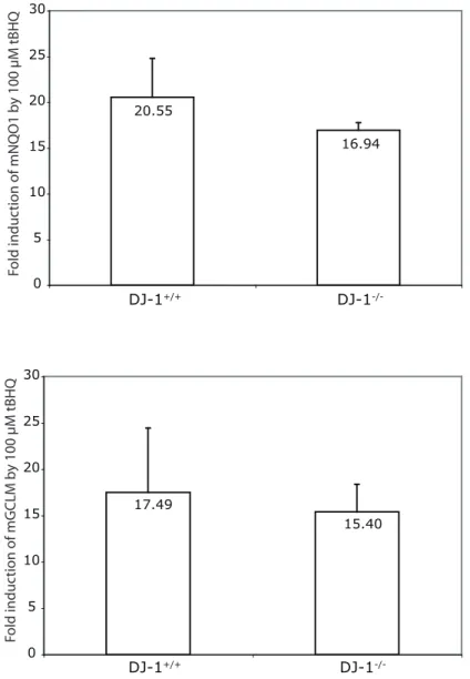
To use a more physiologic measurement, the effect of DJ-1 on the expression of Nrf2-regulated detoxification enzymes: NQO1 and Glutathione Cysteine Ligase, modifier subunit
(GCLM) was tested (Figure 2.10D). Based on the microarray analysis (Figure 2.4), siDJ-1 reduced GCLM expression by 1.478 fold, hence we selected it in addition to NQO1 for further analysis. Induction of MEF cultures with 25 M tBHQ led to a substantial increase of mNQO1 in DJ-1+/+ MEFs but this increase was drastically reduced in DJ-1-/- cells. This pattern is also found for mGCLM. However, at higher (100 μM) dosage, even though differences in mNrf2 protein
2.4 DISCUSSION:
In summary, this work describes functional effects of the DJ-1 protein via Nrf2, a master regulator of anti-oxidant gene responses. Cancer and PD lie at opposite ends of a spectrum defined by dysfunctions in cell death. Our finding may explain how DJ-1 plays an important role in both diseases. One of the hallmarks of PD is the loss of substantia nigra dopaminergic neurons, leading to motor deficits (Kish et al. 1988). DJ-1-/- mice did not exhibit widespread neuronal loss in a PD disease model (Goldberg et al. 2005; Kim et al. 2005), but these neurons were more susceptible to death following toxic insults (Kim et al. 2005). Likewise, human neuronal cell lines with DJ-1 knockdown are more sensitive to toxic compounds (Yokota et al. 2003; Taira et al. 2004). The loss of anti-oxidant gene transcription could account for these phenotypes that are only evident after environmental harm.
It is noteworthy that we initially identified DJ-1’s effect on Nrf2 in lung tumor cells. Studies of Nrf2 knockout mice show that it plays a significant role in lung biology (reviewed in (Cho et al. 2006)). In our studies we found that the H157 lung tumor cells did not consistently induce Nrf2 activity following tBHQ treatment, but instead had a very high basal level of activity that was not inducible by treatment (data not shown). High basal NQO1 expression allowed us to confidently quantify changes in NQO1 expression and implicated the broader effect of DJ-1 on Nrf2. In order to study gene induction, we then used liver cell line models, which are highly inducible. These models allowed us to first identify the effects of DJ-1 on Nrf2 that has heretofore remained unrecognized.
treatment targets in tumors. For example, NQO1, an obligate two-electron reductase, can reduce anti-tumor quinones leading to their bioactivation. Mitomycin C (MMC), and a novel anti-tumor compound, 2,5-diaziridinyl-3- (hydroxymethyl)-6-methyl-1,4-benzoquinone, are activated by NQO1 activity and NQO1 is shown to increase the efficacy of MMC in vivo (Begleiter et al. 2004). It is possible that tumors with high DJ-1 might be more susceptible to therapies that rely on
2.5 MATERIALS AND METHODS:
Cell culture, treatments, and plasmid constructs - Huh7 cells were grown in DMEM (Sigma) with 7% FCS. H157 cells were grown in RPMI-1640 (Gibco) +10% FCS. All mammalian cell culture was grown in the presence of penicillin and streptomycin to minimize contamination effects.
Tert-Butylhydroquinone (Fluka) was dissolved in DMSO (final concentration on cells of 0.0001%) and cells were treated for 18-24 hours. Dexamethasone and forskolin (MP Biochemicals) were dissolved in DMSO and ethanol respectively. Dexamethasone was used at a final
concentration of 100 M, and forskolin at 10 M. In experiments determining Nrf2 protein
stability, cells were treated with cyclohexamide (Sigma) in DMSO at a concentration of 75 g/mL for up to two (2) hours. The peptide proteosome inhibitor MG132 (Calbiochem), was used at 25 M for 4-6 hours for ubiquitination studies.
Other investigators generously provided flag-DJ-1 (Hod 2004), flag-Nrf2 (Furukawa et al. 2005), and hNQO1-ARE-pGL2 (Dhakshinamoorthy et al. 2000) plasmids. SV40-Luciferase [pGL3-control], GRE-Luciferase [pGRE-Luc] (Clontech), and CRE-Luciferase [pCRE-Luc] (Clontech) were all purchased from commercial sources. We directionally cloned human Keap1 into the V5/His containing pcDNA3.1D-Topo plasmid (Invitrogen) by amplifying the Keap1 ORF with the primers 5’ CACCATGCAGCCAGATCCCAGGCCTAGC 3’ and 5’
siRNA knockdown of DJ-1 – Cell lines were transfected with siDJ-1#1 5’
NNGACCCAGUACAGUGUAGCC 3’, siDJ-1#2 5’ NNUGGAGACGGUCAUCCCUGU 3’, scrambled control oligomer (Xeragon), siCONTROL (siCTL) non-targetting siRNA #1
(Dharmacon), or transfection reagent alone (siMock) using Oligofectamine (Invitrogen) for H157 cells, or Lipofectamine 2000 (Invitrogen) for Huh7 cells as per manufacturers protocols. Cells were transfected on consecutive days for 2-3 days in a row, and lysates were taken for RNA and protein analysis ninety-six (96) hours after the first transfection.
Generation of anti-DJ-1 antibody – DJ-1 was cloned into 6x Histidine-tagged E. coli over-expression vector, QE82L (Qiagen), by standard methodology. Expression of DJ-1 was induced with 1 mM IPTG (Isopropyl--D-Thiogalactopyranoside)in the E. coli strain BL21 (DE3). Cells were lysed in PBS plus EDTA-free protease inhibitor cocktail (Roche) and DJ-1 was purified to greater than 95% homogeneity with Ni-NTA (Qiagen) according to the manufacturer’s instructions. Recombinant DJ-1 was sent to Proteintech Group Inc. for the production of the anti-DJ-1 rabbit polyclonal serum.
Affymetrix GeneChip® analysis – Total RNA isolated from H157 cells, was DNAse I treated and column purified (Promega). The quality of the RNA was determined by formamide-agarose
(5X fragmentation buffer: 200mM Tris-acetate, pH8.1, 500mM KOAc, 150mM MgOAc) at 94oC for 35 minutes before the chip hybridization. 15 μg of fragmented cRNA was then added to hybridization cocktail (0.05 μg/μl fragmented cRNA, 50 pM control oligonucleotide B2, BioB,
BioC, BioD, and cre hybridization controls, 0.1 mg/ml herring sperm DNA, 0.5 mg/ml acetylated BSA, 100mM MES, 1M [Na+], 20mM EDTA, 0.01% Tween 20). Ten μg of cRNA was used for hybridization. Arrays were hybridized for 16 hours at 45oC in the GeneChip Hybridization Oven 640. The arrays were washed and stained with R-phycoerythrin streptavidin in the GeneChip Fluidics Station 400. After this, the arrays were scanned with the Hewlett Packard GeneArray Scanner. Affymetrix GeneChip Microarray Suite 5.0 software was used for washing, scanning, and basic analysis. Sample quality was assessed by examination of 3’ to 5’ intensity ratios of certain genes.
These data were then further analyzed, filtered, and compared using GeneSpring software (Silicon Genetics). Gene defined at ‘changed’ were filtered to include those differing greater than three (3) fold between both siCTL chips and siDJ-1 chips, with a raw fluorescence intensity of at least five hundred (500) in both of the highly-expressed (present) arrays. Both siDJ-1 arrays were transfected with siDJ-1 #1, and then verified by real-time PCR using both siDJ-1 #1 and siDJ-1 #2.
Realtime quantitative PCR – Reactions were carried out in an ABI 7900HT (Applied Biosciences) using 15 l, 384 well format and master-mixes from ABGene. Taqman PCR primer/probe sets were designed for human DJ-1: primer1 5’ CCATATGATGTGGTGGTTCTAC 3’, primer2 5’
ACTTCCACAACCTATTTCATGAG 3’, probe 5’
[6-FAM]ACATGGAGCCACTGCCACCA[Tamra-Q] 3’. SYBR green realtime PCR primers were designed for human Nrf2: primer1 5’ AGTGGATCTGCCAACTACTC 3’, primer2 5’
CATCTACAAACGGGAATGTCTG 3’. We used previously published mouse G3PDH primers designed to be used with SYBR green quantitation (Engelbrecht et al. 2003). Pre-designed Taqman PCR primer and probe sets were purchased from Applied Biosystems for mouse NQO1, and GCLM.
Luciferase reporter gene assays – Cells were grown and transfected, as described above, in 6-well plates (Falcon). Cultures were lysed in reporter lysis buffer (Promega) using a single round of freeze-thaw at -80 C. Luciferase assays were then performed as previously described (Piskurich et al. 1999).
Western blot analysis and immunoprecipitation – For all western blot analyses, cells were lysed in RIPA buffer (10 mM NaPO4 ph 7.4, 300 mM NaCl, 0.1% SDS, 1% NP-40, 1% deoxycholic acid, 2 mM EDTA) with protease inhibitors (Roche), diluted with SDS loading buffer and boiled in the presence of the reducing agent dithiothreitol (DTT). Proteins were then separated by molecular weight using SDS-PAGE through polyacrylimide gels ranging from 6% - 12%. Proteins were electrophoretically transferred to nitrocellulose membranes, and blocked using 5% non-fat dry milk in TBS with 0.1% Tween 20. Antibodies used for blotting were anti-Nrf2 H-300 (Santa Cruz Biotechnology), rabbit polyclonal DJ-1, Actin-HRP (Santa Cruz Biotechnology), anti-G3PDH, anti-Hemagglutanin (HA)-HRP (Roche Diagnostics), and anti-Flag (M2)-HRP (Sigma).
incubation with Protein A/G agarose (Pierce Biotechnologies). Protein A/G-antibody-protein complexes were washed extensively and eluted by boiling in loading buffer with reducing equivalents. Eluates and input lysate controls were then western blotted to assay for protein expression and isolation.
Ubiquitination assays were performed in Huh7 cells transfected with epitope tagged Nrf2 and Ubiquitin grown in 100 mm2 plates. The cells were lysed in 200 l SDS lysis buffer (50 mM Tris-HCl pH7.5, 0.5 mM EDTA, 1% SDS, 1 mM DTT) and boiled for 10 minutes. Cellular debris was pelleted and SDS concentrations were diluted by the addition of 1200 l 0.5% NP-40 lysis buffer with added protease inhibitors. Anti-Flag (M2) agarose was then added and incubated for 14-16 hours. The agarose matrix was washed extensively with 0.5% NP-40 lysis buffer and the proteins were eluted by boiling in 2x loading buffer with DTT. The eluates were then analyzed by western blot analysis for the expression of the epitope tags.
DJ-1 Knockout mice and embryonic fibroblast culture: DJ-1 knockout mice and wild type littermates (Kim et al. 2005), backcrossed 6 generations onto the C57BL6 strain were housed according to the guidelines of the National Institutes of Health under an approved IACUCprotocol at
G3PDH DJ-1
NTC siCTL siDJ-1 #1siDJ-1 #2
A
B
siCTL siDJ-1 #1 DJ-1
Actin
siDJ-1 #2 Figure 2.1
C
0 1000
siCTL siDJ1
#1 siDJ1 #2 siMOCK
Relative DJ-1
mRNA
rDJ-1
195.7 99.2 58.7 41.3
27.8 20.4 15.0
6.4
-mDJ-1
+/+
mDJ-1
-/-hDJ-1
+siCTL +siDJ-1 #1 +siDJ-1 #2
195.7 99.2 58.7 41.3
27.8 20.4 15.0
Figure 2.3.Summary of Affymetrix GeneChip analysis - Genes shown represent changes of greater than three (3) fold between siCTL and siDJ-1 transfected samples, where fluorescence in the present (P) state is greater than five hundred (500) in all samples. Green indicates decreased