Recent population studies suggest that gastroesophageal refl ux disease (GERD) is increasing in prevalence, both in the United States and worldwide ( 1,2 ). Th e diagnosis of GERD is associated with a 10–15% risk of Barrett’s esophagus (BE), a change of the normal squamous epithelium of the distal esophagus to a co-lumnar-lined intestinal metaplasia (IM). Risk factors associated with the development of BE include long-standing GERD, male gender, central obesity ( 3 ), and age over 50 years ( 4,5 ). Th e goal of a screening and surveillance program for BE is to identify in-dividuals at risk for progression to esophageal adenocarcinoma (EAC), a malignancy that has been increasing in incidence since the 1970s ( 6,7 ).
Th e purpose of this guideline is to review the defi nition and epidemiology of BE, available screening modalities for BE detec-tion, rationale and methods for surveillance, and available treat-ment modalities including medical, endoscopic, and surgical
techniques. In order to evaluate the level of evidence and strength of recommendations, we used the GRADE (Grading of Recom-mendations Assessment, Development and Evaluation) system ( 8 ). Th e level of evidence ranged from “high” (implying that fur-ther research was unlikely to change the authors’ confi dence in the estimate of the eff ect) to “moderate” (further research would be likely to have an impact on the confi dence in the estimate of eff ect) to “low” (further research would be expected to have an important impact on the confi dence in the estimate of the eff ect and would be likely to change the estimate) or “very low” (any estimate of eff ect is very uncertain). Th e strength of a recommendation was graded as “strong” when the desirable eff ects of an intervention clearly outweighed the undesirable eff ects and as “conditional” when there was uncertainty about the tradeoff s. We used meta-analyses or systematic reviews when available, followed by clinical trials and cohort and case–control studies. In order to determine the level
ACG Clinical Guideline: Diagnosis and Management of
Barrett’s Esophagus
Nicholas J. Shaheen , MD, MPH, FACG 1 , Gary W. Falk , MD, MS, FACG 2 , Prasad G. Iyer , MD, MSc, FACG 3 and
Lauren B. Gerson , MD, MSc, FACG 4
Barrett’s esophagus (BE) is among the most common conditions encountered by the gastroenterologist. In this document, the American College of Gastroenterology updates its guidance for the best practices in caring for these patients. These guidelines continue to endorse screening of high-risk patients for BE; however, routine screening is limited to men with refl ux symptoms and multiple other risk factors. Acknowledging recent data on the low risk of malignant progression in patients with nondysplastic BE, endoscopic surveillance intervals are attenuated in this population; patients with nondysplastic BE should undergo endoscopic surveillance no more frequently than every 3–5 years. Neither routine use of biomarker panels nor advanced endoscopic imaging techniques (beyond high-defi nition endoscopy) is recommended at this time. Endoscopic ablative therapy is recommended for patients with BE and high-grade dysplasia, as well as T1a esophageal adenocarcinoma. Based on recent level 1 evidence, endoscopic ablative therapy is also recommended for patients with BE and low-grade dysplasia, although endoscopic surveillance continues to be an acceptable alternative. Given the relatively common recurrence of BE after ablation, we suggest postablation endoscopic surveillance intervals. Although many of the recommendations provided are based on weak evidence or expert opinion, this document provides a pragmatic framework for the care of the patient with BE.
SUPPLEMENTARY MATERIAL is linked to the online version of the paper at http://www.nature.com/ajg
Am J Gastroenterol 2016; 111:30–50; doi: 10.1038/ajg.2015.322; published online 3 November 2015
1 Division of Gastroenterology and Hepatology, University of North Carolina at Chapel Hill , Chapel Hill , North Carolina , USA ; 2 Division of Gastroenterology, University
of Pennsylvania Perelman School of Medicine , Philadelphia , Pennsylvania , USA ; 3 Division of Gastroenterology and Hepatology, Mayo Clinic Minnesota , Rochester ,
Minnesota , USA ; 4 Division of Gastroenterology, California Pacifi c Medical Center and Department of Medicine, University of California, San Francisco , San
Francisco , California , USA . Correspondence: Nicholas J. Shaheen, MD, MPH, FACG, Division of Gastroenterology and Hepatology, University of North Carolina School of Medicine, University of North Carolina at Chapel Hill , CB 7080 , Chapel Hill , North Carolina 27599-7080 , USA . E-mail: [email protected] Received 19 March 2015 ; accepted 28 August 2015
of evidence, we entered data from the papers of highest evidence into the GRADE program (accessible at www.gradepro.org ). For each recommendation, a GRADE table was constructed, and the evidence rated. Recommendation statements were structured in the “PICO” format (patient population involved, intervention or Indicator assessed, comparison group, and patient-relevant out-come achieved) when possible. Th e aggregate recommendation statements are in Table 1 .
As part of this guideline preparation, a literature search was conducted using Ovid MEDLINE from 1946 to present, EMBASE 1988 to present, and SCOPUS from 1980 to present using major search terms and subheadings including “Barrett esophagus,” “Barrett oesophagus,” “epithelium,” “goblet cells,” “metaplasia,” “dysplasia,” “precancerous conditions,” “adenocarcinoma,” “radio-frequency,” “catheter ablation,” “early detection of cancer,” “mass screening,” and/or “esophagoscopy,” Th e full literature search strat-egy is demonstrated in Supplementary Appendix 1 online.
DIAGNOSIS OF BE
Recommendations
1 . BE should be diagnosed when there is extension of salmon-colored mucosa into the tubular esophagus extending ≥1 cm proximal to the gastroesophageal junction (GEJ) with biopsy confi rmation of IM (strong recommendation, low level of evidence).
2 . Endoscopic biopsy should not be performed in the presence of a normal Z line or a Z line with <1 cm of variability (strong recommendation, low level of evidence).
3 . In the presence of BE, the endoscopist should describe the extent of metaplastic change including circumferential and maximal segment length using the Prague classifi cation (conditional recommendation, low level of evidence). 4 . Th e location of the diaphragmatic hiatus, GEJ, and
squa-mocolumnar junction should be reported in the endoscopy report (conditional recommendation, low level of evidence). 5 . In patients with suspected BE, at least 8 random biopsies
should be obtained to maximize the yield of IM on histology. In patients with short (1–2 cm) segments of suspected BE in whom 8 biopsies may be unobtainable, at least 4 biopsies per cm of circumferential BE, and one biopsy per cm in tongues of BE, should be obtained (conditional recommendation, low level of evidence).
6 . In patients with suspected BE and lack of IM on histology, a repeat endoscopy should be considered in 1–2 years of time to rule out BE (conditional recommendation, very low level of evidence).
Summary of evidence
Establishing a diagnosis of BE . BE has been traditionally defi ned
as the presence of at least 1 cm of metaplastic columnar epithelium that replaces the stratifi ed squamous epithelium normally lining the distal esophagus. Th e reason why such segments <1 cm have been classifi ed as “specialized IM of the esophagogastric junction” (SIM-EGJ) and not BE is because of high interobserver variability,
as well as the low risk for EAC. Patients with SIM-EGJ have not demonstrated an increase in the development of dysplasia or EAC in large cohort studies aft er long-term follow-up, in contrast with patients with segments of IM >1 cm ( 9 ).
Th e defi nition of BE has varied depending upon the require-ment for the presence of IM on endoscopic biopsy. Th e presence of IM has traditionally been a requirement for the diagnosis of BE in the United States. On the other hand, guidelines from the United Kingdom have considered BE to be present if there was visual evi-dence of columnar-lined epithelium (CLE) on endoscopic exami-nation and biopsies demonstrated columnar metaplasia, regardless of the presence of IM ( 10 ). Th e debate regarding the requirement of IM on biopsy from CLE segments has derived from the appar-ently diff erential risk of developing EAC in CLE containing IM compared with non-IM CLE. Large population-based cohort stud-ies have demonstrated a substantially lower EAC risk in subjects with columnar metaplasia without IM compared with those with IM ( 11 ). However, not all studies have corroborated this fi nding ( 12 ). Although DNA content abnormalities appear to be compara-ble in both metaplastic epithelium without gocompara-blet cells compared with metaplastic epithelium with goblet cells, other studies sug-gest that cancer most commonly occurs in columnar metaplasia with goblet cells compared with columnar metaplasia without gob-let cells ( 11,13,14 ). Even if the rate of EAC is markedly higher in CLE containing IM, another complicating factor is sampling error leading to misclassifi cation of IM-containing CLE as non-IM CLE. Th e yield for IM correlates directly with the number of endoscopic biopsies obtained. In a large retrospective study, the yield for IM was 35% if 4 biopsies were obtained, and up to 68% aft er 8 biopsies were performed ( 15 ). Despite the incompletely elucidated risk of EAC in non-IM CLE, and acknowledging the potential for sam-pling error, we continue to suggest that only CLE containing IM be defi ned as BE, given the apparent diff erential cancer risk between CLE containing IM and CLE without IM. Until and unless fur-ther work substantiates a markedly elevated risk of EAC in non-IM CLE patients, it is unwise to give these patients a disease diagnosis that has a documented negative impact on insurance status and quality of life ( 16,17 ).
IM of cardia is very common, being described in up to 20% of asymptomatic subjects presenting for routine open access endo-scopic examinations ( 18 ). Studies have suggested that IM of the cardia is not more common in BE patients compared with con-trols ( 19 ), and that the natural history of IM at the EGJ is asso-ciated with Helicobacter pylori infection and not associated with EAC ( 20 ). Based on this information, biopsy of a normal or slightly irregular EGJ is not recommended.
Table 1 . Recommendation statements
Diagnosis of BE
1. BE should be diagnosed when there is extension of salmon-colored mucosa into the tubular esophagus extending ≥1 cm proximal to the gastroesopha-geal junction with biopsy confi rmation of IM (strong recommendation, low level of evidence).
2. Endoscopic biopsy should not be performed in the presence of a normal Z line or a Z line with <1 cm of variability (strong recommendation, low level of evidence).
3. In the presence of BE, the endoscopist should describe the extent of metaplastic change including circumferential and maximal segment length using the Prague classifi cation (conditional recommendation, low level of evidence).
4. The location of the diaphragmatic hiatus, gastroesophageal junction, and squamocolumnar junction should be reported in the endoscopy report (condi-tional recommendation, low level of evidence).
5. In patients with suspected BE, at least 8 random biopsies should be obtained to maximize the yield of IM on histology. In patients with short (1–2 cm) segments of suspected BE in whom 8 biopsies are unattainable, at least 4 biopsies per cm of circumferential BE, and one biopsy per cm in tongues of BE, should be taken (conditional recommendation, low level of evidence).
6. In patients with suspected BE and lack of IM on histology, a repeat endoscopy should be considered in 1–2 years of time to rule out BE (conditional recommendation, very low level of evidence).
Screening for BE
7. Screening for BE may be considered in men with chronic (>5 years) and/or frequent (weekly or more) symptoms of gastroesophageal refl ux (heartburn or acid regurgitation) and two or more risk factors for BE or EAC. These risk factors include: age >50 years, Caucasian race, presence of central obesity (waist circumference >102 cm or waist–hip ratio (WHR) >0.9), current or past history of smoking, and a confi rmed family history of BE or EAC (in a fi rst-degree relative) (strong recommendation, moderate level of evidence).
8. Given the substantially lower risk of EAC in females with chronic GER symptoms (when compared with males), screening for BE in females is not recommended. However, screening could be considered in individual cases as determined by the presence of multiple risk factors for BE or EAC (age >50 years, Caucasian race, chronic and/or frequent GERD, central obesity: waist circumference >88 cm, WHR >0.8, current or past history of smoking, and a confi rmed family history of BE or EAC (in a fi rst-degree relative)) (strong recommendation, low level of evidence).
9. Screening of the general population is not recommended (conditional recommendation, low level of evidence).
10. Before screening is performed, the overall life expectancy of the patient should be considered, and subsequent implications, such as the need for peri-odic endoscopic surveillance and therapy, if BE with dysplasia is diagnosed, should be discussed with the patient (strong recommendation, very low level of evidence).
11. Unsedated transnasal endoscopy (uTNE) can be considered as an alternative to conventional upper endoscopy for BE screening (strong recommenda-tion, low level of evidence).
12. If initial endoscopic evaluation is negative for BE, repeating endoscopic evaluation for the presence of BE is not recommended. If endoscopy reveals esophagitis (Los Angeles Classifi cation B, C, D), repeat endoscopic assessment after PPI therapy for 8–12 weeks is recommended to ensure healing of esophagitis and exclude the presence of underlying BE (conditional recommendation, low level of evidence).
Surveillance of BE
13. Patients should only undergo surveillance after adequate counseling regarding risks and benefi ts of surveillance (strong recommendation, very low level of evidence).
14. Surveillance should be performed with high-defi nition/high-resolution white light endoscopy (strong recommendation, low level of evidence).
15. Routine use of advanced imaging techniques other than electronic chromoendoscopy is not recommended for endoscopic surveillance at this time (conditional recommendation, very low level of evidence).
16. Endoscopic surveillance should employ four-quadrant biopsies at 2 cm intervals in patients without dysplasia and 1 cm intervals in patients with prior dysplasia (strong recommendation, low level of evidence).
17. Mucosal abnormalities should be sampled separately, preferably with endoscopic mucosal resection. Inability to perform endoscopic mucosal resection in the setting of BE with nodularity should lead to consideration to referral to a tertiary care center (strong recommendation, low level of evidence).
18. Biopsies should not be obtained in mucosal areas with endoscopic evidence of erosive esophagitis until after intensifi cation of antirefl ux therapy to induce mucosal healing (strong recommendation, very low level of evidence).
19. For BE patients with dysplasia of any grade, review by two pathologists, at least one of whom has specialized expertise in GI pathology, is warranted because of interobserver variability in the interpretation of dysplasia (strong recommendation, moderate level of evidence).
20. Use of additional biomarkers for risk stratifi cation of patients with BE is currently not recommended (strong recommendation, low level of evidence).
21. For BE patients without dysplasia, endoscopic surveillance should take place at intervals of 3 to 5 years (strong recommendation, moderate level of evidence).
22. Patients diagnosed with BE on initial examination do not require a repeat endoscopy in 1 year for dysplasia surveillance (conditional recommendation, very low level of evidence).
23. For patients with indefi nite for dysplasia, a repeat endoscopy after optimization of acid suppressive medications for 3–6 months should be performed. If the indefi nite for dysplasia reading is confi rmed on this examination, a surveillance interval of 12 months is recommended (strong recommendation, low level of evidence).
24. For patients with confi rmed low-grade dysplasia and without life-limiting comorbidity, endoscopic therapy is considered as the preferred treatment modality, although endoscopic surveillance every 12 months is an acceptable alternative (strong recommendation, moderate level of evidence).
Table 1 . Continued
25. Patients with BE and confi rmed high-grade dysplasia should be managed with endoscopic therapy unless they have life-limiting comorbidity (strong recommendation, high level of evidence).
Therapy
Chemoprevention
26. Patients with BE should receive once-daily PPI therapy. Routine use of twice-daily dosing is not recommended, unless necessitated because of poor control of refl ux symptoms or esophagitis (strong recommendation, moderate level of evidence).
27. Aspirin or NSAIDs should not be routinely prescribed to patients with BE as an antineoplastic strategy. Similarly, other putative chemopreventive agents currently lack suffi cient evidence and should not be administered routinely (conditional recommendation, high level of evidence).
Endoscopic therapy
28. Patients with nodularity in the BE segment should undergo endoscopic mucosal resection of the nodular lesion(s) as the initial diagnostic and therapeutic maneuver (see point 17 above). Histologic assessment of the EMR specimen should guide further therapy. In subjects with EMR specimens demonstrating HGD, or IMC, endoscopic ablative therapy of the remaining BE should be performed (strong recommendation, high level of evidence).
29. In patients with EMR specimens demonstrating neoplasia at a deep margin, residual neoplasia should be assumed, and surgical, systemic, or ad-ditional endoscopic therapies should be considered (strong recommendation, low level of evidence).
30. Endoscopic ablative therapies should not be routinely applied to patients with nondysplastic BE because of their low risk of progression to EAC (strong recommendation, very low level of evidence). Endoscopic eradication therapy is the procedure of choice for patients with confi rmed LGD, and confi rmed HGD, as noted above (see points 24 and 25).
31. In patients with T1a EAC, endoscopic therapy is the preferred therapeutic approach, being both effective and well tolerated (strong recommendation, moderate level of evidence).
32. In patients with T1b EAC, consultation with multidisciplinary surgical oncology team should occur before embarking on endoscopic therapy. In such patients, endoscopic therapy may be an alternative strategy to esophagectomy, especially in those with superfi cial (sm1) disease with a well-differentiated neoplasm lacking lymphovascular invasion, as well as those who are poor surgical candidates (strong recommendation, low level of evidence).
33. Routine staging of patients with nodular BE with EUS or other imaging modalities before EMR has no demonstrated benefi t. Given the possibility of over- and understaging, fi ndings of these modalities should not preclude the performance of EMR to stage-early neoplasia (Strong recommendation, moderate level of evidence).
34. In patients with known T1b disease, EUS may have a role in assessing and sampling regional lymph nodes, given the increased prevalence of lymph node involvement in these patients compared with less advanced disease (strong recommendation, moderate level of evidence).
35. In patients with dysplastic BE who are to undergo endoscopic ablative therapy for nonnodular disease, radiofrequency ablation is currently the preferred endoscopic ablative therapy (strong recommendation, moderate level of evidence).
Surgical therapy
36. Antirefl ux surgery should not be pursued in patients with BE as an antineoplastic measure. However, this surgery should be considered in those with incomplete control of refl ux symptoms on optimized medical therapy (strong recommendation, high level of evidence).
37. In cases of EAC with invasion into the submucosa, especially those with invasion to the mid or deep submucosa (T1b, sm2–3), esophagectomy, with consideration of neoadjuvant therapy, is recommended in the surgical candidate (strong recommendation, low level of evidence).
38. In patients with T1a or T1b sm1 adenocarcinoma, poor differentiation, lymphovascular invasion, or incomplete endoscopic mucosal resection should prompt consideration of surgical and/or multimodality therapies (strong recommendation, low level of evidence).
Management of BE after endoscopic therapy
39. Following successful endoscopic therapy and complete elimination of intestinal metaplasia (CEIM), endoscopic surveillance should be continued to detect recurrent IM and/or dysplasia (strong recommendation, low level of evidence).
40. Endoscopic surveillance following CEIM, for patients with HGD or IMC before ablation, is recommended every 3 months for the fi rst year following CEIM, every 6 months in the second year, and annually thereafter (conditional recommendation, low level of evidence).
41. In patients with LGD before ablation, endoscopic surveillance is recommended every 6 months in the fi rst year following CEIM, and annually thereafter (conditional recommendation, low level of evidence).
42. During endoscopic surveillance after CEIM, careful inspection of the tubular esophagus and gastroesophageal junction (in antegrade and retrograde views) should be performed with high-resolution white light imaging and narrow band imaging to detect mucosal abnormalities that may refl ect recurrent IM and/or dysplasia (strong recommendation, low level of evidence).
43. Treatment of recurrent metaplasia and/or dysplasia should follow guidelines for the treatment of metaplasia/dysplasia in BE before ablation (strong recommendation, low level of evidence).
44. Following CEIM, the goal of medical antirefl ux therapy should be control of refl ux as determined by absence of frequent refl ux symptoms (more than once a week) and/or esophagitis on endoscopic examination (conditional recommendation, very low level of evidence).
Endoscopic eradication therapy: training and education
45. Endoscopists who plan to practice endoscopic ablative procedures should additionally offer endoscopic mucosal resection (strong recommendation, very low level of evidence).
be lower than the EGJ in the majority of patients, translating to short segments of CLE without IM. In a comparative study of the two methods performed in Japan, investigators concluded that the proximal extent of the gastric folds was more accurate compared with the palisade vessels ( 22 ). Th e diaphragmatic hiatus is identi-fi ed as an indentation of the gastric folds that is apparent during upper endoscopy with inspiration.
Any segment of BE measuring >3 cm has been classifi ed as long-segment BE, with segments <3 cm classifi ed as short-seg-ment BE ( 23 ). It is recommended that a uniform classifi cation be used to facilitate diagnosis, but to date usage of a standard classifi -cation system has not been demonstrated to change patient man-agement. Th e Prague classifi cation, described initially in 2006, uses assessment of the circumferential and maximum extent of the endoscopically visualized BE segment as well as endoscopic landmarks ( Figure 1 ) ( 24 ). Applying this system prospectively, there were high reliability coeffi cients (RCs) for recognition of BE segments > 1 cm (RC 0.72), locations of the EGJ (RC 0.88), and diaphragmatic hiatus (RC 0.85), but not for BE segments <1 cm (RC 0.22). In addition to usage of the Prague classifi cation, it is recommended that all three landmarks, including the diaphrag-matic hiatus, EGJ, and squamocolumnar junction, be mentioned in every endoscopic report. Isolated islands of columnar mucosa were not included in the Prague classifi cation and should be reported separately in the endoscopy report. Th ere are no data to suggest that a confi rmatory endoscopic examination is of utility in 1 year aft er diagnosis, as long as a suffi cient number (up to 8) of biopsies are obtained during the initial examination from the Barrett’s segment ( 15 ). Th erefore, in situations where BE is suspected, we recommend acquiring 4 biopsies every 2 cm of segment length, or a total of at least 8 biopsies if the segment is <2 cm, at the initial exam.
In patients with suspected BE on endoscopy without con-fi rmation of IM despite adequate number of biopsies, a repeat examination could be considered in 1–2 years of time based on a longitudinal cohort study demonstrating that ∼ 30% of these
patients can be expected to demonstrate IM on a repeat examina-tion ( 25 ).
EPIDEMIOLOGY AND NATURAL HISTORY OF BE Summary statements
What are the risk factors for BE?
1 . Th e known risk factors for the presence of BE include the following:
a . Chronic (>5 years) GERD symptoms b . Advancing age (>50 years)
c . Male gender d . Tobacco usage e . Central obesity f . Caucasian race
2 . Alcohol consumption does not increase risk of BE. Wine drinking may be a protective factor.
3 . BE is more common in fi rst-degree relatives of subjects with known BE.
What are the risk factors associated with dysplasia and develop-ment of EAC in patients with BE?
1 . Th e known risk factors for the development of neoplasia in BE include:
a . Advancing age b . Increasing length of BE c . Central obesity d . Tobacco usage
e . Lack of nonsteroidal anti-infl ammatory agent use f . Lack of PPI use
g . Lack of statin use.
What is the cancer risk in BE, based on degree of dysplasia?
1 . Th e risk of cancer progression for patients with nondysplastic is ∼ 0.2–0.5% per year.
2 . For patients with low-grade dysplasia (LGD) the annual risk of progression to cancer is ∼ 0.7% per year.
3 . For patients with high-grade dysplasia (HGD), the annual risk of neoplastic progression is ∼ 7% per year.
4 . Th e majority (>90%) of patients diagnosed with BE die of causes other than EAC.
Summary of evidence
Risk factors for BE . BE has been detected in ∼ 15% of patients
with chronic GERD ( 26 ) and in ∼ 1–2% of population subjects ( Table 2 ) ( 27,28 ). In a population-based study from Sweden, the authors found that severe and chronic GERD were risk factors for the development of EAC; however, 40% of the cohort with esophageal cancer reported no prior history of GERD symp-toms ( 29 ). In subjects with GERD, symptom duration has been shown to be a risk factor for the presence of BE. In a cohort study Figure 1 . Illustration of Prague Classifi cation for Barrett’s esophagus (BE)
where C indicates circumferential extent of metaplasia and M indicates maximal extent of metaplasia. Schema shows a C2M5 segment with identifi cation of the gastroesophageal junction (GEJ) below the squamo-columnar junction. Reprinted with permission ( 24 ).
8
6
Distance (cm) from GEJ
Maximal extent of metaplasia: M = 5.0 cm
Circumferential extent of metaplasia: C = 2.0 cm
True position of GEJ: Origin = 0.0 cm 4
2
of onset of GERD symptoms may also be associated with BE. In a VA study, patients reporting frequent (defi ned as at least weekly) GERD symptoms starting before the age of 30 years had the high-est risk of BE (OR 15.1, 95% CI 7.91–28.8), and risk increased linearly with earlier age at onset of symptoms ( P =0.001). Th e risk of BE also increased with cumulative GERD symptom duration ( P =0.002) ( 32 ).
Male gender has been consistently identifi ed as a risk factor for BE and EAC. A meta-analysis demonstrated an overall pooled male/ female ratio of 2:1 (95% CI 1.8–2.2) ( 33 ). Th e risk of development of EAC is also signifi cantly higher in men. In a study using the SEER (Th e Surveillance, Epidemiology, and End Results) database, women composed only 12% of all EACs. In this study, the risk of EAC in women with GERD symptoms was approximately equivalent to the risk of breast cancer in men (3.9 per 100,0000 at age 60 years) ( 34 ).
Tobacco usage has been demonstrated to be a risk factor for BE in a recent meta-analysis based on 39 studies and 7,069 BE examining duration of GERD symptoms and risk for BE ( 30 ), 77
(11%) of 701 patients with GERD symptoms were found to have BE on upper endoscopy. Compared with patients with GERD symptoms for <1 year, the odds ratio (OR) for BE increased to 3.0 (95% confi dence interval (CI) 1.2–8.0) and 6.4 (95% CI 2.4–17.1) when symptoms were present for >5 and >10 years, respectively. A meta-analysis further demonstrated that the OR for the associ-ation of GERD symptoms and BE was 2.9 (95% CI 1.9–4.5) with signifi cant heterogeneity between studies. When stratifi ed by length of BE, the heterogeneity resolved, demonstrating a strong association between GERD and long-segment BE (OR 4.9, 95% CI 2–12) but no association with short-segment BE (OR 1.2, 95% CI 0.8–1.7) ( 31 ).
Increasing age is a risk factor for BE. In a retrospective study using the CORI (Clinical Outcomes Research Initiative) data-base, the yield of BE in white men with GERD was 2% in the third decade of life, but increased to 9% in the sixth decade ( 4 ). Early age
Table 2 . Risk factors for BE (estimates drawn from meta-analyses where available)
Risk factor OR (95% CI) Reference
Age (per 10-year increment) 1.53 (1.05–2.25) Rubenstein et al. ( 5 ) a
1.96 (1.77–2.17) Cook et al. ( 33 )
Race/ethnicity
AA vs. Caucasian ethnicity 0.34 (0.12–0.97) Abrams et al. ( 49 )
Hispanic vs. Caucasian ethnicity 0.38 (0.18–0.84) Abrams et al. ( 49 ) b
Hispanic vs. Caucasian ethnicity 1.1 (0.4–2.7) Keyashian et al. ( 50 ) c
GERD symptoms
Frequency (weekly vs. less frequent) 2.33 (1.34–4.05) Rubenstein et al. ( 5 ) a
Duration (>5 years vs. <1 year) 3.0 (1.2–8.0) Lieberman et al. ( 30 )
Age of onset (weekly symptoms, <30 years vs. later) 31.4 (13.0–75.8) Thrift et al. ( 32 )
Obesity
Overall 1.98 (1.52–2.57) Singh et al. ( 3 ) d
Increased WC 1.58 (1.25–1.99) Singh et al. ( 3 )
Increased WHR 2.04 (1.49–2.81) Singh et al. ( 3 )
Smoking
Current/past use vs. never 1.44 (1.20–1.74) Andrici et al. ( 35 )
Pack years of cigarette use 1.99 (1.21–3.29) Cook et al. ( 196 )
Family history
(BE, EAC, or GEJAC in fi rst- or second-degree relative) 12.23 (3.34–44.76) Chak et al. ( 42 )
Hiatal hernia (overall) 3.94 (3.02–5.13) Andrici et al. ( 197 )
Short-segment BE 2.87 (1.75–4.7) Andrici et al. ( 197 )
Long-segment BE 12.67 (8.33–19.25) Andrici et al. ( 197 )
AA, African American; BE, Barrett’s esophagus; CI, confi dence interval; EAC, esophageal adenocarcinoma; GEJAC, gastroesophageal junction adenocarcinoma; GERD, gastroesophageal refl ux disease; OR, odds ratio; WC, waist circumference; WHR, waist–hip ratio.
a In men only.
b In Hispanics from Dominican Republic.
c In Hispanics from Mexico.
Table 3 . Cancer risk based on degree of dysplasia
Dysplasia type Studies/patients Incidence 95% CI References
ND to EAC 57 Studies, 11,434 patients 50 Studies, 14,109 patients
3.3/1,000 person-years 6.3/1,000 person-years
2.8–3.8 4.7–8.4
( 60 ) ( 65 )
ND to EAC or HGD 602 patients 4.8/1,000 person-years 0.3–7.8 ( 198 )
LGD to EAC 24 Studies, 2,694 patients 5.4/1,000 person-years 3–8 ( 61 )
LGD to EAC or HGD 17 Studies, 1,064 patients 173/1,000 person-years 100–250 ( 61 )
HGD to EAC 4 Studies, 236 patients 7/100 patient-years 5–8 ( 62 )
CI, confi dence interval; EAC, esophageal adenocarcinoma; HGD, high-grade dysplasia; LGD, low-grade dysplasia; ND, nondysplastic.
patients. Any smoking during a patient’s lifetime was associated with a greater risk for BE compared with non-GERD controls (OR 1.4, 95% CI 1.2–1.7), but not when compared with patients with chronic GERD (OR 1.2, 95% CI 0.8–1.9), suggesting that the increased risk of BE associated with tobacco usage may be medi-ated via increasing GERD ( 35 ).
In contrast to tobacco usage, alcohol consumption has not been demonstrated to be signifi cantly associated with the risk for devel-opment of BE ( 36,37 ). In fact, there are data suggesting a possible protective eff ect of wine consumption, with ORs ranging from 0.44 (95% CI 0.2–0.99) to 0.71 (95% CI 0.52–0.98) ( 37,38 ).
Th e presence of obesity is an independent risk factor for BE and EAC ( 39 ). However, it appears that a central pattern of obe-sity, rather than overall body fat content (measured by BMI), is the primary risk factor for BE. In a meta-analysis ( 3 ), patients with central adiposity had a higher risk for BE compared with patients with normal body habitus (OR 2.0, 95% CI 1.5–2.6) and this rela-tionship persisted aft er adjustment for BMI and GERD, suggesting a refl ux independent role for central obesity in BE pathogenesis. Indeed, overall body fat content is not associated with BE risk ( 40 ). Central obesity is a risk factor for BE in both men and women ( 41 ).
Th e presence of a family history of BE has been identifi ed as another potential risk factor for BE ( 42 ). A cohort study demon-strated that BE was markedly more common in fi rst- or second-degree relatives of subjects with BE compared with controls (24% vs. 5%, P <0.005). Aft er adjusting for age, gender, and body mass index, the presence of family history was strongly associated with BE (OR 12, 95% CI 3.3–44.8) ( 42 ). In a subsequent study, endo-scopic screening was off ered to fi rst-degree previously uninvesti-gated relatives of subjects with BE. Th e overall diagnostic yield was 20% ( 43 ). Single-nucleotide polymorphisms on gene loci, which may confer increased susceptibility to BE development, have recently been described ( 44–47 ).
Caucasian race appears to be a strong risk factor for BE. Although the evidence for lower prevalence of BE in African Americans compared with Caucasians is consistent ( 48,49 ), the results of studies comparing BE incidence in Hispanics and non-Hispanic whites are inconsistent, likely refl ecting the heterogeneity of the Hispanic population ( 49,50 ).
Other risk factors for BE have also been reported. Disease conditions such as metabolic syndrome ( 51 ), type 2 diabetes
mellitus ( 52 ), and sleep apnea ( 53 ) have been identifi ed as poten-tial BE risk factors. H. pylori infection, particularly infection with Cag A+ strains, is associated with a decreased risk of BE in some studies ( 54,55 ).
Risk factors associated with dysplasia and EAC in patients with BE . Advancing age and increasing BE segment length are known risk factors for the presence of dysplasia in patients with BE. In a multicenter study of 309 BE patients (5 with cancer, 11 with HGD, and 29 with LGD), the risk factors for prevalent dysplasia included age (3.3% increase in dysplasia per year and BE segment length over 3 cm (risk increase of 14% per cm of BE present) ( 56 ).
In patients with known BE, a variety of medications have been associated with reduced risk of progression to dysplasia and/or esophageal cancer including proton pump inhibitors (PPIs), aspi-rin, nonsteroidal anti-infl ammatory agents, and statins. A meta-analysis based on 7 studies with 2,813 patients demonstrated a 71% reduced risk of HGD and/or EAC with PPI users (OR 0.3, 95% CI 0.1–0.8). No signifi cant eff ect was shown for H 2 RA usage in two studies ( 57 ). In another meta-analysis of 9 observational studies of 5,446 participants (605 with HGD or EAC), usage of cyclooxyge-nase inhibitors, aspirin, and nonaspirin cyclooxygecyclooxyge-nase inhibitors was associated with reduced risk for HGD and EAC independent of duration of therapy ( 58 ). By means of their antiproliferative, proa-poptotic, antiangiogenic, and immunomodulatory eff ects, statins may prevent cancer development and growth. In a meta-analysis of 5 studies including 2,125 BE patients (312 EAC cases), statin usage was associated with a 41% reduction in EAC risk (adjusted OR 0.6, 95% CI 0.45–0.78) with the number needed to treat of 389 to prevent 1 case of EAC ( 59 ).
Cancer risk in BE based on degree of dysplasia . A recent
meta-analysis published in 2012 demonstrated lower risk for progres-sion of nondysplastic BE than previously reported ( Table 3 ) ( 60 ). It included 57 studies and demonstrated that the pooled annual incidence of EAC was 0.33% (95% CI 0.28–0.38%). In patients with short-segment BE reported from 16 studies, the annual can-cer risk was 0.19%.
implications, such as the need for periodic endoscopic surveillance and therapy, if BE with dysplasia is diagnosed, should be discussed with the patient (strong recommenda-tion, very low level of evidence).
11 . Unsedated transnasal endoscopy (uTNE) can be considered as an alternative to conventional upper endoscopy for BE screening (strong recommendation, low level of evidence). 12 . If initial endoscopic evaluation is negative for BE, repeating
endoscopic evaluation for the presence of BE is not recom-mended. If endoscopy reveals esophagitis (Los Angeles Classifi cation B, C, D), repeat endoscopic assessment aft er PPI therapy for 8–12 weeks is recommended to ensure heal-ing of esophagitis and exclude the presence of underlyheal-ing BE (conditional recommendation, low level of evidence).
Summary of evidence
Survival of subjects diagnosed with EAC with regional or distant disease remains dismal, at <20% at 5 years ( 7 ). Th e concept of metaplasia–dysplasia–carcinoma progression sequence in BE has led to the hypothesis that screening for BE, institution of endoscopic surveillance to detect dysplasia, followed by endo-scopic intervention, will lead to a decreased incidence of EAC ( 66 ). In addition to detecting BE, screening also detects preva-lent dysplasia or carcinoma that may be treated with endoscopic therapy. Th e available evidence to support this hypothesis, however, consists of retrospective studies that may be subject to biases. Indeed, >90% of EACs are diagnosed in patients without a prior BE diagnosis, despite the increasing use of endoscopy ( 67,68 ).
Given the number of patients involved, a widely embraced population screening eff ort could lead to substantial economic costs (from diagnostic tests and need for subsequent surveillance). Economic modeling studies ( 69 ) have found BE screening (done by endoscopy) followed by surveillance in hypothetical popula-tions (50-year-old male subjects with GERD symptoms) to be cost eff ective, with acceptable incremental cost-eff ectiveness ratios ranging from $10,000 to 50,000/quality-adjusted life-year gained ( 70,71 ). Estimates vary among studies, likely because of diff er-ences in assumptions ( Supplementary Table S1 ). Th ree of these studies found that screening with video capsule endoscopy ( 72,73 ) or uTNE ( 74 ) was cost eff ective compared with no screening, but that standard endoscopy was preferred over capsule endoscopy. All assumed participation rates of almost 100% and accuracy rates of 100%. Th is is likely an overestimate with lower participation rates (18–49%) ( 75–77 ), and lower accuracy rates for endoscopy (80%) being reported in prior studies ( 78 ). Of note, a substantial propor-tion of BE diagnoses in the community are reversed, likely because of incorrect landmark identifi cation and incorrect targeting of biopsies ( 79 ). In addition, the yield of a repeat endoscopy following an initial negative endoscopy for BE is low (2.3%), with esophagitis and male gender being predictors of BE being diagnosed at sub-sequent endoscopy ( 80 ). However, studies report a BE prevalence of 9–12% on repeat endoscopy following treatment of esophagitis with PPIs, making a repeat endoscopy aft er healing of more severe erosive esophagitis advisable ( 81,82 ).
or EAC combined ( 61 ). However, there was considerable hetero-geneity in these results and when stratifi ed by the LGD/BE ratio as a surrogate for pathology quality, the incidence rate for EAC was 0.76% per year for a ratio of <0.15 and 0.32% per year for a ratio of >0.15. Th is fi nding suggests that in settings where the diagnosis of LGD is made more liberally, and perhaps overcalled, there is a lower risk of progression.
Th e risk of EAC for patients with HGD was examined in a meta-analysis of 4 studies and 236 patients. Th e weighted annual incidence rate was 7% (95% CI 5–8) ( 62 ). However, the AIM-Dys-plasia trial that randomized 127 patients with dysAIM-Dys-plasia to ablation therapy compared with surveillance reported a much higher yearly progression rate of 19% in the HGD surveillance arm ( 63 ). Th is rate is similar to a second randomized trial that also required con-fi rmation of HGD by a second expert pathologist, again suggesting that the rigor with which the histology is validated likely predicts the subsequent EAC risk ( 64 ).
What are the common causes of death in subjects with BE?
Most BE patients die of other causes than EAC. A meta-analysis reported mortality rates from 19 studies in 7,930 patients ( 65 ). Th ere were 88 deaths because of EAC and 1,271 deaths because of other causes, resulting in a pooled incidence rate of fatal EAC of 3/1,000 person-years (95% CI 2–4). In 12 studies reporting cause-specifi c mortality, 7% of deaths (64/921) were from EAC, and 93% (857/921) because of other causes. Th e most common causes in-cluded cardiac disease in 35%, followed by pulmonary disease in 20% and other malignancies in 16% of the cohort.
SCREENING FOR BE
Recommendations
7 . Screening for BE may be considered in men with chronic (>5 years) and/or frequent (weekly or more) symptoms of gastroesophageal refl ux (heartburn or acid regurgitation) and two or more risk factors for BE or EAC. Th ese risk factors include: age >50 years, Caucasian race, presence of central obesity (waist circumference >102 cm or waist–hip ratio >0.9), current or past history of smoking, and a confi rmed family history of BE or EAC (in a fi rst-degree relative) (strong recommendation, moderate level of evidence). 8 . Given the substantially lower risk of EAC in females with
chronic GER symptoms (when compared with males), screening for BE in females is not recommended. However, screening could be considered in individual cases as de-termined by the presence of multiple risk factors for BE or EAC (age >50 years, Caucasian race, chronic and/or fre-quent GERD, central obesity: waist circumference >88 cm, waist–hip ratio >0.8, current or past history of smoking, and a confi rmed family history of BE or EAC (in a fi rst-degree relative)). (strong recommendation, low level of evidence). 9 . Screening of the general population is not recommended
(conditional recommendation, low level of evidence). 10 . Before screening is performed, the overall life expectancy
SURVEILLANCE OF BE
Recommendations
13 . Patients should only undergo surveillance aft er adequate counseling regarding risks and benefi ts of surveillance (strong recommendation, very low level of evidence). 14 . Surveillance should be performed with high-defi
nition/high-resolution white light endoscopy (strong recommendation, low level of evidence).
15 . Routine use of advanced imaging techniques other than electronic chromoendoscopy is not recommended for endo-scopic surveillance at this time (conditional recommenda-tion, very low level of evidence).
16 . Endoscopic surveillance should employ four-quadrant biop-sies at 2 cm intervals in patients without dysplasia and 1 cm intervals in patients with prior dysplasia (strong recommen-dation, low level of evidence).
17 . Mucosal abnormalities should be sampled separately, prefer-ably with endoscopic mucosal resection (EMR). Inability to perform EMR in the setting of BE with nodularity should lead to referral to a tertiary care center (strong recommenda-tion, low level of evidence).
18 . Biopsies should not be obtained in mucosal areas with endo-scopic evidence of erosive esophagitis until aft er intensifi ca-tion of antirefl ux therapy to induce mucosal healing (strong recommendation, very low level of evidence).
19 . For BE patients with dysplasia of any grade, review by two pathologists, at least one of whom has specialized expertise in gastrointestinal (GI) pathology, is warranted because of interobserver variability in the interpretation of dysplasia (strong recommendation, moderate level of evidence). 20 . Use of additional biomarkers for risk stratifi cation of patients
with BE is currently not recommended (strong recommenda-tion, low level of evidence).
21 . For BE patients without dysplasia, endoscopic surveillance should take place at intervals of 3 to 5 years (strong recom-mendation, moderate level of evidence).
22 . Patients diagnosed with BE on initial examination with adequate surveillance biopsies do not require a repeat endo-scopy in 1 year for dysplasia surveillance (conditional recommendation, very low level of evidence).
23 . For patients with indefi nite for dysplasia, a repeat endoscopy aft er optimization of acid suppressive medications for 3–6 months should be performed. If the indefi nite for dysplasia reading is confi rmed on the repeat examination, a surveil-lance interval of 12 months is recommended (strong recom-mendation, low level of evidence).
24 . For patients with confi rmed LGD and without life-limiting comorbidity, endoscopic therapy is considered as the preferred treatment modality, although endoscopic surveil-lance every 12 months is an acceptable alternative (strong recommendation, moderate level of evidence).
25 . Patients with BE and confi rmed HGD should be managed with endoscopic therapy unless they have life-limiting comorbidity (strong recommendation, high level of evidence).
BE screening has several challenges. Although symptomatic GERD is a risk factor for BE and EAC, it is neither a sensitive nor specifi c marker ( 29,31 ). Only 5–15% of subjects with chronic (>5 years) and frequent (weekly or more frequent) refl ux have BE ( 83 ), and as many as 50% of subjects with BE or EAC do not report chronic refl ux symptoms ( 31,84 ). Several studies have reported a substantial prevalence of BE in those without refl ux symptoms ( 27,85,86 ). Indeed, although refl ux symptoms are associated with long-segment BE, they may not be consistently associated with short-segment BE ( 31 ). Hence, a BE screening strategy based solely on GERD symptoms is likely to be unsuccessful. Women (even those with daily or weekly refl ux symptoms) have a low inci-dence of EAC comparable to that of men without refl ux symptoms ( 34 ). Th is may relate to the lower risk of progression to EAC in women with BE compared with men with BE ( 60,87 ) and should likely infl uence the threshold of BE screening in women.
Recent reports have described the creation of prediction or risk scores for BE using a combination of risk factors ( 5,88 ). Th is may enable the synthesis of multiple risk factors into a single clinically applicable parameter and make BE screening more effi cient by targeting a high-risk target population. Accuracy for BE predic-tion, though improved from GERD-only models, remains modest (area under the curve 0.73–81), but is likely to be improved by the addition of other variables such as circulating cytokine levels ( 89 ). Validation in larger unselected populations will be critical before widespread use.
Summary of the evidence
Rationale for surveillance . Survival in EAC is stage dependent
and early spread before the onset of symptoms is characteristic of this tumor. Lymph node metastases are a clear prognostic factor for decreased survival ( 95 ). Th us, the best hope for improved sur-vival of patients with EAC remains detection of cancer at an early and potentially curable stage.
A number of observational studies suggest that patients with BE in whom EAC was detected in a surveillance program have their cancers detected at an earlier stage with markedly improved survival compared with similar patients not undergoing routine endoscopic surveillance ( 96–99 ). Furthermore, nodal involvement is far less likely in surveyed patients compared with nonsurveyed patients. As esophageal cancer survival is stage dependent, these studies suggest that survival may be enhanced by endoscopic surveillance. Recent work from a large Dutch population-based cohort study confi rmed that there is a survival advantage for EAC in patients who received adequate endoscopic surveillance com-pared with patients who were not participating in endoscopic surveillance ( 100 ). Similarly, a large Northern Ireland population-based study found that in patients with EAC and a prior diagnosis of BE, survival was enhanced, tumor stage was lower, and tumor grade was lower compared with patients without a prior diagno-sis ( 101 ). Importantly, these fi ndings were maintained, although attenuated, aft er attempting to correct for both lead time and length time bias. On the other hand, a case–control study from the Northern California Kaiser Permanente population found no evi-dence that endoscopic surveillance improved survival from EAC ( 102 ). Although there are no prospective clinical trial data that demonstrate a benefi t of endoscopic surveillance, the considerable heterogeneity of available evidence makes it prudent to continue to perform endoscopic surveillance of BE patients.
It is important to recognize, however, that endoscopic surveil-lance, as currently practiced, has numerous shortcomings. Dys-plasia may not be visible endoscopically and the distribution of dysplasia and cancer is highly variable. Even the most thorough biopsy surveillance program has the potential for sampling error. Current surveillance programs are expensive and time consuming. It is well known that adherence to practice guidelines is problem-atic at best and worsens with longer segment lengths ( 103 ). All of these shortcomings likely diminish any benefi t from these pro-grams, and eff orts to adhere to published standards for the per-formance of various elements of surveillance are recommended.
Counseling for surveillance . Before entering into a
surveil-lance program, patients should be counseled about the risks and benefi ts of this program, including the limitations of surveillance endoscopy as well as the importance of adhering to appropriate surveillance intervals. Other considerations include age, likeli-hood of survival over the next 5 years, and ability to tolerate interventions including endoscopic therapy, surgery, and medical or radiation oncologic treatments for EAC.
Until recently, the concept of early outpatient consultation to review the signifi cance of BE has not been a point of emphasis in prior practice guidelines ( 10 ). Why is this important? First, wide
access to the Internet allows patients to obtain information about BE and EAC in an unfi ltered manner. Studies to date suggest that patients both over- and under-estimate their cancer risk ( 16,104 ). Given the low risk of progression to cancer for most patients with BE and the data suggesting that most BE patients die of causes other than EAC, such counseling should now be part of the ongo-ing care of these patients to help inform decision makongo-ing regardongo-ing therapeutic options ( 65 ).
Surveillance technique . Endoscopic surveillance should utilize
high-resolution/high-defi nition white light endoscopy to opti-mize visualization of mucosal detail. Recent work suggests that this is superior to standard-defi nition white light endoscopy for the detection of dysplastic lesions ( 105 ). Th is should be accom-panied by removal of any mucosal debris and careful insuffl ation and desuffl ation of the lumen. Part of the examination should also incorporate a retrofl exed view of the GEJ. Data demonstrate a di-rect correlation between inspection time of the Barrett’s segment and detection of patients with HGD/EAC ( 106 ). Inspection of the Barrett’s segment should also involve careful attention to the right hemisphere of the segment, extending from the 12 o’clock to 6 o’clock location where early cancer appears to have a predilection to develop ( 107,108 ).
Th e aim of surveillance is detection of dysplasia. Th e descrip-tion of dysplasia should use a standard fi ve-tier system: (i) nega-tive for dysplasia, (ii) indefi nite for dysplasia, (iii) LGD, (iv) HGD, and (v) carcinoma ( 109 ). Active infl ammation makes it more dif-fi cult to distinguish dysplasia from reparative changes. As such, surveillance biopsies should only be performed aft er any active infl ammation related to GERD is controlled with antisecretory therapy. Th e presence of ongoing erosive esophagitis is a relative contraindication to performing surveillance biopsies. Once any infl ammation related to GERD is controlled with antisecretory therapy, systematic four-quadrant biopsies at 2 cm intervals along the entire length of the Barrett’s segment remains the standard for endoscopic surveillance of nondysplastic BE.
the most advanced histology found on the combination of targeted and random biopsies.
Advanced endoscopic imaging techniques . A wide variety of
enhancements to endoscopic imaging with white light endoscopy have been studied in recent years to allow for detailed inspection of the Barrett’s segment. Electronic chromoendoscopy allows for detailed imaging of the mucosal and vascular surface patterns in BE without the need for chromoendoscopy dye sprays. Th is may be accomplished with either narrow band imaging that uses opti-cal fi lters to narrow the band width of white light to blue light or by postprocessing soft ware systems to accomplish similar visu-alization. Most of the published literature to date have examined narrow band imaging in conjunction with magnifi cation endos-copy. A randomized clinical trial of narrow band imaging vs. high-defi nition white light endoscopy demonstrated no diff erence in the number of patients detected with dysplasia or neoplasia. However, fewer biopsies were required for narrow band imaging ( 119 ). A recent meta-analysis also suggests that electronic chro-moendoscopy may increase detection of dysplasia ( 120 ). A wide variety of other image enhancement techniques have been stud-ied including methylene blue staining, acetic acid staining, indigo carmine staining, autofl uorescence endoscopy, confocal laser en-domicroscopy, volumetric laser enen-domicroscopy, spectroscopy, and molecular imaging, but none of these methods appear ready for widespread clinical use at present.
Importance of confi rmation of dysplasia . Dysplasia remains
the best clinically available marker of cancer risk in patients with BE. However, there is considerable interobserver variability in the interpretation of dysplasia in both the community and academic settings. Th at being said, there is reasonable interobserver agree-ment among GI pathologists for the extremes of dysplasia, namely IM without dysplasia and HGD/EAC ( 109 ). Th ere is considerably more diffi culty in the interpretation of indefi nite for dysplasia and LGD ( 121 ). Th e importance of the confi rmation of the diagnosis of LGD comes from two recent studies from the Netherlands. Review by two GI pathologists, with extensive experience in the diagnosis of BE-related neoplasia, found that of 147 patients diag nosed with LGD in the community, 85% of the patients were downgraded to a diagnosis of no dysplasia ( 122 ). Further work by that group examined 293 additional patients with LGD diagnosed in the community who had biopsies reviewed by at least 2 GI pathologists and 73% of the cases were downgraded to indefi -nite for dysplasia or nondysplastic BE ( 123 ). Other studies sug-gest that community-based pathologists have diffi culties in the interpretation of both nondysplastic BE and dysplasia ( 124 ). Th erefore, current evidence supports the importance of having all readings of dysplasia confi rmed by a second pathologist with extensive experience in the interpretation of Barrett’s associated neoplasia.
Surveillance intervals . Surveillance intervals are determined by
the presence and grade of dysplasia and are currently governed by expert opinion. Given the low risk of progression of BE to
EAC, surveillance at 3- to 5-year intervals remains reasonable in patients without dysplasia.
Th ere is a paucity of data to guide the management of BE patients with biopsies indefi nite for dysplasia. It is reasonable to use double-dose PPI therapy to decrease any ongoing infl am-mation. A retrospective study found that indefi nite for dyspla-sia was associated with a similar risk of progression to cancer as was LGD ( 125 ). More recent data suggest an especially high risk of progression to higher grades of dysplasia within the fi rst year of diagnosis but a risk comparable to nondysplastic BE aft er the fi rst year ( 126 ). Th e progression risk may be more pronounced in multifocal indefi nite for dysplasia (defi ned as indefi nite for dysplasia in biopsies from more than one level of the esophagus) than in focal indefi nite for dysplasia ( 127 ). Th us, surveillance in these patients should follow the recommendations for LGD as described below.
If LGD is found, the diagnosis should fi rst be confi rmed by a second pathologist with expertise in BE. Th ese patients should also receive aggressive antisecretory therapy for refl ux disease with a PPI to decrease the changes associated with regeneration or infl ammation. A repeat endoscopy aft er optimization of acid sup-pressant therapy may result in downgrading of the LGD reading. If LGD is confi rmed and endoscopic therapy not performed, annual surveillance is recommended until two examinations in a row are negative for dysplasia, aft er which time surveillance intervals for nondysplastic BE can be followed. A protocol of four-quadrant biopsies at 1 cm intervals is advisable, given that anatomic studies suggest that dysplasia can occur in a mosaic pattern and involve small portions of the overall surface area of the esophagus. EMR should be performed if any mucosal abnormality is present in these patients.
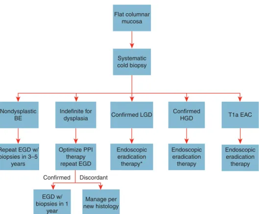
If HGD is found, the diagnosis should fi rst be confi rmed by a second pathologist with experience in GI pathology. Th e presence of any mucosal abnormality warrants EMR in an eff ort to maximize staging accuracy. If HGD is confi rmed, endoscopic intervention is warranted as described below. Figure 2 demon-strates the recommended actions for surveillance endoscopy of nonnodular BE.
Biomarkers of increased risk . Given the limitations of endoscopic
surveillance and histologic dysplasia as a risk stratifi cation tool, molecular markers to identify patients at increased risk for pro-gression have been studied. Abnormalities including DNA con-tent abnormalities, chromosomal abnormalities, gene mutations, methylation changes, and clonal diversity measurements defi ne patients at increased risk for progression to cancer ( 128–132 ). Th ese genetic abnormalities appear to occur early in disease development ( 133 ).
apy. In subjects with EMR specimens demonstrating HGD, or intramucosal carcinoma, endoscopic ablative therapy of the remaining BE should be performed (strong recommen-dation, high level of evidence).
29 . In patients with EMR specimens demonstrating neoplasia at a deep margin, residual neoplasia should be assumed, and sur-gical, systemic, or additional endoscopic therapies should be considered (strong recommendation, low level of evidence). 30 . Endoscopic ablative therapies should not be routinely applied
to patients with nondysplastic BE because of their low risk of progression to EAC (strong recommendation, very low level of evidence). Endoscopic eradication therapy is the procedure of choice for patients with confi rmed LGD, and confi rmed HGD, as noted above (see points 24 and 25). 31 . In patients with T1a EAC, endoscopic therapy is the preferred
therapeutic approach, being both eff ective and well tolerated (strong recommendation, moderate level of evidence). 32 . In patients with T1b EAC, consultation with
multidiscipli-nary surgical oncology team should occur before embarking on endoscopic therapy. In such patients, endoscopic therapy may be an alternative strategy to esophagectomy, especially in those with superfi cial (sm1) disease with a well-diff erentiated neoplasm lacking lymphovascular invasion, as well as those who are poor surgical candidates (strong recommendation, low level of evidence).
stratifi cation. At the present time, no biomarkers or panels of bio-markers are ready for clinical practice. In order to become part of the clinical armamentarium, biomarkers will have to be validated in large prospective cohorts. Such studies will be challenging given the low overall progression of BE to HGD/EAC.
THERAPY
Recommendations
Chemoprevention .
26 . Patients with BE should receive once-daily PPI therapy. Routine use of twice-daily dosing is not recommended, unless necessi-tated because of poor control of refl ux symptoms or esophagitis (strong recommendation, moderate level of evidence). 27 . Aspirin or nonsteroidal anti-infl ammatory drugs should not
be routinely prescribed to patients with BE as an antineo plastic strategy. Similarly, other putative chemopreventive agents cur-rently lack suffi cient evidence and should not be administered routinely (conditional recommendation, high level of evidence).
Endoscopic therapy .
28 . Patients with nodularity in the BE segment should undergo EMR of the nodular lesion(s) as the initial diagnostic and therapeutic maneuver (see point 17 above). Histologic assessment of the EMR specimen should guide further
ther-Figure 2 . Management of nonnodular Barrett’s esophagus (BE). *Although endoscopic eradication therapy is associated with a decreased rate of progression, surveillance upper endoscopy at 1-year intervals is an acceptable alternative. The above schema assumes that the T1a esophageal adeno-carcinoma (EAC) displays favorable characteristics for endoscopic therapy, including well-differentiated histology and lack of lymphovascular invasion. EGD, esophagogastroduodenoscopy; HGD, high-grade dysplasia; LGD, low-grade dysplasia; PPI, proton pump inhibitor.
Flat columnar mucosa
Systematic cold biopsy
Nondysplastic BE
Repeat EGD w/ biopsies in 3–5
years
Indefinite for dysplasia
Optimize PPI therapy repeat EGD
Confirmed
EGD w/ biopsies in 1
year
Manage per new histology Discordant
Endoscopic eradication therapy*
Endoscopic eradication therapy
Endoscopic eradication therapy Confirmed LGD Confirmed
33 . Routine staging of patients with nodular BE with endoscopic ultrasound (EUS) or other imaging modalities before EMR has no demonstrated benefi t. Given the possibility of over-staging and underover-staging, fi ndings of these modalities should not preclude the performance of EMR to stage early neoplasia (strong recommendation, moderate level of evidence). 34 . In patients with known T1b disease, EUS may have a role
in assessing and sampling regional lymph nodes, given the increased prevalence of lymph node involvement in these patients compared with less advanced disease (strong recom-mendation, moderate level of evidence).
35 . In patients with dysplastic BE who are to undergo endoscopic ablative therapy for nonnodular disease, radiofrequency abla-tion is currently the preferred endoscopic ablative therapy (strong recommendation, moderate level of evidence).
Surgical therapy .
36 . Antirefl ux surgery should not be pursued in patients with BE as an antineoplastic measure. However, this surgery should be considered in those with incomplete control of refl ux on optimized medical therapy (strong recommendation, high level of evidence).
37 . In cases of EAC with invasion into the submucosa, especially those with invasion to the mid or deep submucosa (T1b, sm2–3), esophagectomy, with consideration of neoadjuvant therapy, is recommended in the surgical candidate (strong recommendation, low level of evidence).
38 . In patients with T1a or T1b sm1 EAC, poor diff erentiation, lymphovascular invasion, or incomplete EMR should prompt consideration of surgical and/or multimodality therapies (strong recommendation, low level of evidence).
Summary of evidence
No aspect of these guidelines has evolved more since the last guideline iteration than therapeutic aspects of BE ( 135 ). Most profound of these changes is our markedly augmented ability to provide eff ective endoscopic therapy for subjects with neoplastic BE. Aspects of chemoprevention, endoscopic intervention, and surgical evaluation are discussed below.
Chemoprevention . Data substantiating a chemopreventive eff ect
in the setting of BE are sparse. In part, this paucity of data refl ects the low rate of progression to neoplasia in BE ( 65,136 ), making in-tervention studies diffi cult to perform. In addition, patients who might have previously been considered for chemoprevention, such as those with BE and LGD, are now considered for endo-scopic ablative therapy, making the pool of patients who would gain markedly from a chemopreventive agent even smaller.
PPI therapy is common in patients with BE, in part because of the high proportion of those patients who also have symptomatic GERD. In these cases, the use of PPIs is substantiated by the need for symptom control, making consideration of chemoprevention secondary. However, even in patients without refl ux symptoms, in whom BE is incidentally found during evaluation of other symp-toms and/or signs, the use of PPIs deserves consideration. Several
cohort studies now suggest that subjects with BE maintained on PPI therapy have a decreased risk of progression to neoplastic BE compared with those with either no acid suppressive therapy or those maintained on H 2 RA therapy ( 57,137–139 ). In addition, the risk profi le of these medications is favorable in most patients, and the cost of this class of drugs has diminished substantially in recent years because of the availability of generic forms of the medica-tions. Th ese factors, combined with the theoretical consideration that the same infl ammation that may be in part be responsible for pathogenesis of BE may also promote progression of BE, make the use of PPIs in this patient population appear justifi ed, even in those without GERD symptoms ( 57 ). Given the low probability of a randomized study of PPI use in BE, decisions regarding this intervention will likely rely on these retrospective data and expert opinion.
Some indirect evidence also supports consideration of acetylsali-cyclic acid (ASA) as a chemopreventive agent in BE. Patients taking ASA appear less likely to develop esophageal cancer in epidemio-logical studies ( 140,141 ). In additionally, ASA and nonsteroidal anti-infl ammatory drugs may inhibit several pathways important in oncogenesis. However, unlike the case with PPIs, the side-eff ect profi le of ASA is not benign, and adverse events including cerebral and GI hemorrhage may be catastrophic. Also, given recent level 1 evidence demonstrating markedly diminished cancer risk in sub-jects with LGD undergoing endoscopic therapy ( 142 ), it is likely that more patients with confi rmed LGD will undergo this therapy, as opposed to surveillance endoscopy. If so, these patients will likely not need chemoprevention. Given that the risk of progres-sion in patients with nondysplastic BE is so low, any chemopre-ventive agent in this group of patients must be very safe to justify its use. While we await results from a trial randomizing patients with BE to ASA or placebo ( 143 ), the current data do not justify the routine use of ASA or other nonsteroidal anti-infl ammatory drugs in chemoprevention in BE. However, in the substantial pro-portion of subjects with BE who are also candidates for ASA use for cardioprotection, additional benefi t may be derived from any chemoprotective eff ect of ASA on their BE.
Endoscopic therapy . Advances in endoscopic therapy in the past
decade have broadened the pool of patients with BE who may be considered for intervention as well as diminished the need for esophagectomy in this patient population. Given the rapid evolution of these technologies, it is important that endoscopists apply evidence-based decision making with respect to the utiliza-tion of these technologies.