MECHANISMS OF FRANCISELLA TULARENSIS VIRULENCE AS REVEALED BY RIPA AND ICLR
Brittany Lynne Mortensen
A dissertation submitted to the faculty of the University of North Carolina at Chapel Hill in partial fulfillment of the requirements for the degree of Doctor of Philosophy in the
Department of Microbiology and Immunology
Chapel Hill, NC 2011
ii ABSTRACT
BRITTANY LYNNE MORTENSEN: Mechanisms of Francisella tularensis virulence as revealed by RipA and IclR
(Under the direction of Thomas H. Kawula)
F. tularensis is a Gram-negative coccobacillus that is the etiologic agent of the zoonotic disease tularemia. With a low infectious dose via the inhalational route and the ability to cause a potentially severe disease in humans, F. tularensis is a very successful pathogen. While it is known that F. tularensis depends on intracellular replication and immune suppression of the host, little is known about the specific mechanisms of virulence. Using a screen to identify genes required for intracellular replication, we identified a locus FTL_1914 which was subsequently named ripA. A mutant containing a deletion of ripA (LVS∆ripA) escaped the phagosome; however, it failed to replicate intracellularly in the
iii
greater understanding of innate immune signaling pathways that are being suppressed by F. tularensis. Finally, biochemical determination of the unique topology of RipA and
iv
ACKNOWLEDGEMENTS
I would like to first acknowledge Tom Kawula, who has been an excellent graduate student advisor. I credit Tom with much of my success being due to the supportive
environment in which I was able to work for my PhD. His door is always open, and not only that, you are always greeted with a smile and you are guaranteed to leave feeling better. Tom has also been generous in providing me with opportunities to attend both national and even international meetings, and the experiences in travel, networking, and scientific presentation have been invaluable. Finally, I am thankful to Tom for being more than supportive and accommodating when life outside school and work got overwhelming.
I also want to acknowledge another important group of people, and that is the Kawula lab. Sharon, Eric, Jim, Todd, Cheryl, Jason and Shaun have all been great to work with, and I think each of them have helped me in multiple ways throughout my graduate career when our time in the lab overlapped. I am also thankful for the opportunity to get to know them all outside of lab too, including the weekly track workout lead by Cheryl, conferences, parties, basketball game watching, and even visiting me in the hospital after my accident involving the incubator and helping take care of me when I needed it.
v
TABLE OF CONTENTS
List of Tables ... viii
List of Figures ... ix
List of Abbreviations ... xi
Chapter 1: Introduction ...1
Francisella classification and epidemiology ...2
Pathogenesis and intracellular lifecycle ...6
Host immune response to Francisella infection ...9
Innate immune response ...9
Adaptive immune response ...13
Francisella response to the host ...14
Francisella pathogenicity island ...15
Regulation of virulence factors ...16
Iron acquisition ...18
Protection from oxidative stress...19
The capsule ...21
Type IV pili ...22
Protein secretion...24
Identification and initial characterization of RipA ...25
vi
Chapter 2: Effects of the putative transcriptional regulator IclR
on Francisella tularensis pathogenesis ...55
Abstract ...56
Introduction ...57
Materials and Methods ...58
Results ...64
Discussion ...74
Figures...78
References ...87
Chapter 3: Deletion of ripA alleviates suppression of the inflammasome and MAP kinase by Francisella tularensis ...93
Abstract ...94
Introduction ...95
Materials and Methods ...98
Results ...101
Discussion ...112
Figures...118
References ...125
Chapter 4: Francisella tularensis RipA protein topology and identification of functional domains ...130
Abstract ...131
Introduction ...132
Materials and Methods ...134
Results ...142
vii
Figures...155
References ...167
Chapter 5: Summary and Future Directions ...171
viii
LIST OF TABLES
ix
LIST OF FIGURES
Figure 2.1: Comparison of iclR in three Francisella strains ...78 Figure 2.2: Intracellular replication of LVS∆iclR and SchuS4∆iclR
in murine macrophages or lung epithelial cells ...79 Figure 2.3: Recovery of LVS∆iclR mutant in mice following i.n. or i.d. inoculation ...80 Figure 2.4: Recovery of the SchuS4∆iclR mutant in mice following i.n. inoculation ...81
Figure 2.5: Recovery of the U112 iclR transposon mutant in mice following i.n.
inoculation...82 Figure 2.6: IL-1β release and cytotoxicity in murine bone marrow-derived
macrophages infected with LVS∆iclR ...83 Figure 2.7: Transcript levels of genes found significantly changed in
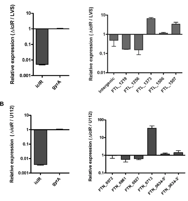
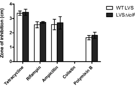
microarray analysis comparing LVS and LVS∆icR ...84 Figure 2.8: Antibiotic sensitivity of LVS∆iclR...85
Figure 3.1: F. tularensis LVS∆ripA fails to suppress pro-inflammatory
cytokine release by primary mouse macrophages ...118 Figure 3.2: LVS∆ripA induced pro-inflammatory cytokines are regulated
by inflammasome components and MyD88 ...120 Figure 3.3: LVS ripA is necessary for suppression of pro-inflammatory
cytokine release by human THP-1 cells...121 Figure 3.4: F. tularensis LVS suppresses IL-1β response in macrophages
by interfering with IL-1b synthesis and processing ...122 Figure 3.5: F. tularensis LVS suppresses pro-inflammatory cytokine
release by disrupting MAPK signaling pathways ...123 Figure 3.6: LVS∆ripA fails to suppress pro-inflammatory cytokine
x
Figure 4.1: Mapping of RipA within the cytoplasmic membrane using
C-terminal fusion reporter proteins in Escherichia coli ...155 Figure 4.2: In vivo protein crosslinking of RipA ...156 Figure 4.3: Characterization of two RipA cytoplasmic domain deletion mutants ...157 Figure 4.4: Characterization of RipA alanine substitution mutants
for intracellular replication and suppression of the
pro-inflammatory response ...159 Figure 4.5: Further analysis of amino acids K114, E122 and E150 to RipA function ...161 Figure 4.6: Identification and characterization of an intragenic
xi
LIST OF ABBREVIATIONS
AIM2 Absent in melanoma 2
ASC Apoptotic speck-containing protein with a CARD BALF Broncheolar lavage fluid
BHI Brain heart infusion
BM(D)M Bone marrow-derived macrophages CARD Caspase recruitment domain
CDM Chamberlain’s defined medium CFU Colony forming units
DC Dendritic cell
DSP Dithiobis (succinimidyl propionate) EEA1 Early endosomal antigen 1
ELISA Enzyme-linked immunosorbent assay
ERK1/2 Extracellular signal-regulated protein kinases 1 and 2 FCV Francisella-containing vacuole
FPI Francisella Pathogenicity Island FRET Fluorescence resonance energy transfer i.d. Intradermal
IFN Interferon
IκBα Nuclear factor of kappa light polypeptide gene enhancer in B-cells inhibitor, alpha
IL Interleukin
xii i.p. Intraperitoneal
JNK c-Jun NH2-terminal kinases
LAMP Lysosomal-associated membrane protein LB Lysogeny broth
LD50 Median lethal dose LD100 Lethal dose
LPS Lipopolysaccharide LVS Live vaccine strain
MAPK Mitogen-activated protein kinase MCP-1 Monocyte chemotactic protein-1 MOI Multiplicity of infection
MyD88 Myeloid differentiation primary response gene 88 NADPH Nicotinamide adenine dinucleotide phosphate
NF-κB Nuclear factor kappa-light-chain-enhancer of activated B cells NLR Nucleotide binding domain-leucine rich repeats containing NBD Nucleotide Binding domain
ORF Open reading frame
P/MAMPs Pathogen/microbe- associated molecular patterns PGE2 Prostaglandin E2
PhoA Alkaline phosphatase PFA Paraformaldehyde
xiii ppGpp Guanosine-tetraphosphate
PTEN Phosphatase and tensin homolog PYCARD Pyrin-CARD
RIG-I Retinoic acid-inducible gene 2 RNAP RNA polymerase
SAM Significance analysis for microarrays s.c. Subcutaneous
SOE Splice overlap extension SQ Starting quantity
TGF-β Transforming growth factor beta
Th T helper
TLR Toll-like receptor
TMS1 Target of methylation-induced silencing TNF-α Tumor necrosis factor alpha
CHAPTER 1
2
FRANCISELLA CLASSIFICATION AND EPIDEMIOLOGY
Francisella tularensis is a Gram-negative, coccobacillus belonging to the family Francisellaceae of the γ-Proteobacteria. Francisellaceae are found throughout the
environment and commonly isolated from marine and fresh water (30, 37, 135); however, the environmental reservoir is not known. Additionally, Francisellaceae have been isolated from a wide-range of animal species. The most common mammalian hosts are lagomorphs such as rabbits, but Francisellaceae have been isolated from over 500 other species including rodents (188), birds (211), cats (238), dogs (125, 126), sheep and oxen (13), fish (214), and even crayfish (8). F. tularensis is also frequently recovered from arthropod vectors, mainly ticks (188), flies (155), and mosquitoes (86). Certain species within Francisellaceae have been reported to infect amoeba, including Acanthamoeba castellani (1, 85, 164, 277) and Hartmannella vermiformis (250) suggesting that amoeba may serve as an environmental reservoir; however, F. tularensis has not yet been isolated from amoeba in nature.
F. tularensis infection in many of the animal hosts, including humans, results in the development of the zoonotic disease tularemia. In fact, F. tularensis was first identified as Bacterium tularense, the organism causing a disease in ground squirrels in Tulare County, California in 1911 (196, 197). Soon after, a febrile disease in humans called “deer-fly fever” was reported in Utah (219). Dr. Edward Francis is attributed with the identification and naming of Bacterium tularense as the agent causing “deer-fly fever” in the Utah patients
(105, 106), and in 1947 the organism was renamed to Francisella tularensis in recognition of his contributions (215, 221). F. noatunensis (203, 204), F. philomiragia (214), and F.
3
near-drowning victims (107, 262, 285). The diverse host range and geographic distribution of Franciselleaceae reflects the success of the organism to adapt to many environments, including humans. Based on the ability to infect and cause disease in so many hosts, there is a repertoire of animal models, including non-human primates (287), mice (27), rabbits (27), guinea pigs (27), rats (144, 233, 289), zebrafish (279), A. castellani (1), H. vermiformis (250), and Drosophila melanogaster (280), that have been used for studying the pathogenesis in F. tularensis infections.
There are three main subspecies of F. tularensis, the species that causes tularemia in humans: tularensis, holarctica, and mediasiastica. F. tularensis subspecies tularensis has only been reported in North America and is considered the most highly infectious subspecies for humans (269). Subspecies tularensis is also known as Type A and the primary strain used in research is the Schu S4 strain, which requires biosafety level 3 conditions to handle in the laboratory. F. tularensis subspecies holarctica is found throughout the northern hemisphere, and while holarctica still infects humans, this subspecies has a higher infectious dose (<1000 organisms) and usually causes a less severe form of tularemia (269). Nonetheless, subspecies holarctica is a source of significant endemic disease in European countries (269). Subspecies holarctica was also used to generate the live vaccine strain (LVS) which is one of the
frequently used strains in laboratories due to the ability to use this strain in under biosafety level 2 conditions and retained virulence for mice (269). F. tularensis subspecies
4
for humans; however, it has been reported to cause disease, predominantly in immune-compromised patients (29, 32, 58, 94, 135, 165, 286). The Utah 112 (U112) strain, which was isolated from a water sample in Utah in 1955 (161), is a frequently-used laboratory strain for F. novicida. Within the field, some consider F. novicida as a subspecies of F. tularensis; however, based on divergence realized by whole genome single-nucleotide polymorphism analysis as well as differences in virulence and in at least 11 metabolic processes, novicida is considered a separate species by others (143, 147, 152, 162, 163). Nevertheless, due to journal policies and the current classification debate in the field (45, 147), chapters 2-4 refer to F. novicida as F. tularensis subspecies novicida.
In humans, tularemia is manifested in several different forms depending on the route of transmission of F. tularensis. General symptoms can include fever, headache, chills, joint pain, muscle aches, cough, and diarrhea, which begin to appear usually 3-5 days post
infection (252, 253). Regardless of the route of inoculation, inflammation and swelling of the lymph nodes is also typical (269). The most common transmission route is contact with F. tularensis through the skin either through bites from arthropod vectors or handling
contaminated animals, tissues or indirectly through items such as tools or bedding (269). There have been multiple cases of tularemia resulting from scratches or bites from house pets, commonly cats (205, 255). Dermal contact results in the ulceroglandular form of tularemia and the characteristic formation of a lesion at the site of infection (269). Transmission via the eye is rare but results in the oculoglandular form of tularemia
5
when the source of infection is not known (269). The most severe form of tularemia is pneumonic tularemia that can be a result of complications from another form of tularemia or from inhalation of aerosolized bacteria. Often aerosols are generated by disruption of
materials such as hay, soil, or grass contaminated with F. tularensis and therefore farmers and lawncare workers are often infected in this way (269). A famous set of cases in the United States occurred in Martha’s Vineyard where multiple lawn care workers were
diagnosed with pneumonic tularemia (95, 273). There have also been at least two cases of pneumonic tularemia resulting from transmission by dogs (241, 263). Historically, lab workers were infected via inhalation, although these incidences have decreased due to increased biosafety in handling the organism. Surprisingly, there is no evidence that F. tularensis is transmitted human to human (269).
F. tularensis subspecies tularensis has a very low infectious dose via the inhalational route, with as few as 10 organisms sufficient to cause disease (252, 253), and pneumonic tularemia has a mortality rate as high as 60% if left untreated (80). Infection can be controlled with prompt antibiotic treatment, and there is partial protection by previous vaccination using LVS (80). Due to the lack of knowledge on why LVS is attenuated, only being partially protective as a vaccine, and the inability to vaccinate immune-compromised individuals with a live strain, LVS is not licensed in the United States. The preferred
6
mortality rate after pulmonary infection, F. tularensis subspecies tularensis is designated as a Select Agent by the Centers for Disease Control (80).
PATHOGENESIS AND INTRACELLULAR LIFECYCLE
In a pulmonary infection, once in the lung, F. tularensis travels to the alveolus where it infects a number of cell types including macrophages, dendritic cells (DCs), neutrophils, and Type II alveolar epithelial cells (21, 36, 129, 130). The distribution of infected cells changes over the course of infection and among subspecies (130). F. tularensis has also been reported to infect erythrocytes (140), fibroblasts (108), kidney epithelial cells (138), and hepatocytes (68), cell types that could presumably be infected in systemic disease. Through the use of a uracil biosynthesis mutant (∆pyrF), it has been suggested that intracellular
replication in non-macrophage cells is sufficient to cause infection (139). There is also
evidence that F. tularensis undergoes an extracellular phase during an infection (21, 99, 292). Overall, the ability to infect and manipulate so many cell types reflects the success of F. tularensis as a pathogen.
7
receptor with additional contributions by class A scavenger receptors and nucleolin (20, 115, 223, 258). After phagocytosis, F. tularensis escapes the phagosome as early as 20 minutes post invasion, and by approximately 1 hour, begins replicating within the cytoplasm. The kinetics of phagosomal escape depends on opsonization status of the bacteria. when
compared to non-opsonizing conditions, serum opsonization of bacteria delays phagosomal escape and thereby impairs intracellular growth (115).
After uptake of bacteria and prior to phagosomal escape, the phagosome undergoes a maturation process in which the phagosome acquires early endosomal markers such as EEA1 and late endosomal markers such as CD63, LAMP-1, and LAMP-2 (59, 119). Studies using lysosomotropic agents revealed that these late-stage phagosomes are not acidified, even by 3 hours post infection (59). On the other hand, additional studies demonstrated that there is an initial, transient acidification as well as acquisition of the vacuolar ATPase that drops by 1 hour post infection, and escape is delayed if acidification is blocked (57, 102, 247). In
contrast, another study suggests that phagosomal acidification is not required for phagosomal escape or intracellular replication (61). Therefore, the requirement for phagosomal
acidification for phagosomal escape by the bacteria remains under debate. Once in the cytoplasm, F. tularensis can replicate up to several logs by 24 hours post infection (10). At this point, some bacteria are located in a membranous vacuole shown to be autophagic vacuoles, as is discussed below, and it is not yet clear how bacteria exit host cells to spread and infect other host cells.
8
surface ligand required for entry, but the host cell receptors involved are not known (75). Entry into epithelial cells also requires actin polymerization, microtubule rearrangement, and associated upstream signaling by phosphatidylinositol 3-kinase (PI3K) and tyrosine kinase pathways (75). Once inside the epithelial cell, F. tularensis escapes the phagosome to replicate within the cytoplasm following the same endocytic pathway and with similar kinetics as in the macrophage (75). DCs are thought to engulf F. tularensis via a mechanism that is dependent on serum opsonization of bacteria and the DC integrins CR3 and CR4 (28). Like DCs, neutrophils also take up F. tularensis in a serum-dependent manner (177, 227). After entry, F. tularensis bacteria escape the phagosome to persist in the cytoplasm, though no reports demonstrate any intracellular replication in neutrophils (194). In fact, F. tularensis has been shown to actively recruit neutrophils to the lung at least in part through the activity of matrix metalloproteinase-9, suggesting that neutrophils may provide an additional in vivo niche for F. tularensis (186). Finally, both complement-dependent and independent
mechanisms are involved in uptake into erythrocytes, but nothing else is known about the lifecycle of F. tularensis in this cell type (140). We still have much to learn about the life cycle of F. tularensis within non-macrophage cell types.
9
that FCV formation also occurs with LVS and that other autophagy-associated molecules such as cathepsin D, phosphatase and tensin homolog (PTEN), and p53 may have altered expression in infected cells (141). Finally, microarray studies at 24 hours post infection demonstrated that infection with Schu S4 or U112 results in decreased expression of other autophagic response proteins and the autophagy-related PI3K signaling pathway (46, 76). This suggests that Francisella may be suppressing and thereby delaying the autophagic response to infection; however, it is not known what bacterial effectors may be involved.
Related to this, some host cell death is induced by F. tularensis, even with bacteria-mediated suppression of the process. Several reports have shown that F. tularensis induces caspase-3 activation and cell death in macrophages and in vivo using F. novicida, LVS, and Type A strains KU49 and KU54 (33, 159, 230, 251, 288). To build on this idea, in two studies using F. novicida, it was demonstrated that caspase-3 activation and cell death are downregulated at early time points as a result of activation of Ras signaling pathways and that although there is eventually upregulation of caspase-3, apoptosis is delayed further, potentially as a result of anti-apoptotic signaling (6, 251). Therefore, although apoptotic pathways are eventually activated, the delayed kinetics suggest the possibility that the bacteria are downregulating these apoptotic pathways, yet the specifics of caspase-3 and apoptosis signaling pathways during F. tularensis infections are not completely clear.
HOST IMMUNE RESPONSE TO FRANCISELLA INFECTION
Innate immune response
10
holarctica and tularensis subspecies. Whereas F. novicida stimulates the innate immune system, F. tularensis subspecies holarctica and tularensis does so to a much lesser extent and in a delayed manner due to active suppression on the part of these organisms (9, 35, 36, 46, 53, 176, 270, 271). In fact, it has also been reported that both LVS and Schu S4 upregulate expression of the immunosuppressive cytokine TGF-β (35, 36). The pathways involved in response to F. tularensis, and which F. tularensis is suppresses, have only been partially elucidated. In terms of extracellular recognition, unlike many other Gram-negative bacteria, F. tularensis lipopolysaccharide (LPS) does not stimulate the host cell sensor protein toll-like receptor (TLR) 4 (2, 62, 128, 270). This low stimulatory effect is likely due in part to
changes in lipid A, specifically the lack of a 4’-phosphate, which is removed by LpxF, and
hypoacylation of the fatty acid chains (222, 278, 281, 282). There has been a fair bit of controversy on the exact structure of lipid A between the strains of Francisella (25, 222, 261, 278, 282). Despite the inability of F. tularensis LPS to activate the host response, F.
tularensis does stimulate TLR2, which recognizes lipoproteins, and whether this recognition is specific to TLR1/TLR2 or TLR6/TLR2 heterodimers is not clear, but F. tularensis may possibly stimulate both heterodimers with different ligands (2, 150, 168, 187, 274). Following stimulation of TLR2, several downstream pathways are required for immune activation. Overall, signaling occurs through MyD88 with a partial contribution of the
11
signaling pathways may be involved in the suppression of the immune response observed for F. tularensis (199) . The specific downstream signaling pathways involved in suppression of pro-IL-1β transcription need further elucidation.
Processing of pro-IL-1β to mature IL-1β for secretion requires a two-step process. First, there must be transcription and synthesis of pro-IL-1β as described above. A second signal is required for processing into mature IL-1β in which a cytoplasmic sensor molecule recognizes a pathogen and then recruits additional protein components to form a complex called the inflammasome. The most well-characterized families of cytoplasmic sensors are the nucleotide binding domain leucine rich repeats containing proteins (NLRs) (145); however, there are reports of inflammasomes containing the cytoplasmic sensors absent in melanoma 2 (AIM2) or retinoic acid-inducible gene 2 (RIG-I) (44, 96, 97, 137, 225, 239). The respective sensor molecule will recruit procaspase-1, and then depending on the sensor molecule involved, also recruit the adaptor protein PYCARD/ASC (Pyrin-CARD/apoptotic speck-containing protein with a CARD) (71, 193). The formation of the complete
inflammasome complex results in the auto-catalytic cleavage of pro-1 into caspase-1, which can then process pro-IL-1β into mature IL-1β for secretion.
12
Several groups have published that a non-NLR sensor AIM2 recognizes F. tularensis DNA, which leads to inflammasome activation (97, 149, 232, 276); however, it is likely that this is not the only mechanism in play, and whether AIM2 is important for recognition by human cells is not clear. Type I interferon (IFN-α/β) is secreted in response to F. tularensis in a TLR-independent, interferon regulatory factor (IRF) 3-dependent manner and is necessary for caspase-1 processing, macrophage cell death, and IL-1β secretion (133). While Type I interferon secretion does not require the AIM2 inflammasome activation, Type I interferon and IRF-3 are required for activation of the AIM2 inflammasome in response to F. tularensis infection (97, 149). In contrast to these studies, another report argues that IFN-β is not
required for inflammasome activation in DC’s or control of replication in this cell type but
rather is important for the production of IL-12p40 (24). There is still a lot to learn about the inflammasome pathways activated in response to F. tularensis infection, and if and how F. tularensis is suppressing this arm of the pro-inflammatory pathway is not completely clear.
Many immunological studies with F. tularensis have been done in vivo or in
13
blunted responsiveness (98, 210). The above data demonstrating downregulation of
cytokines and chemokines makes sense in light of a study showing that both LVS and Schu S4 actively suppress the pro-inflammatory response of endothelial cells by acting through the anti-inflammatory endothelial protein C receptor (EPCR) (41). Type II epithelial cells
exposed to F. tularensis secrete several cytokines and chemokines including IL-8, MCP-1, and GRO-α, and this secretion is NF-κB-dependent and promotes recruitment of neutrophils (116). Overall, these studies support the idea that F. tularensis also modulates the pro-inflammatory response by altering chemokine release, leukocyte adhesion molecule expression, and subsequent recruitment of neutrophils, albeit less successfully.
Adaptive immune response
14
tularensis T-cell epitopes identified, with the most prominent epitope being a peptide from the outer membrane protein Tul4 (104, 120, 264, 265).
In addition to suppressing the innate immune response, Francisella also modulates the adaptive immune response to promote infection. As mentioned above, F. tularensis has been shown to suppress IFN-γ secretion (218). Another mechanism of suppression is through the induction of infected macrophages to secrete prostaglandin E2 (PGE2), which skews the T-cell response to an anti-inflammatory, Th2-like response by increasing IL-5 production (291). PGE2 has also been shown to be important in vivo following intranasal inoculation where it delays production of IFN-γ-positive T-cells and promotes IL-17 secretion and Th17 cell production (74, 192, 290). Blocking IL-12 and producing IL-23, both of which result in an increase of Th17 cells, are also important in pulmonary infections of F. tularensis (47, 82, 88).
FRANCISELLA RESPONSE TO THE HOST
15
acquisition systems, and the Francisella Pathogenicity Island (FPI), all of which are
discussed below. Other Francisella virulence factors include mviN (276), citrulline ureidase (182), a potassium uptake protein TrkH (7), minD (11), valAB (198), an alanine racemase (123), a purine biosynthetic enzyme with homology to glutamine
phosphoribosylpyrophosphate amidotransferases (123), purMCD (220), purF (229), an AcrB RND efflux pump (31), OppB oligopeptide transporter (40), and FTL_0200 encoding a putative AAA+ ATPase of the MoxR subfamily (81). Finally, numerous hypothetical
proteins were identified as virulence factors in the screens mentioned above and remain to be characterized.
Francisella pathogenicity island
16
required for virulence including pdpD (181, 213), pdpA (213, 256, 257), iglA and iglB (78), iglD (248), pmcA/anmK (181), FTT_1347 (22), iglI (22, 38), and iglG (38). Interestingly, PdpE is not required for virulence (38). Of the FPI proteins, at least IglC (34, 170, 249), PdpA (256), IglD (34), IglG (38) and IglI (38), are important for phagosomal biogenesis and/or escape. Additionally, IglA and IglB have been shown to interact (78), but otherwise, the function of the FPI proteins are unknown.
There is some evidence that the FPI may encode a secretion system that is distantly related to the Type VI secretion system (T6SS) found in several other bacterial species (22, 78). One study in F. novicida shows that the VgrG homolog (FTT_1347), is secreted into macrophages and in culture supernatants as observed for VgrG in other T6SS; however, unlike other T6SS, secretion of VgrG is independent of the known FPI T6SS homologs (22). Also unlike other characterized T6SS, the Hcp-homolog PdpE is not secreted in F. novicida (22). Curiously, using deletion mutants of either the FPI, vgrG, or the icmF homolog pdpB in F. novicida, it was shown that IglI is secreted and that this secretion is dependent on the FPI, VgrG, and PdpB (22). Overall, the studies on the FPI suggest that the proteins are part of a secretion system, potentially a novel secretion system similar but distinct from the T6SS.
Regulation of virulence factors
Regulation of virulence factors in F. tularensis has been largely focused on the FPI genes. MglA is one of the key regulators of FPI gene transcription as well as at least 100 other genes, including ones encoding secreted proteins PepO and BglX and genes involved in oxidative and general stress response (40, 124). MglA is also required for phagosomal
17
mosquito and D. melanogaster cells (235, 246). In order to function as a transcriptional regulator, MglA binds to the RNA polymerase (RNAP) to direct transcription of the subset of genes mentioned above (52). This interaction with RNAP is dependent on MglA first binding to a protein called SspA and the MglA-SspA heterodimer then binding to RNAP; thus, the SspA regulon overlaps with that of MglA (52). Another regulator, MigR, regulates the FPI and other genes in the MglA/SspA regulon (39). FevR (also called PigR) has also been shown to regulate both FPI genes and other genes, and this regulation involves the small alarmone guanosine-tetraphosphate (ppGpp) which promotes the interaction between FevR and MglA to coordinate regulation of the same set of genes (42, 51).
Another distinctive aspect of F. tularensis is that the genome does not appear to encode any classic paired two-component regulatory systems, but does contain orphaned members (163). The orphan response regulator PmrA has been shown regulate the FPI as well as numerous other virulence genes and is required for both intracellular replication and in vivo pathogenesis (207, 244). More recently it was shown that PmrA is phosphorylated by a kinase KdpD and may interact with MglA and SspA (26). QseC is another orphan two-component system member, which its partner, QseB, was only recently identified, and both genes were required for biofilm formation, typical of QseBC in other bacteria (84).
Another important F. tularensis regulator is Hfq, which traditionally acts as a post-transcriptional regulator that facilitates the interaction between small non-coding RNAs in and RNA transcripts, which in turn decreases or enhances translation of the RNA transcripts. Hfq is not only valuable as a global regulator for stress tolerance, but also for its contribution to the regulation of virulence genes, including some genes found in the FPI (50, 200).
18
non-coding small RNAs that are involved in gene regulation, although interactions with Hfq were not determined (226). In addition to virulence regulators, F. tularensis encodes the major sigma-70 factor (σ70), as well as a σ32 factor RpoH that regulates numerous genes required for heat-shock response (122).
Regulation of both gene and protein expression occurs in response to numerous environmental stimuli including inside the host macrophage (121, 283), different culture medias (43, 132, 293), heat shock (91, 122), osmotic and membrane stress (200), starvation and oxidative stress (91, 124, 167), mammalian body temperature (91, 138), iron limitation (79, 167), and glucose (138). A surprising result from a recent study demonstrated that F. tularensis alters gene expression in response to spermine and that this gene regulation is largely mediated through adjacent insertion sequence elements (48).
Iron acquisition
As mentioned, F. tularensis gene expression changes in response to iron levels, which is not unexpected considering the iron is an essential metal that bacteria require for normal metabolism and is actively sequestered from pathogens by the host. The role of iron in F. tularensis infection was first observed in the context of iron acquisition upon phagosomal acidification in F. tularensis-infected cells (102). A common mechanism of iron uptake by bacteria is the use of siderophores, secreted iron-binding molecules. In F. tularensis, the fslABCDEF operon, is required for production of a siderophore similar to the
19
Fur protein that regulates genes involved in iron acquisition (79, 92, 93, 131). Later, fslE was also shown to be required for siderophore utilization, growth in iron-limiting conditions, and was under control of the Fur promoter as part of the fsl operon in Schu S4 (231).
Interestingly, the F. tularensis genome does not appear to have tonB, exbB, or exbD, three genes encoding proteins that are typically required for siderophore uptake (163). Another protein recently described as being involved in both siderophore-dependent and -independent iron uptake by Schu S4 is FupA (FTT_0918) (171). In LVS, the FupA homolog (FTL_0439), which is a fusion protein of two adjacent genes in Schu S4, FupA/B, is required for
siderophore-mediated iron uptake, and unlike Schu S4, FslE in LVS is considered a
secondary player in siderophore-mediated iron uptake (260). FupA is required for virulence for both LVS and Schu S4 (171, 260, 275). These studies display some differences in iron uptake between Francisella strains, and both these differences as well as specific
mechanisms of iron uptake need to be explained.
Protection from oxidative stress
20
cells and mice deficient for production of reactive nitrogen and oxygen species, another group demonstrated more definitively that there are distinctive roles of reactive nitrogen and oxygen species to controlling both F. tularensis replication in host cells and an infection in vivo (170, 174) Macrophages infected with F. tularensis have also been shown to
upregulate proteins involved in the oxidative stress response of the macrophage (9, 283). Despite this host response, F. tularensis replicates within macrophages, as discussed above. In neutrophils, F. tularensis survives intracellularly, at least in part due to blocking the oxidative response through prevention of the nicotinamide adenine dinucleotide phosphate (NADPH) oxidase formation and gp91/p22phox or p47/p67phox acquisition on the phagosome, and this blockage leads to a failure to control infection in vivo as well (83, 178, 179, 194, 227).
21
tularensis and holarctica, AcpA is not required for inhibition of the oxidative burst, and AcpABC proteins are not required for Schu S4 virulence in vitro or in vivo (56, 195). Together these studies highlight a key difference between the human-virulent F. tularensis species and F. novicida.
The capsule
A polysaccharide capsule is one common virulence factor found on many but not all pathogenic bacteria, likely including Francisella. The presence and type of capsule expressed by F. tularensis is a subject of debate. F. tularensis was originally shown to have a capsule-like substance surrounding the bacterium over thirty years ago (55, 60, 136, 245). Mutants lacking this capsule structure demonstrate increased sensitivity to serum but reduced
sensitivity to killing by neutrophils and are also avirulent in mouse models of tularemia (19, 55, 245, 266). Furthermore, the composition and expression of the capsule can change depending on the growth conditions (55, 293). The F. tularensis capBCA locus has partial homology to the cap locus in B. anthracis that encodes for proteins needed to synthesize the poly-D-glutamic acid capsule (163, 185). Interestingly, while F. tularensis capBCA is required for intracellular growth and in mouse models of tularemia for both Schu S4 and LVS (69, 146, 202, 267), F. tularensis capsule has not been reported to consist of poly-D-glutamic acid (202, 234). Early studies suggest that the capsule consists of carbohydrates, including mannose, rhamnose, and two other unidentified sugars, amino acids, and fatty acids (136). A more recent study found that the F. tularensis capsule is made up of O-antigen capsular polysaccharide and its production is dependent on the wbt O-antigen
22
required for O-antigen and/or O-antigen capsule formation (169). Besides the O-antigen capsular polysaccharide, higher molecular weight carbohydrate complex has also been identified in isolated capsule and consists of glucose, galactose, mannose, as well as Proteinase K-resistant protein, and this complex synthesis is dependent on the
glycosyltransferase genes FTT_1422 and FTT_1423 (19). So although it is largely accepted that Francisella has a capsule, the composition, biosynthesis, and regulation of the capsule are not well defined.
Type IV Pili
The use of type IV pili is a common way bacteria adhereto host cells and/or use for motility, yet the presence, composition, and function of type IV pili in Francisella are also subjects of debate. Type IV pili-like structures were first observed on LVS using microscopy (117) and later observed on F. novicida (117, 295) and Schu S4 (14). Pili formation in LVS and F. novicida is dependent on both the assembly ATPase PilB as well as the disassembly ATPase PilT (49, 295). Surprisingly, in F. novicida pili formation is not dependent on PilC or PilQ, which are proteins typically required for pilus assembly in other bacteria, though it has yet to be determined whether this is the case for other Francisella strains (295).
23
subspecies tularensis and holarctica and F. novicida strains that do encode for PilA, the only difference is found in the 3’ end of pilA in F. novicida (295). The role of these individual
proteins in pilus composition has yet to be determined, and there are differences between subspecies. There is some evidence that the major pilin for F. novicida is PilE/PilE2 (295). Using heterologous expression of Schu S4 and F. novicida pilA in Neisseria gonnorhea, another group suggested that pilA is sufficient for pilus formation (242). Another recent study showed that in both LVS and Schu S4, pilE4 was required for pilus formation (14).
Interestingly, pilE4 was not required for virulence; however, pilE5 and pilE6 were not required for pilus formation but were required for virulence in LVS only, again highlighting subspecies and species differences (14).
24
Protein secretion
To date, all bacteria have (or are predicted to have/encode) mechanisms for secretion of effector molecules and proteins into the environment in order to support survival and replication, and Francisella is no exception. Although no toxins or other well-characterized secreted effectors have been identified in Francisella, several secreted proteases have been identified including ClpB heat shock protease (123), FTT1209c metallopeptidase family M13 protein (40), PepO zinc protease (127), BglX beta-glucosidase (127), and hemolysins (160). Unlike many other pathogens, Francisella does not encode any classic Type III, Type IV, or Type V secretion system; however, there are Type I and Type II secretion systems as well as the cytoplasmic membrane Sec translocon (163, 189). For Type I secretion, TolC is the prototypical outer membrane efflux pump that interacts with inner membrane ATPases for secretion of toxic products. F. tularensis encodes a functional TolC protein involved with multidrug resistance and is required for intracellular replication and virulence in vivo (118, 224). Francisella also encodes a putative secretion system in the FPI, as described above.
The Type II secretion system in Francisella is thought to be synonymous with Type IV pili encoded by the pil genes. The key difference is the use of GspC, GspL, and GspM as well as several periplasmic proteins in Type II secretion; however, Francisella does not encode GspC, GspL, and GspM, so it’s possible that the Type IV pili system is sufficient. Evidence of Type II secretion being functional in Francisella was determined by the observation that secretion of seven F. novicida proteins was dependent on pilA/pilE1, pilB/pilF, pilC/pilG, and pilQ, although these genes were not required for intracellular replication, they were important in vivo (127, 128, 295). Furthermore, pilE4, which is
25
and pilE6 were not required for pilus formation but were required for virulence and the authors suggest a role for these proteins in secretion (14). Clearly, much remains to be elucidated in regards to mechanism and secreted effectors of Type II secretion as well as its relation to type IV pili and virulence.
DISCOVERY AND INITIAL CHARACTERIZATION OF RIPA
Using a LVS transposon mutant library screen for intracellular replication in epithelial cells, our lab identified the locus FTL_1914. Deletion of this locus via allelic exchange resulted in an LVS mutant (LVS∆ripA) that is deficient for intracellular replication in both epithelial cells and macrophages and therefore was named ripA, required for
intracellular proliferation, factor A (110). Interestingly, LVS∆ripA escapes the phagosome and re-enters the autophagosome-like vacuoles with the same kinetics as wild-type (110). Both ripA gene expression and RipA protein expression are upregulated at higher pH 7.5 and ripA expression is also upregulated upon host cell infection, especially between 1-6 hours post invasion when bacteria have begun to enter the cytoplasm and replicate (111).
Furthermore, deletion of the FPI transcriptional regulators mglA and sspA in LVS results in increased ripA expression, suggesting that these regulators inhibit ripA transcription. LVS∆ripA is also attenuated in a mouse model of pulmonary tularemia (110). A ∆ripA
mutant in the highly virulent Schu S4 strain is also attenuated in a pulmonary mouse model and defective for intracellular replication (our unpublished results). Altogether these data suggest that ripA is required for virulence of F. tularensis.
26
functional motifs or homologs with known functions, it revealed that there are RipA-like proteins found in select strains of a few randomly-distributed bacterial species (110). None of these RipA-like proteins have any known function. Interestingly, in a recent study RipA was identified as a glycosylated protein in a screen using lectin affinity chromatography (18). However, despite the importance of ripA to F. tularensis pathogenesis, the function of this protein is unknown. To address the question of RipA function, our lab began a search for RipA-interacting proteins. Since RipA was predicted to have two large cytoplasmic domains, we hypothesized that RipA likely interacts with cytoplasmic proteins. The search for RipA-interacting proteins was initiated by means of immunoprecipitation using HA-tagged RipA and LVS lysates (109). These studies revealed a list of putative RipA-interacting proteins, one of which was confirmed using reciprocal pull-down experiments (109). This RipA-interacting protein was named IclR due to its homology to the IclR family of transcriptional regulators (109). The F. novicida U112 IclR homolog had previously been described as being required for F. novicida virulence (284). Due to the role of IclR for the virulence of F.
novicida and our results demonstrating an interaction with RipA, we considered that its role in virulence may be at least in part through its interactions with RipA.
27
Together these studies help to elucidate RipA’s role in virulence and function, as well as
28
REFERENCES
1. Abd, H., T. Johansson, I. Golovliov, G. Sandstrom, and M. Forsman. 2003. Survival and growth of Francisella tularensis in Acanthamoeba castellanii. Appl. Environ. Microbiol.
69:600-606.
2. Abplanalp, A. L., I. R. Morris, B. K. Parida, J. M. Teale, and M. T. Berton. 2009. TLR-dependent control of Francisella tularensis infection and host inflammatory responses. PLoS One. 4:e7920.
3. Ahlund, M. K., P. Ryden, A. Sjostedt, and S. Stoven. 2010. Directed screen of
Francisella novicida virulence determinants using Drosophila melanogaster. Infect. Immun.
78:3118-3128.
4. Akimana, C., S. Al-Khodor, and Y. Abu Kwaik. 2010. Host factors required for modulation of phagosome biogenesis and proliferation of Francisella tularensis within the cytosol. PLoS One. 5:e11025.
5. Alibek, K. 1999. Biohazard, p. 29-38. In Anonymous . Random House, New York, NY. 6. Al-Khodor, S., and Y. Abu Kwaik. 2010. Triggering Ras signalling by intracellular Francisella tularensis through recruitment of PKCalpha and beta to the SOS2/GrB2 complex is essential for bacterial proliferation in the cytosol. Cell. Microbiol. 12:1604-1621.
7. Alkhuder, K., K. L. Meibom, I. Dubail, M. Dupuis, and A. Charbit. 2010.
Identification of trkH, encoding a potassium uptake protein required for Francisella tularensis systemic dissemination in mice. PLoS One. 5:e8966.
8. Anda, P., J. Segura del Pozo, J. M. Diaz Garcia, R. Escudero, F. J. Garcia Pena, M. C. Lopez Velasco, R. E. Sellek, M. R. Jimenez Chillaron, L. P. Sanchez Serrano, and J. F. Martinez Navarro. 2001. Waterborne outbreak of tularemia associated with crayfish fishing. Emerg. Infect. Dis. 7:575-582.
9. Andersson, H., B. Hartmanova, P. Ryden, L. Noppa, L. Naslund, and A. Sjostedt.
2006. A microarray analysis of the murine macrophage response to infection with Francisella tularensis LVS. J. Med. Microbiol. 55:1023-1033.
10. Anthony, L. D., R. D. Burke, and F. E. Nano. 1991. Growth of Francisella spp. in rodent macrophages. Infect. Immun. 59:3291-3296.
11. Anthony, L. S., S. C. Cowley, K. E. Mdluli, and F. E. Nano. 1994. Isolation of a Francisella tularensis mutant that is sensitive to serum and oxidative killing and is avirulent in mice: correlation with the loss of MinD homologue expression. FEMS Microbiol. Lett.
29
12. Apicella, M. A., D. M. Post, A. C. Fowler, B. D. Jones, J. A. Rasmussen, J. R. Hunt, S. Imagawa, B. Choudhury, T. J. Inzana, T. M. Maier, D. W. Frank, T. C. Zahrt, K. Chaloner, M. P. Jennings, M. K. McLendon, and B. W. Gibson. 2010. Identification, characterization and immunogenicity of an O-antigen capsular polysaccharide of Francisella tularensis. PLoS One. 5:e11060.
13. Arata, A., H. Chamsa, A. Farhang-Azad, O. Mescerjakova, V. Neronov, and S. Saidi. 1973. First detection of tularaemia in domestic and wild mammals in Iran. Bull. World Health Organ. 49:597-603.
14. Ark, N. M., and B. J. Mann. 2011. Impact of Francisella tularensis pilin homologs on pilus formation and virulence. Microb. Pathog. 51:110-120.
15. Asare, R., and Y. Abu Kwaik. 2010. Molecular complexity orchestrates modulation of phagosome biogenesis and escape to the cytosol of macrophages by Francisella tularensis. Environ. Microbiol. 12:2559-2586.
16. Bakshi, C. S., M. Malik, K. Regan, J. A. Melendez, D. W. Metzger, V. M. Pavlov, and T. J. Sellati. 2006. Superoxide dismutase B gene (sodB)-deficient mutants of Francisella tularensis demonstrate hypersensitivity to oxidative stress and attenuated virulence. J.
Bacteriol. 188:6443-6448.
17. Balagopal, A., A. S. MacFarlane, N. Mohapatra, S. Soni, J. S. Gunn, and L. S. Schlesinger. 2006. Characterization of the receptor-ligand pathways important for entry and survival of Francisella tularensis in human macrophages. Infect. Immun. 74:5114-5125. 18. Balonova, L., L. Hernychova, B. F. Mann, M. Link, Z. Bilkova, M. V. Novotny, and J. Stulik. 2010. Multimethodological approach to identification of glycoproteins from the proteome of Francisella tularensis, an intracellular microorganism. J. Proteome Res. 9: 1995-2005.
19. Bandara, A. B., A. E. Champion, X. Wang, G. Berg, M. A. Apicella, M. McLendon, P. Azadi, D. S. Snyder, and T. J. Inzana. 2011. Isolation and mutagenesis of a capsule-like complex (CLC) from Francisella tularensis, and contribution of the CLC to F. tularensis virulence in mice. PLoS One. 6:e19003.
20. Barel, M., A. G. Hovanessian, K. Meibom, J. P. Briand, M. Dupuis, and A. Charbit.
2008. A novel receptor - ligand pathway for entry of Francisella tularensis in monocyte-like THP-1 cells: interaction between surface nucleolin and bacterial elongation factor Tu. BMC Microbiol. 8:145.
30
22. Barker, J. R., A. Chong, T. D. Wehrly, J. J. Yu, S. A. Rodriguez, J. Liu, J. Celli, B. P. Arulanandam, and K. E. Klose. 2009. The Francisella tularensis pathogenicity island encodes a secretion system that is required for phagosome escape and virulence. Mol. Microbiol. 74:1459-1470.
23. Baron, G. S., and F. E. Nano. 1999. An erythromycin resistance cassette and mini-transposon for constructing transcriptional fusions to cat. Gene. 229:59-65.
24. Bauler, T. J., J. C. Chase, and C. M. Bosio. 2011. IFN-beta Mediates Suppression of IL-12p40 in Human Dendritic Cells following Infection with Virulent Francisella tularensis. J. Immunol. 187:1845-1855.
25. Beasley, A. S., R. J. Cotter, S. N. Vogel, T. J. Inzana, A. A. Qureshi, and N. Qureshi.
2011. A variety of novel lipid a structures obtained from Francisella tularensis live obtained from Francisella tularensis live. Innate Immun. doi: 10.1177/1753425911401054.
26. Bell, B. L., N. P. Mohapatra, and J. S. Gunn. 2010. Regulation of virulence gene transcripts by the Francisella novicida orphan response regulator PmrA: role of
phosphorylation and evidence of MglA/SspA interaction. Infect. Immun. 78:2189-2198. 27. Bell, J. F., C. R. Owen, and C. L. Larson. 1955. Virulence of Bacterium tularense. I. A study of the virulence of Bacterium tularense in mice, guinea pigs, and rabbits. J. Infect. Dis.
97:162-166.
28. Ben Nasr, A., J. Haithcoat, J. E. Masterson, J. S. Gunn, T. Eaves-Pyles, and G. R. Klimpel. 2006. Critical role for serum opsonins and complement receptors CR3
(CD11b/CD18) and CR4 (CD11c/CD18) in phagocytosis of Francisella tularensis by human dendritic cells (DC): uptake of Francisella leads to activation of immature DC and
intracellular survival of the bacteria. J. Leukoc. Biol. 80:774-786.
29. Bernard, K., S. Tessier, J. Winstanley, D. Chang, and A. Borczyk. 1994. Early recognition of atypical Francisella tularensis strains lacking a cysteine requirement. J. Clin. Microbiol. 32:551-553.
30. Berrada, Z. L., and S. R. Telford. 2011. Survival of Francisella tularensis Type A in brackish-water. Arch. Microbiol. 193:223-226.
31. Bina, X. R., C. L. Lavine, M. A. Miller, and J. E. Bina. 2008. The AcrAB RND efflux system from the live vaccine strain of Francisella tularensis is a multiple drug efflux system that is required for virulence in mice. FEMS Microbiol. Lett. 279:226-233.
31
33. Bokhari, S. M., K. J. Kim, D. M. Pinson, J. Slusser, H. W. Yeh, and M. J. Parmely.
2008. NK cells and gamma interferon coordinate the formation and function of hepatic granulomas in mice infected with the Francisella tularensis live vaccine strain. Infect. Immun. 76:1379-1389.
34. Bonquist, L., H. Lindgren, I. Golovliov, T. Guina, and A. Sjostedt. 2008. MglA and Igl proteins contribute to the modulation of Francisella tularensis live vaccine
strain-containing phagosomes in murine macrophages. Infect. Immun. 76:3502-3510.
35. Bosio, C. M., H. Bielefeldt-Ohmann, and J. T. Belisle. 2007. Active suppression of the pulmonary immune response by Francisella tularensis Schu4. J. Immunol. 178:4538-4547. 36. Bosio, C. M., and S. W. Dow. 2005. Francisella tularensis induces aberrant activation of pulmonary dendritic cells. J. Immunol. 175:6792-6801.
37. Broman, T., J. Thelaus, A. C. Andersson, S. Backman, P. Wikstrom, E. Larsson, M. Granberg, L. Karlsson, E. Back, H. Eliasson, R. Mattsson, A. Sjostedt, and M.
Forsman. 2011. Molecular Detection of Persistent Francisella tularensis Subspecies holarctica in Natural Waters. Int. J. Microbiol. 2011:851946. Epub 2010 Sep 8.
38. Broms, J. E., M. Lavander, L. Meyer, and A. Sjostedt. 2011. IglG and IglI of the Francisella Pathogenicity Island Are Important Virulence Determinants of Francisella tularensis LVS. Infect. Immun. 79:3683-3696.
39. Brotcke, A., and D. M. Monack. 2008. Identification of fevR, a novel regulator of virulence gene expression in Francisella novicida. Infect. Immun. 76:3473-3480. 40. Brotcke, A., D. S. Weiss, C. C. Kim, P. Chain, S. Malfatti, E. Garcia, and D. M. Monack. 2006. Identification of MglA-regulated genes reveals novel virulence factors in Francisella tularensis. Infect. Immun. 74:6642-6655.
41. Bublitz, D. C., C. E. Noah, J. L. Benach, and M. B. Furie. 2010. Francisella tularensis suppresses the proinflammatory response of endothelial cells via the endothelial protein C receptor. J. Immunol. 185:1124-1131.
42. Buchan, B. W., R. L. McCaffrey, S. R. Lindemann, L. A. Allen, and B. D. Jones.
2009. Identification of migR, a regulatory element of the Francisella tularensis live vaccine strain iglABCD virulence operon required for normal replication and trafficking in
macrophages. Infect. Immun. 77:2517-2529.
43. Buchan, B. W., M. K. McLendon, and B. D. Jones. 2008. Identification of
differentially regulated francisella tularensis genes by use of a newly developed Tn5-based transposon delivery system. Appl. Environ. Microbiol. 74:2637-2645.
32
orthogonal proteomic-genomic screen identifies AIM2 as a cytoplasmic DNA sensor for the inflammasome. Nat. Immunol. 10:266-272.
45. Busse, H. J., B. Huber, P. Anda, R. Escudero, H. C. Scholz, E. Seibold, W. D. Splettstoesser, and P. Kampfer. 2010. Objections to the transfer of Francisella novicida to the subspecies rank of Francisella tularensis - response to Johansson et al. Int. J. Syst. Evol. Microbiol. 60:1718-1720.
46. Butchar, J. P., T. J. Cremer, C. D. Clay, M. A. Gavrilin, M. D. Wewers, C. B. Marsh, L. S. Schlesinger, and S. Tridandapani. 2008. Microarray analysis of human monocytes infected with Francisella tularensis identifies new targets of host response subversion. PLoS One. 3:e2924.
47. Butchar, J. P., M. V. Rajaram, L. P. Ganesan, K. V. Parsa, C. D. Clay, L. S.
Schlesinger, and S. Tridandapani. 2007. Francisella tularensis induces IL-23 production in human monocytes. J. Immunol. 178:4445-4454.
48. Carlson, P. E., Jr, J. Horzempa, D. M. O'Dee, C. M. Robinson, P. Neophytou, A. Labrinidis, and G. J. Nau. 2009. Global transcriptional response to spermine, a component of the intramacrophage environment, reveals regulation of Francisella gene expression through insertion sequence elements. J. Bacteriol. 191:6855-6864.
49. Chakraborty, S., M. Monfett, T. M. Maier, J. L. Benach, D. W. Frank, and D. G. Thanassi. 2008. Type IV pili in Francisella tularensis: roles of pilF and pilT in fiber assembly, host cell adherence, and virulence. Infect. Immun. 76:2852-2861
50. Chambers, J. R., and K. S. Bender. 2011. The RNA chaperone Hfq is important for growth and stress tolerance in Francisella novicida. PLoS One. 6:e19797.
51. Charity, J. C., L. T. Blalock, M. M. Costante-Hamm, D. L. Kasper, and S. L. Dove.
2009. Small molecule control of virulence gene expression in Francisella tularensis. PLoS Pathog. 5:e1000641.
52. Charity, J. C., M. M. Costante-Hamm, E. L. Balon, D. H. Boyd, E. J. Rubin, and S. L. Dove. 2007. Twin RNA polymerase-associated proteins control virulence gene expression in Francisella tularensis. PLoS Pathog. 3:e84.
53. Chase, J. C., J. Celli, and C. M. Bosio. 2009. Direct and indirect impairment of human dendritic cell function by virulent Francisella tularensis Schu S4. Infect. Immun. 77:180-195. 54. Checroun, C., T. D. Wehrly, E. R. Fischer, S. F. Hayes, and J. Celli. 2006.
33
55. Cherwonogrodzky, J. W., M. H. Knodel, and M. R. Spence. 1994. Increased encapsulation and virulence of Francisella tularensis live vaccine strain (LVS) by subculturing on synthetic medium. Vaccine. 12:773-775.
56. Child, R., T. D. Wehrly, D. Rockx-Brouwer, D. W. Dorward, and J. Celli. 2010. Acid phosphatases do not contribute to pathogenesis of Type A Francisella tularensis. Infect. Immun. 78:59-67.
57. Chong, A., T. D. Wehrly, V. Nair, E. R. Fischer, J. R. Barker, K. E. Klose, and J. Celli. 2008. The early phagosomal stage of Francisella tularensis determines optimal phagosomal escape and Francisella pathogenicity island protein expression. Infect. Immun.
76:5488-5499.
58. Clarridge, J. E., 3rd, T. J. Raich, A. Sjosted, G. Sandstrom, R. O. Darouiche, R. M. Shawar, P. R. Georghiou, C. Osting, and L. Vo. 1996. Characterization of two unusual clinically significant Francisella strains. J. Clin. Microbiol. 34:1995-2000.
59. Clemens, D. L., B. Y. Lee, and M. A. Horwitz. 2004. Virulent and avirulent strains of Francisella tularensis prevent acidification and maturation of their phagosomes and escape into the cytoplasm in human macrophages. Infect. Immun. 72:3204-3217.
60. Clemens, D. L., B. Y. Lee, and M. A. Horwitz. 2005. Francisella tularensis enters macrophages via a novel process involving pseudopod loops. Infect. Immun. 73:5892-5902. 61. Clemens, D. L., B. Y. Lee, and M. A. Horwitz. 2009. Francisella tularensis phagosomal escape does not require acidification of the phagosome. Infect. Immun. 77:1757-1773. 62. Cole, L. E., K. L. Elkins, S. M. Michalek, N. Qureshi, L. J. Eaton, P. Rallabhandi, N. Cuesta, and S. N. Vogel. 2006. Immunologic consequences of Francisella tularensis live vaccine strain infection: role of the innate immune response in infection and immunity. J. Immunol. 176:6888-6899.
63. Cole, L. E., M. H. Laird, A. Seekatz, A. Santiago, Z. Jiang, E. Barry, K. A. Shirey, K. A. Fitzgerald, and S. N. Vogel. 2010. Phagosomal retention of Francisella tularensis results in TIRAP/Mal-independent TLR2 signaling. J. Leukoc. Biol. 87:275-281.
64. Cole, L. E., A. Santiago, E. Barry, T. J. Kang, K. A. Shirey, Z. J. Roberts, K. L. Elkins, A. S. Cross, and S. N. Vogel. 2008. Macrophage proinflammatory response to Francisella tularensis live vaccine strain requires coordination of multiple signaling pathways. J. Immunol. 180:6885-6891.
34
66. Collazo, C. M., A. I. Meierovics, R. De Pascalis, T. H. Wu, C. R. Lyons, and K. L. Elkins. 2009. T cells from lungs and livers of Francisella tularensis-immune mice control the growth of intracellular bacteria. Infect. Immun. 77:2010-2021.
67. Conlan, J. W., R. KuoLee, H. Shen, and A. Webb. 2002. Different host defences are required to protect mice from primary systemic vs pulmonary infection with the facultative intracellular bacterial pathogen, Francisella tularensis LVS. Microb. Pathog. 32:127-134. 68. Conlan, J. W., and R. J. North. 1992. Early pathogenesis of infection in the liver with the facultative intracellular bacteria Listeria monocytogenes, Francisella tularensis, and Salmonella typhimurium involves lysis of infected hepatocytes by leukocytes. Infect. Immun.
60:5164-5171.
69. Conlan, J. W., H. Shen, I. Golovliov, C. Zingmark, P. C. Oyston, W. Chen, R. V. House, and A. Sjostedt. 2010. Differential ability of novel attenuated targeted deletion mutants of Francisella tularensis subspecies tularensis strain SCHU S4 to protect mice against aerosol challenge with virulent bacteria: effects of host background and route of immunization. Vaccine. 28:1824-1831.
70. Conlan, J. W., A. Sjostedt, and R. J. North. 1994. CD4+ and CD8+ T-cell-dependent and -independent host defense mechanisms can operate to control and resolve primary and secondary Francisella tularensis LVS infection in mice. Infect. Immun. 62:5603-5607. 71. Conway, K. E., B. B. McConnell, C. E. Bowring, C. D. Donald, S. T. Warren, and P. M. Vertino. 2000. TMS1, a novel proapoptotic caspase recruitment domain protein, is a target of methylation-induced gene silencing in human breast cancers. Cancer Res. 60: 6236-6242.
72. Cowley, S. C., and K. L. Elkins. 2003. Multiple T cell subsets control Francisella tularensis LVS intracellular growth without stimulation through macrophage interferon gamma receptors. J. Exp. Med. 198:379-389.
73. Cowley, S. C., E. Hamilton, J. A. Frelinger, J. Su, J. Forman, and K. L. Elkins. 2005. CD4-CD8- T cells control intracellular bacterial infections both in vitro and in vivo. J. Exp. Med. 202:309-319.
74. Cowley, S. C., A. I. Meierovics, J. A. Frelinger, Y. Iwakura, and K. L. Elkins. 2010. Lung CD4-CD8- double-negative T cells are prominent producers of IL-17A and IFN-gamma during primary respiratory murine infection with Francisella tularensis live vaccine strain. J. Immunol. 184:5791-5801.
35
77. Culkin, S. J., T. Rhinehart-Jones, and K. L. Elkins. 1997. A novel role for B cells in early protective immunity to an intracellular pathogen, Francisella tularensis strain LVS. J. Immunol. 158:3277-3284.
78. de Bruin, O. M., J. S. Ludu, and F. E. Nano. 2007. The Francisella pathogenicity island protein IglA localizes to the bacterial cytoplasm and is needed for intracellular growth. BMC Microbiol. 7:1.
79. Deng, K., R. J. Blick, W. Liu, and E. J. Hansen. 2006. Identification of Francisella tularensis genes affected by iron limitation. Infect. Immun. 74:4224-4236.
80. Dennis, D. T., T. V. Inglesby, D. A. Henderson, J. G. Bartlett, M. S. Ascher, E. Eitzen, A. D. Fine, A. M. Friedlander, J. Hauer, M. Layton, S. R. Lillibridge, J. E. McDade, M. T. Osterholm, T. O'Toole, G. Parker, T. M. Perl, P. K. Russell, K. Tonat, and Working Group on Civilian Biodefense. 2001. Tularemia as a biological weapon: medical and public health management. JAMA. 285:2763-2773.
81. Dieppedale, J., D. Sobral, M. Dupuis, I. Dubail, J. Klimentova, J. Stulik, G. Postic, E. Frapy, K. L. Meibom, M. Barel, and A. Charbit. 2011. Identification of a putative chaperone involved in stress resistance and virulence in Francisella tularensis. Infect. Immun.
79:1428-1439.
82. Duckett, N. S., S. Olmos, D. M. Durrant, and D. W. Metzger. 2005. Intranasal interleukin-12 treatment for protection against respiratory infection with the Francisella tularensis live vaccine strain. Infect. Immun. 73:2306-2311.
83. Dunaeva, T. N., and K. N. Shlygina. 1975. Phagocytic activity of the neutrophils in tularemia in animals with varying infective sensitivity. Zh. Mikrobiol. Epidemiol.
Immunobiol. 10:22-26.
84. Durham-Colleran, M. W., A. B. Verhoeven, and M. L. van Hoek. 2010. Francisella novicida forms in vitro biofilms mediated by an orphan response regulator. Microb. Ecol.
59:457-465.
85. El-Etr, S. H., J. J. Margolis, D. Monack, R. A. Robison, M. Cohen, E. Moore, and A. Rasley. 2009. Francisella tularensis type A strains cause the rapid encystment of
Acanthamoeba castellanii and survive in amoebal cysts for three weeks postinfection. Appl. Environ. Microbiol. 75:7488-7500.
86. Eliasson, H., J. Lindback, J. P. Nuorti, M. Arneborn, J. Giesecke, and A. Tegnell.
2002. The 2000 tularemia outbreak: a case-control study of risk factors in disease-endemic and emergent areas, Sweden. Emerg. Infect. Dis. 8:956-960.
36
88. Elkins, K. L., A. Cooper, S. M. Colombini, S. C. Cowley, and T. L. Kieffer. 2002. In vivo clearance of an intracellular bacterium, Francisella tularensis LVS, is dependent on the p40 subunit of interleukin-12 (IL-12) but not on IL-12 p70. Infect. Immun. 70:1936-1948. 89. Elkins, K. L., T. R. Rhinehart-Jones, S. J. Culkin, D. Yee, and R. K. Winegar. 1996. Minimal requirements for murine resistance to infection with Francisella tularensis LVS. Infect. Immun. 64:3288-3293.
90. Ericsson, M., M. Kroca, T. Johansson, A. Sjostedt, and A. Tarnvik. 2001. Long-lasting recall response of CD4+ and CD8+ alphabeta T cells, but not gammadelta T cells, to heat shock proteins of francisella tularensis. Scand. J. Infect. Dis. 33:145-152.
91. Ericsson, M., A. Tarnvik, K. Kuoppa, G. Sandstrom, and A. Sjostedt. 1994.
Increased synthesis of DnaK, GroEL, and GroES homologs by Francisella tularensis LVS in response to heat and hydrogen peroxide. Infect. Immun. 62:178-183.
92. Ernst, J. F., R. L. Bennett, and L. I. Rothfield. 1978. Constitutive expression of the iron-enterochelin and ferrichrome uptake systems in a mutant strain of Salmonella
typhimurium. J. Bacteriol. 135:928-934.
93. Escolar, L., J. Perez-Martin, and V. de Lorenzo. 1998. Binding of the fur (ferric uptake regulator) repressor of Escherichia coli to arrays of the GATAAT sequence. J. Mol. Biol. 283:537-547.
94. Escudero, R., M. Elia, J. A. Saez-Nieto, V. Menendez, A. Toledo, G. Royo, M. Rodriguez-Vargas, M. J. Whipp, H. Gil, I. Jado, and P. Anda. 2010. A possible novel Francisella genomic species isolated from blood and urine of a patient with severe illness. Clin. Microbiol. Infect. 16:1026-1030.
95. Feldman, K. A., R. E. Enscore, S. L. Lathrop, B. T. Matyas, M. McGuill, M. E. Schriefer, D. Stiles-Enos, D. T. Dennis, L. R. Petersen, and E. B. Hayes. 2001. An outbreak of primary pneumonic tularemia on Martha's Vineyard. N. Engl. J. Med. 345: 1601-1606.
96. Fernandes-Alnemri, T., J. W. Yu, P. Datta, J. Wu, and E. S. Alnemri. 2009. AIM2 activates the inflammasome and cell death in response to cytoplasmic DNA. Nature.
458:509-513.
97. Fernandes-Alnemri, T., J. W. Yu, C. Juliana, L. Solorzano, S. Kang, J. Wu, P. Datta, M. McCormick, L. Huang, E. McDermott, L. Eisenlohr, C. P. Landel, and E. S. Alnemri. 2010. The AIM2 inflammasome is critical for innate immunity to Francisella tularensis. Nat. Immunol. 11:385-393.
37
99. Forestal, C. A., M. Malik, S. V. Catlett, A. G. Savitt, J. L. Benach, T. J. Sellati, and M. B. Furie. 2007. Francisella tularensis has a significant extracellular phase in infected mice. J. Infect. Dis. 196:134-137.
100. Forslund, A. L., K. Kuoppa, K. Svensson, E. Salomonsson, A. Johansson, M. Bystrom, P. C. Oyston, S. L. Michell, R. W. Titball, L. Noppa, E. Frithz-Lindsten, M. Forsman, and A. Forsberg. 2006. Direct repeat-mediated deletion of a type IV pilin gene results in major virulence attenuation of Francisella tularensis. Mol. Microbiol. 59: 1818-1830.
101. Forslund, A. L., E. N. Salomonsson, I. Golovliov, K. Kuoppa, S. Michell, R. Titball, P. Oyston, L. Noppa, A. Sjostedt, and A. Forsberg. 2010. The type IV pilin, PilA, is required for full virulence of Francisella tularensis subspecies tularensis. BMC Microbiol.
10:227.
102. Fortier, A. H., D. A. Leiby, R. B. Narayanan, E. Asafoadjei, R. M. Crawford, C. A. Nacy, and M. S. Meltzer. 1995. Growth of Francisella tularensis LVS in macrophages: the acidic intracellular compartment provides essential iron required for growth. Infect. Immun.
63:1478-1483.
103. Fortier, A. H., T. Polsinelli, S. J. Green, and C. A. Nacy. 1992. Activation of macrophages for destruction of Francisella tularensis: identification of cytokines, effector cells, and effector molecules. Infect. Immun. 60:817-825.
104. Fortier, A. H., M. V. Slayter, R. Ziemba, M. S. Meltzer, and C. A. Nacy. 1991. Live vaccine strain of Francisella tularensis: infection and immunity in mice. Infect. Immun.
59:2922-2928.
105. Francis, E. 1921. The occurrence of tularemia in nature as a disease of man. Public Health Rep. 36:731-738.
106. Francis, E. 1925. Tularemia. JAMA. 84:1243-1250.
107. Friis-Moller, A., L. E. Lemming, N. H. Valerius, and B. Bruun. 2004. Problems in identification of Francisella philomiragia associated with fatal bacteremia in a patient with chronic granulomatous disease. J. Clin. Microbiol. 42:1840-1842.
108. Fujita, H., Y. Watanabe, T. Sato, Y. Ohara, and M. Homma. 1993. The entry and intracellular multiplication of Francisella tularensis in cultured cells: its correlation with virulence in experimental mice. Microbiol. Immunol. 37:837-842.