Humanized mice: models for evaluating NeuroHIV
and cure strategies
Jenna B. Honeycutt1&J. Victor Garcia1
Received:23May2017/ Revised:29July2017/ Accepted:8August2017/ Publishedonline:22August 2017
Abstract While the human immunodeficiency virus (HIV) epidemic was initially characterized by a high prevalence of severe and widespread neurological pathologies, the develop-ment of better treatdevelop-ments to suppress viremia over years and even decades has mitigated many of the severe neurological pathologies previously observed. Despite effective treatment, mild neurocognitive impairment and premature cognitive ag-ing are observed in HIV-infected individuals, suggestag-ing a changing but ongoing role of HIV infection in the central nervous system (CNS). Although current therapies are effec-tive in suppressing viremia, they are not curaeffec-tive and patients must remain on life-long treatment or risk recrudescence of virus. Important for the development and evaluation of a cure for HIV will be animal models that recapitulate critical aspects of infection in vivo. In the following, we seek to summarize some of the recent developments in humanized mouse models and their usefulness in modeling HIV infection of the CNS and HIV cure strategies.
Keywords Humanized mice . HIV/AIDS . CNS . Antiretroviral therapy (ART) . NeuroHIV
Introduction
Infection with human immunodeficiency virus (HIV), the un-derlying cause of acquired immune deficiency syndrome
(AIDS), results in system-wide manifestations of disease. The central nervous system (CNS) is one of the affected sys-tems, and in the absence of treatment, infection can result in severe brain pathology and cognitive/behavioral abnormalities. These neurological deficiencies are grouped under the umbrel-la term of HIV-1-associated neurocognitive disorders (HAND). Effective treatment of HIV with combination antire-troviral therapy (cART) has led to a decrease in the frequency of severe HAND (HIV-associated dementia or HAD) from 10– 15 to 2% (Heaton et al.2010). However, asymptomatic forms of HAND exist and HIV infection is associated with premature cognitive aging, despite effective cART (Cohen et al.2015). The asymptomatic form of HAND, termed asymptomatic neurocognitive impairment (ANI), is included for research pur-poses but not clinical diagnosis, and is detected via direct neu-ropsychological assessment (Heaton et al.2010).
The recent and dogma-redefining discovery that the brain has a lymphatic system, consisting of lymphatic vessels adja-cent to the dura that empty into the periphery (Aspelund et al.
2015; Louveau et al.2015), suggests that better systems to study the interactions between the CNS and peripheral im-mune system will be key to modeling HIV infection in the brain. A better understanding of the mechanisms involved in viral trafficking and cART penetration into the brain and whether or not the brain represents a tissue reservoir of HIV that cannot be cleared with cART (or where HIV persists despite prolonged cART) is all important for developing more effective treatments to target HIV infection in the CNS.
Humanized mouse models
Mice that are reconstituted with human cells offer a unique small animal model for studying HIV infection, as all strains of mice are refractory to HIV infection. Humanized mice are
* Jenna B. Honeycutt [email protected]
1 Division of Infectious Diseases, Center for AIDS Research,
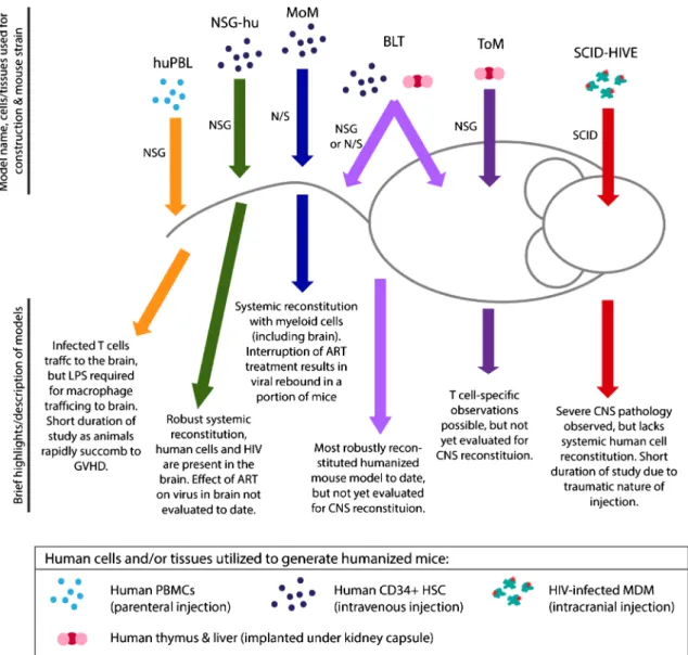
created by implanting human tissues and/or injecting human cells into various strains of immunodeficient mice (Fig.1); these animals have been extensively used successfully to study HIV pathogenesis, transmission, and persistence in vivo.
SCID-HIVE model To determine whether or not HIV-infected cells directly contribute to HIV-associated encephali-tis (HIVE), HIV-infected human myeloid cells have been di-rectly injected into the brains of severe combined immune deficient (SCID) mice, which lack murine B and T cells (Tyor et al.1993; Persidsky and Gendelman1997; Persidsky et al.1996,1997,1999). This direct injection model results in pathologic changes in about a week; pathology includes
encephalitis, astrogliosis (activated astrocytes), and increased levels of mononuclear phagocyte trafficking.
Recently, this model has been used to investigate strategies to mitigate HIV-induced inflammation in the CNS. CEP-1347, an inhibitor of mixed multilineage kinases, has shown promise in animal models of Huntington’s disease (Apostol et al. 2008; Conforti et al.2008). CEP-1347 has also been shown to reduce microglia inflammation in vitro (Lund et al.
2005). In SCID-HIVE mice, CEP-1347 was able to reduce the levels of microglia activation (microgliosis), as measured by decreases in Iba-1 expression (Eggert et al.2010). An IFN-alpha inhibitor, B18R, was shown to effectively cross the blood-brain barrier (BBB) and was able to resolve some of the pathologies present in the SCID-HIVE model (Fritz-French et al. 2014). Decreased presence of mouse
Fig. 1 Schematic of humanized mouse models for evaluating NeuroHIV infection and/or cure strategies. The name of each model is followed by the human cells/tissues and immunodeficient mouse strains used in the
mononuclear phagocytes, decreased expression of IFN-stimulated genes (ISGs), and decreased levels of mouse microtubule-associated protein-2 (MAP-2) were noted in B18R-treated mice, compared with untreated SCID-HIVE mice. However, the levels of astrogliosis were unaffected in mice treated with B18R, which remained significantly higher than those seen in mice injected with uninfected cells. A Jak/ STAT inhibitor, ruxolitnib, was also evaluated for its ability to reduce HIV-associated inflammation in the SCID-HIVE mod-el (Haile et al.2016). Dosing with ruxolitnib (20 or 50 mg/kg twice per day) was able to significantly reduce astrogliosis in treated mice, as measured by a reduction in glial fibrillary acidic protein (GFAP) expression.
A number of the pathologies associated with the SCID-HIVE model are mitigated or reduced in the presence of cART (Cook et al. 2005; Cook-Easterwood et al. 2007; Koneru et al.2014), including astrogliosis, mononuclear cell infiltration, and restored neuronal integrity (reduced MAP-2 levels). This suggests that HIV replication may directly con-tribute to the extent of brain pathology in this model. They also demonstrate that ART consisting of atazanavir, tenofovir, and emtricitabine was able to effectively penetrate into the brain within 1 h of injection (Koneru et al.2014). A significant decrease, approximately 4-fold, in the numbers of p24+ cells in treated animals compared with saline-treated mice was ob-served. The authors conclude that this regimen was insuffi-cient to eradicate virus from the brain, as p24+ cells were present in the brains of cART-treated mice; however, animals were treated for a period of 10 days, a relatively short time. Nevertheless, this study provides unique information regard-ing drug penetrance into the brain and demonstrates partial control of infection in the brains of SCID-HIVE mice on cART.
PBMC-transplanted NSG mouse model or NSG-huPBL or Hu-PBMC-NSGThe NSG-huPBL model is generated by intraperitoneal injection of healthy human donor PBMCs into NSG mice, and systemic distribution of human cells is observed in these mice (Kim et al.2016; Wu et al.2016). After intraperitoneal exposure to virus, cell-free HIV-RNA and p24 antigen can be detected in the peripheral blood of animals, and the ratio of CD4+ to CD8+ T cells declines rapidly. HIV-infected human T cells (p24+/CD3+ dual positive) were pres-ent in the meninges and cortex of infected animals (Wu et al.
2016). Cell-free HIV-RNA and p24 were detectable in brain homogenates of infected mice. Microgliosis, astrogliosis, and neuronal dropout were noted in the brains of mice infected with HIV-1JR-FL, although this was not noted in mice infected
with HIV-1BJZ57, compared with uninfected controls. The
lev-el of microgliosis observed was corrlev-elated with the amount of p24 present in the brain (Wu et al.2016). While macrophages are absent in the brains of huPBL mice, administration of
lipopolysaccharide (LPS) to these mice induces macrophage trafficking into the CNS (Miura et al.2003).
Treatment with cART consisting of azidotymidine, indina-vir, and atazanavir suppressed plasma viremia in HIV-infected NSG-huPBL mice (Kim et al.2016). Interruption of treatment resulted in rebound of virus in the plasma in a group of ani-mals. Furthermore, treatment with a neutralizing antibody, 068P, prevented the decline in the CD4:CD8 ratio, suggesting that HIV infection could be inhibited with this antibody (Kim et al.2016). However, direct evidence of viral control (p24 production or HIV-RNA levels) was not shown.
CD34-transplanted NSG mouse model or hu-NSGThe hu-NSG model is generated by intravenously or intrahepatically injecting human CD34+ hematopoietic stem cells, derived from the cord blood, bone marrow, or fetal liver, into pre-conditioned NSG mice (Gorantla et al. 2010; Choi et al.
2011; Dash et al.2011, 2012). In this model, human cells are present systemically as well as in the CNS, including the cortex, meninges, and brain stem (Gorantla et al.2010). As these animals are reconstituted systemically, they are suscep-tible to HIV transmission via parenteral routes and support long-term HIV infection (Denton and Garcia 2011; Krisko et al.2013). In hu-NSG mice infected intraperitoneally with a macrophage-tropic HIV isolate (HIV-1ADA), multi-spliced
and unspliced HIV-RNA was detected by semi-nested qPCR in the brains of a portion of animals (Arainga et al.2016). However, total and integrated DNA levels in the brains of infected mice were below the limit of detection.
A recent manuscript described a macrophage (Bmature macrophage^HLA-DR+/CD14++/CD16+, typically referred to as intermediate phenotype) reservoir for HIVafter treatment with long-acting cART (Arainga et al.2017). In this paper, hu-NSG mice were infected with HIV-1ADAand were either
un-treated, treated with a 2-drug ART regimen (cabotegravir and rilpivirine), or a 4-drug ART regimen (cabotegravir, rilpivirine, lamivudine, and abacavir). Animals were treated for 4 weeks prior to necropsy. Various T cell populations, monocytes/macrophages, and CD34 HSCs were sorted from the blood, spleen, and bone marrow. The frequency of viral RNA (multi-spliced and unspliced) and viral DNA (total and integrated) in the sorted monocytes/macrophages (CD14+/ CD16+) was greatest in the spleens of animals treated with the 4-drug ART regimen (Arainga et al.2017). This finding seems unusual as one would expect to find the most virus in the untreated animals or animals treated with only two drugs. This could be the result of poor cART penetration or contam-ination with T cells in the sorted monocyte/macrophage fraction.
mice (Honeycutt et al. 2016, 2017). These animals are reconstituted with human myeloid and B cells and permit for the systemic analysis of HIV infection in tissue macrophages. A major benefit of this model is that the presence of virus in macrophages cannot be attributed to phagocytosis of infected T cells (Baxter et al.2014; Calantone et al.2014). Using flow cytometry, both classical (CD14+/CD16−) and intermediate (CD14+/CD16+) macrophages were present in the brains of MoM. HIV-infected cells were clearly detected in the brains of infected MoM (Honeycutt et al.2016). HIV infection was associated with higher numbers of human hematopoietic cells and macrophages present in the brain compared with uninfect-ed controls. Notably, human macrophages were present in the cerebellum, caudate putamen, cerebral cortex, ventral stria-tum, and brain stem (Honeycutt et al.2016). HIV-RNA was detected by PCR analysis in 57% of samples analyzed, dem-onstrating that human T cells are not necessary to traffic virus to or maintain infection in the brain. Further analysis demon-strated the presence of HIV-infected cells throughout the brains of MoM. Regions where HIV p24+ cells were observed included the cerebellum, cerebral cortex, corpus callosum, midbrain, brain stem, and caudate putamen (Honeycutt et al.
2016).
While most studies of HIV persistence have focused on T cells, the MoM model offers a unique model to study persistence exclusively in tissue macrophages. This is critically important when developing strategies for targeting HIV infection in the CNS, as the predominant targets for infection of the brain are myeloid-derived cells such as macrophages and microglia. ART treatment rapidly reduced the plasma viral load and levels of cell-associated virus in the tissues of treated MoM, compared to those of untreated animals (Honeycutt et al.2017). The half-life of infected cells in this model was calculated to be approximately a day, suggesting that the number of productively infected cells would decay to approximately 1% after a week of cART treat-ment (Honeycutt et al.2017). After cART interruption, re-establishment of systemic infection was observed in a third of MoM, and this viral rebound was associated with higher pre-ART viral burden. This study suggests a role for macrophages in HIV persistence during treatment and suggests the possibility that persistent HIV infection of brain myeloid-derived cells could re-start infection systemically when therapy is removed.
Humanized bone marrow/liver/thymus mouse model
Humanized BLT mice are created by implanting human thy-mus and liver tissues under the kidney capsule of NSG mice; these animals are also injected with autologous hematopoietic stem cells. The result is a robust small animal model that is reconstituted with a variety of human immune cells including T cells, myeloid cells, B cells, and NK cells. These animals are susceptible to both parenteral and mucosal routes of transmis-sion (Sun et al.2007; Denton et al.2008,2010). There is no peer-reviewed publication detailing the characterization of the
brain of this model system. However, some information re-garding the brains of BLT mice has been reported. Specifically, human hematopoietic cells are present in the brains of BLT mice, including T cells, B cells, and myeloid cells; HIV is detectable in the brains of systemically infected animals (Brew et al.2015).
Implementation of cART consisting of tenofovir, raltegravir, and emtricitabine in HIV-infected BLT mice sup-presses viremia in the plasma and tissues (Denton et al.2012,
2014). Latently infected resting CD4+ memory T cells are present in the BLT mouse model (Denton et al. 2012; Marsden et al.2012). Latently infected resting CD4+ T cells are present at a frequency of about eight infectious units per million cells in this model (Denton et al. 2012). Rapid viral rebound is observed in this model after ART interruption. The BLT mouse model has proved especially useful in the evalu-ation of latency-reversing agents (LRAs). A combinevalu-ation of three LRAs (vorinostat, I-BET151, and CTLA-4) in combina-tion with three antibodies (3BNC117, 10-174, and PG16) was able to prevent viral rebound in over 50% of HIV-infected BLT mice. Additionally, two groups have demonstrated that targeting the interferon response during suppressive cART treatment restored T cell immune function, reduced size of the viral reservoir, and delayed viral rebound after ART inter-ruption (Cheng et al. 2017; Zhen et al.2017). These results indicate the potential usefulness of BLT mice for future study of HIV infection in the CNS.
Humanized T cell-only mouse modelImplantation of human thymus and liver tissues under the kidney capsule of NSG mice leads to systemic reconstitution with human T cells, ex-clusively (Honeycutt et al.2013). These T cell-only mice, or ToM, have been used to study HIV infection in vivo without contribution from human antigen-presenting cells (Honeycutt et al. 2013,2016). T cell-only mice support sustained HIV replication, and institution of cART in these animals leads to undetectable plasma viremia over time. More importantly, analysis of resting memory CD4+ T cells from these mice demonstrates that latency is established at similar levels to that present in patients that initiated therapy during the chronic phase of infection (Honeycutt et al.2013). Like for BLT mice, there is no information published as of this date regarding the repopulation of the brains of ToM with human T cells. However, if T cells are present in the brains of these animals, it will allow for the investigation of the role of T cells in HIV infection and persistence in the CNS.
Strengths and limitations of current humanized
mouse models
the fact that these animals are highly customizable. Systemic or local human reconstitution can be achieved with a variety of cell types, which can shape the types of questions that can be addressed. In humans, studies are limited to imaging (non-invasive), cerebral spinal fluid sampling (invasive and not clear how representative of the brain), and post-mortem anal-ysis. The use of humanized mice allows for controlled exper-iments, which can have defined endpoints with whole brain analysis. Humanized mice are reconstituted with the natural targets of HIV infection, human T cells, and human myeloid cells and allow scientists to mimic natural routes of infection with full-length, replication-competent viruses. By using cells and/or tissues from a variety of donors in experiments, hu-manized mice can also represent some of the genetic varia-tions present in the general population.
The recent observation that macrophages in culture re-main in a G0-terminally differentiated state, as opposed to the G1-like state found in tissue macrophages, further highlights the need for models where human macrophages can be studied in vivo (Mlcochova et al.2017). The G1-like state present in tissue macrophages is thought to al-low HIV-1 to bypass the replication restriction generally imposed by SAMHD1, and likely explains how tissue macrophages are able to sustain HIV replication over time in vivo (Mlcochova et al. 2017). In this respect, human-ized mice reconstituted with myeloid cells, including hu-NSG, huNOG, MoM, and BLT mice, offer a unique ad-vantage over culture systems.
Each humanized mouse model also has its limitations. For the SCID-HIVE model, the major limitation is the traumatic nature of humanization (direct injection into the brain), which leads to brain pathology even when uninfected cells are used, although the pathology is more pronounced in HIV-infected versus that of uninfected cells. Studies in SCID-HIVE mice have generally been limited to 4 weeks or less as a robust graft-versus-host disease (GVHD) occurs, which prevents long-term analysis of HIV persistence in these animals. In the myeloid-only mouse model, there is a time restraint for
study, as NOD/SCID mice succumb to formation of thymic lymphomas which are ultimately lethal (generally 9– 14 months of age) (Prochazka et al.1992). However, infection studies upwards of 22 weeks have been shown in this model (Honeycutt et al.2017). In models reconstituted with human PBMCs, GVHD occurs rapidly and limits the lifespan of the animals (Ali et al.2012).
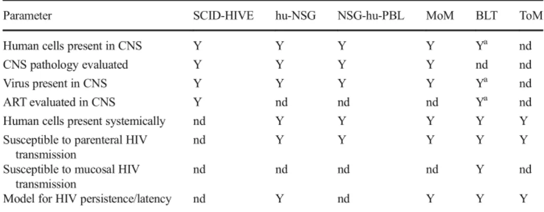
In summary, in all humanized mouse models, the hu-man immune cells exist in a chimeric environment and must interact with the endogenous mous e c ells. However, there is a remarkable correspondence between the human condition and each model is able to recapitu-late specific key aspects of HIV infection, including routes of transmission, response to cART, and establish-ment of viral persistence (Table 1).
Interesting topics for future study
Immune activation in the brains of humanized mice In response to chronic infection with HIV, a state of persistent immune activation occurs systemically. In the CNS, this chronic immune activation can lead to detrimental patholo-gies. While the robust immune response is extremely benefi-cial during early/acute infection, long-term chronic inflamma-tion leads to detrimental effects, including immune exhausinflamma-tion (Deeks et al.2017). By tempering the levels of immune acti-vation via antagonism of the interferon response in humanized BLT mice infected with HIV, improved functioning of CD8+ T cells and a possible decrease in the viral reservoir have been reported (Cheng et al.2017; Zhen et al.2017). Unfortunately, the CNS was not evaluated in these studies. Several studies in SCID-HIVE mice have begun to address this phenomenon in the CNS, including treatment with CEP-1347, B18R, and ruxolitnib (described earlier). However, it is not clear whether the pathology observed in the SCID-HIVE model is represen-tative of the majority of patients in the post-ART era.
Table 1 Comparison of humanized mouse models for studies of NeuroHIV and HIV cure studies
Parameter SCID-HIVE hu-NSG NSG-hu-PBL MoM BLT ToM
Human cells present in CNS Y Y Y Y Ya nd
CNS pathology evaluated Y Y Y Y nd nd
Virus present in CNS Y Y Y Y Ya nd
ART evaluated in CNS Y nd nd nd Ya nd
Human cells present systemically nd Y Y Y Y Y
Susceptible to parenteral HIV transmission
nd Y Y Y Y Y
Susceptible to mucosal HIV transmission
nd nd nd nd Y nd
Model for HIV persistence/latency nd Y nd Y Y Y
Yyes,Nno,ndnot done
a
CD4+ and CD8+ T cell infiltration in the brains of HIV-infected humanized miceEncephalitis in patients on cART was associated with infiltration with CD8+ lymphocytes, sug-gesting these cells may have a role in HAND (Gray et al.
2013). By using systemically reconstituted models, such as hu-NSG, BLT mice, and T cell-only mice (ToM), it may be possible to elucidate the underlying mechanisms driving T cell infiltration into the brain. However, this will be dependent on the integrity of the blood-brain barrier in these models.
Summary
Humanized mouse models reflect key aspects of HIV infec-tion in patients and provide useful research platforms for studying infection in vivo. More widespread inclusion of the CNS into current studies of humanized mouse models, espe-cially with regard to persistence and eradication studies, will advance the field considerably. This will be especially impor-tant in studies using stem cell-transplanted models, which al-low for prolonged observation (> 20 weeks) and could be used to elucidate the effects of long-term cART on the brain. Overall, humanized mice have thus far proved to be valuable in describing the pathogenesis of HIV infection and represent a potentially useful tool for both NeuroHIV and HIV cure research.
Acknowledgements This work has been supported in part by the Virology Training Grant (T32 AI-007419) (JBH) and grants from the National Institute of Mental Health (NIMH) (MH-108179), the National Institute of Allergy and Infectious Diseases (NIAID) (AI-111899), the UNC CFAR (P30 AI050410), and CARE [a Martin Delaney Collaboratory of the NIAID, National Institute of Neurological Disorders and Stroke (NINDS), National Institute On Drug Abuse (NIDA), and the NIMH of the National Institutes of Health, grant number 1UM1AI126619-01] (JVG).
Compliance with ethical standards
Conflict of interest The authors declare that they have no conflict of interest.
References
Ali N, Flutter B, Sanchez Rodriguez R, Sharif-Paghaleh E, Barber LD, Lombardi G, Nestle FO (2012) Xenogeneic graft-versus-host-disease in NOD-scid IL-2Rgammanull mice display a T-effector memory phenotype. PLoS One 7(8):e44219
Apostol BL, Simmons DA, Zuccato C, Illes K, Pallos J, Casale M, Conforti P, Ramos C, Roarke M, Kathuria S, Cattaneo E, Marsh JL, Thompson LM (2008) CEP-1347 reduces mutant huntingtin-associated neurotoxicity and restores BDNF levels in R6/2 mice. Mol Cell Neurosci 39(1):8–20
Arainga M, Edagwa B, Mosley RL, Poluektova LY, Gorantla S, Gendelman HE (2017) A mature macrophage is a principal HIV-1
cellular reservoir in humanized mice after treatment with long acting antiretroviral therapy. Retrovirology 14(1):17
Arainga M, Su H, Poluektova LY, Gorantla S, Gendelman HE (2016) HIV-1 cellular and tissue replication patterns in infected humanized mice. Sci Rep 6:23513
Aspelund A, Antila S, Proulx ST, Karlsen TV, Karaman S, Detmar M, Wiig H, Alitalo K (2015) A dural lymphatic vascular system that drains brain interstitial fluid and macromolecules. J Exp Med 212(7):991–999
Baxter AE, Russell RA, Duncan CJ, Moore MD, Willberg CB, Pablos JL, Finzi A, Kaufmann DE, Ochsenbauer C, Kappes JC, Groot F, Sattentau QJ (2014) Macrophage infection via selective capture of HIV-1-infected CD4+ T cells. Cell Host Microbe 16(6):711–721 Brew BJ, Robertson K, Wright EJ, Churchill M, Crowe SM, Cysique LA,
Deeks S, Garcia JV, Gelman B, Gray LR, Johnson T, Joseph J, Margolis DM, Mankowski JL, Spencer B (2015) HIV eradication symposium: will the brain be left behind? J Neuro-Oncol 21(3):322– 334
Calantone N, Wu F, Klase Z, Deleage C, Perkins M, Matsuda K, Thompson EA, Ortiz AM, Vinton CL, Ourmanov I, Lore K, Douek DC, Estes JD, Hirsch VM, Brenchley JM (2014) Tissue myeloid cells in SIV-infected primates acquire viral DNA through phagocytosis of infected T cells. Immunity 41(3):493–502 Cheng L, Ma J, Li J, Li D, Li G, Li F, Zhang Q, Yu H, Yasui F, Ye C, Tsao
LC, Hu Z, Su L, Zhang L (2017) Blocking type I interferon signal-ing enhances T cell recovery and reduces HIV-1 reservoirs. J Clin Invest 127(1):269–279
Choi B, Chun E, Kim M, Kim SY, Kim ST, Yoon K, Lee KY, Kim SJ (2011) Human T cell development in the liver of humanized NOD/ SCID/IL-2Rgamma(null) (NSG) mice generated by intrahepatic in-jection of CD34(+) human (h) cord blood (CB) cells. Clin Immunol 139(3):321–335
Cohen RA, Seider TR, Navia B (2015) HIV effects on age-associated neurocognitive dysfunction: premature cognitive aging or neurode-generative disease? Alzheimers Res Ther 7(1):37
Conforti P, Ramos C, Apostol BL, Simmons DA, Nguyen HP, Riess O, Thompson LM, Zuccato C, Cattaneo E (2008) Blood level of brain-derived neurotrophic factor mRNA is progressively reduced in ro-dent models of Huntington's disease: restoration by the neuroprotec-tive compound CEP-1347. Mol Cell Neurosci 39(1):1–7
Cook-Easterwood J, Middaugh LD, Griffin WC 3rd, Khan I, Tyor WR (2007) Highly active antiretroviral therapy of cognitive dysfunction and neuronal abnormalities in SCID mice with HIV encephalitis. Exp Neurol 205(2):506–512
Cook JE, Dasgupta S, Middaugh LD, Terry EC, Gorry PR, Wesselingh SL, Tyor WR (2005) Highly active antiretroviral therapy and human immunodeficiency virus encephalitis. Ann Neurol 57(6):795–803 Dash PK, Gendelman HE, Roy U, Balkundi S, Alnouti Y, Mosley RL,
Gelbard HA, McMillan J, Gorantla S, Poluektova LY (2012) Long-acting nanoformulated antiretroviral therapy elicits potent antiretro-viral and neuroprotective responses in HIV-1-infected humanized mice. AIDS 26(17):2135–2144
Dash PK, Gorantla S, Gendelman HE, Knibbe J, Casale GP, Makarov E, Epstein AA, Gelbard HA, Boska MD, Poluektova LY (2011) Loss of neuronal integrity during progressive HIV-1 infection of human-ized mice. J Neurosci 31(9):3148–3157
Deeks SG, Odorizzi PM, Sekaly RP (2017) The interferon paradox: can inhibiting an antiviral mechanism advance an HIV cure? J Clin Invest 127(1):103–105
Denton PW, Estes JD, Sun Z, Othieno FA, Wei BL, Wege AK, Powell DA, Payne D, Haase AT, Garcia JV (2008) Antiretroviral pre-exposure prophylaxis prevents vaginal transmission of HIV-1 in humanized BLT mice. PLoS Med 5(1):e16
Denton PW, Krisko JF, Powell DA, Mathias M, Kwak YT, Martinez-Torres F, Zou W, Payne DA, Estes JD, Garcia JV (2010) Systemic administration of antiretrovirals prior to exposure prevents rectal and intravenous HIV-1 transmission in humanized BLT mice. PLoS One 5(1):e8829
Denton PW, Long JM, Wietgrefe SW, Sykes C, Spagnuolo RA, Snyder OD, Perkey K, Archin NM, Choudhary SK, Yang K, Hudgens MG, Pastan I, Haase AT, Kashuba AD, Berger EA, Margolis DM, Garcia JV (2014) Targeted cytotoxic therapy kills persisting HIV infected cells during ART. PLoS Pathog 10(1):e1003872
Denton PW, Olesen R, Choudhary SK, Archin NM, Wahl A, Swanson MD, Chateau M, Nochi T, Krisko JF, Spagnuolo RA, Margolis DM, Garcia JV (2012) Generation of HIV latency in humanized BLT mice. J Virol 86(1):630–634
Eggert D, Dash PK, Gorantla S, Dou H, Schifitto G, Maggirwar SB, Dewhurst S, Poluektova L, Gelbard HA, Gendelman HE (2010) Neuroprotective activities of CEP-1347 in models of neuroAIDS. J Immunol 184(2):746–756
Fritz-French C, Shawahna R, Ward JE, Maroun LE, Tyor WR (2014) The recombinant vaccinia virus gene product, B18R, neutralizes inter-feron alpha and alleviates histopathological complications in an HIV encephalitis mouse model. J Interf Cytokine Res 34(7):510–517 Gorantla S, Makarov E, Finke-Dwyer J, Castanedo A, Holguin A,
Gebhart CL, Gendelman HE, Poluektova L (2010) Links between progressive HIV-1 infection of humanized mice and viral neuropathogenesis. Am J Pathol 177(6):2938–2949
Gray F, Lescure FX, Adle-Biassette H, Polivka M, Gallien S, Pialoux G, Moulignier A (2013) Encephalitis with infiltration by CD8+ lym-phocytes in HIV patients receiving combination antiretroviral treat-ment. Brain Pathol 23(5):525–533
Haile WB, Gavegnano C, Tao S, Jiang Y, Schinazi RF, Tyor WR (2016) The Janus kinase inhibitor ruxolitinib reduces HIV replication in human macrophages and ameliorates HIV encephalitis in a murine model. Neurobiol Dis 92(Pt B):137–143
Heaton RK, Clifford DB, Franklin DR Jr, Woods SP, Ake C, Vaida F, Ellis RJ, Letendre SL, Marcotte TD, Atkinson JH, Rivera-Mindt M, Vigil OR, Taylor MJ, Collier AC, Marra CM, Gelman BB, McArthur JC, Morgello S, Simpson DM, McCutchan JA, Abramson I, Gamst A, Fennema-Notestine C, Jernigan TL, Wong J, Grant I, Group C (2010) HIV-associated neurocognitive disorders persist in the era of potent antiretroviral therapy: CHARTER study. Neurology 75(23):2087–2096
Honeycutt JB, Thayer WO, Baker CE, Ribeiro RM, Lada SM, Cao Y, Cleary RA, Hudgens MG, Richman DD, Garcia JV (2017) HIV persistence in tissue macrophages of humanized myeloid-only mice during antiretroviral therapy. Nat Med 23:638–643
Honeycutt JB, Wahl A, Archin N, Choudhary S, Margolis D, Garcia JV (2013) HIV-1 infection, response to treatment and establishment of viral latency in a novel humanized T cell-only mouse (TOM) model. Retrovirology 10:121
Honeycutt JB, Wahl A, Baker C, Spagnuolo RA, Foster J, Zakharova O, Wietgrefe S, Caro-Vegas C, Madden V, Sharpe G, Haase AT, Eron JJ, Garcia JV (2016) Macrophages sustain HIV replication in vivo independently of T cells. J Clin Invest 126(4):1353–1366 Kim KC, Choi BS, Kim KC, Park KH, Lee HJ, Cho YK, Kim SI, Kim
SS, Oh YK, Kim YB (2016) A simple mouse model for the study of human immunodeficiency virus. AIDS Res Hum Retrovir 32(2): 194–202
Koneru R, Olive MF, Tyor WR (2014) Combined antiretroviral therapy reduces brain viral load and pathological features of HIV encepha-litis in a mouse model. J Neuro-Oncol 20(1):9–17
Krisko JF, Martinez-Torres F, Foster JL, Garcia JV (2013) HIV restriction by APOBEC3 in humanized mice. PLoS Pathog 9(3):e1003242 Louveau A, Smirnov I, Keyes TJ, Eccles JD, Rouhani SJ, Peske JD,
Derecki NC, Castle D, Mandell JW, Lee KS, Harris TH, Kipnis J (2015) Structural and functional features of central nervous system lymphatic vessels. Nature 523(7560):337–341
Lund S, Porzgen P, Mortensen AL, Hasseldam H, Bozyczko-Coyne D, Morath S, Hartung T, Bianchi M, Ghezzi P, Bsibsi M, Dijkstra S, Leist M (2005) Inhibition of microglial inflammation by the MLK inhibitor CEP-1347. J Neurochem 92(6):1439–1451
Marsden MD, Kovochich M, Suree N, Shimizu S, Mehta R, Cortado R, Bristol G, An DS, Zack JA (2012) HIV latency in the humanized BLT mouse. J Virol 86(1):339–347
Miura Y, Misawa N, Kawano Y, Okada H, Inagaki Y, Yamamoto N, Ito M, Yagita H, Okumura K, Mizusawa H, Koyanagi Y (2003) Tumor necrosis factor-related apoptosis-inducing ligand induces neuronal death in a murine model of HIV central nervous system infection. Proc Natl Acad Sci U S A 100(5):2777–2782
Mlcochova P, Sutherland KA, Watters SA, Bertoli C, de Bruin RA, Rehwinkel J, Neil SJ, Lenzi GM, Kim B, Khwaja A, Gage MC, Georgiou C, Chittka A, Yona S, Noursadeghi M, Towers GJ, Gupta RK (2017) A G1-like state allows HIV-1 to bypass SAMHD1 restriction in macrophages. EMBO J 36(5):604–616 Persidsky Y, Buttini M, Limoges J, Bock P, Gendelman HE (1997) An
analysis of HIV-1-associated inflammatory products in brain tissue of humans and SCID mice with HIV-1 encephalitis. J Neuro-Oncol 3(6):401–416
Persidsky Y, Gendelman HE (1997) Development of laboratory and an-imal model systems for HIV-1 encephalitis and its associated de-mentia. J Leukoc Biol 62(1):100–106
Persidsky Y, Ghorpade A, Rasmussen J, Limoges J, Liu XJ, Stins M, Fiala M, Way D, Kim KS, Witte MH, Weinand M, Carhart L, Gendelman HE (1999) Microglial and astrocyte chemokines regu-late monocyte migration through the blood-brain barrier in human immunodeficiency virus-1 encephalitis. Am J Pathol 155(5):1599– 1611
Persidsky Y, Limoges J, McComb R, Bock P, Baldwin T, Tyor W, Patil A, Nottet HS, Epstein L, Gelbard H, Flanagan E, Reinhard J, Pirruccello SJ, Gendelman HE (1996) Human immunodeficiency virus encephalitis in SCID mice. Am J Pathol 149(3):1027–1053 Prochazka M, Gaskins HR, Shultz LD, Leiter EH (1992) The nonobese
diabetic scid mouse: model for spontaneous thymomagenesis asso-ciated with immunodeficiency. Proc Natl Acad Sci U S A 89(8): 3290–3294
Sun Z, Denton PW, Estes JD, Othieno FA, Wei BL, Wege AK, Melkus MW, Padgett-Thomas A, Zupancic M, Haase AT, Garcia JV (2007) Intrarectal transmission, systemic infection, and CD4+ T cell deple-tion in humanized mice infected with HIV-1. J Exp Med 204(4): 705–714
Tyor WR, Power C, Gendelman HE, Markham RB (1993) A model of human immunodeficiency virus encephalitis in scid mice. Proc Natl Acad Sci U S A 90(18):8658–8662
Wu X, Liu L, Cheung KW, Wang H, Lu X, Cheung AK, Liu W, Huang X, Li Y, Chen ZW, Chen SM, Zhang T, Wu H, Chen Z (2016) Brain invasion by CD4(+) T cells infected with a transmitted/founder HIV-1BJZS7 during acute stage in humanized mice. J NeuroImmune Pharmacol 11(3):572–583