CONTRIBUTIONS OF THE HOST IMMUNE RESPONSE TO CONTROL AND PROTECTION UPON INFECTION WITH A NEUROVIRULENT ALPHAVIRUS
Amy Catherine Wollish
A dissertation submitted to the faculty of the University of North Carolina at Chapel Hill in partial fulfillment of the requirements for the degree of Doctor of Philosophy in the
Department of Microbiology and Immunology.
Chapel Hill 2012
ii ©2012
iii ABSTRACT
AMY CATHERINE WOLLISH: Contributions of the host immune response to control and protection upon infection with a neurovirulent alphavirus.
(Under the direction of Mark Heise, Ph.D.)
Alphaviruses are mosquito-borne viruses within the family Togaviridae. These
positive-sense RNA viruses pose a significant human and equine public health threat due to their ability to cause explosive epidemics of infectious rheumatic disease and encephalitis. The AR86 strain of Sindbis virus (SINV, infectious clone: S300) is neurovirulent in adult mice. A critical viral genetic determinant of neurovirulence within S300 is a Threonine at nonstructural protein (nsP) 1 position 538, whereby introduction of an Isoleucine at this locus is attenuating in vivo. The mutant nsP1 T538I virus induces more type I interferon (IFN) and fails to efficiently block type I and II IFN signaling as compared to S300 virus. Importantly, nsP1 T538I replicates as well as S300 virus at early times post-infection within the central nervous system (CNS) of infected mice, however at late times post-infection, nsP1 T538I is more efficiently controlled and cleared. In this work, we investigated the components of the host innate and adaptive immune system that modulate AR86-induced neurologic disease.
iv
of TLR4 signaling, is moderately protective against the AR86 strain of SINV. IPS-1 and TRIF are adaptor molecules essential for signal transduction that results in the upregulation of interferon stimulated genes (ISGs), which in turn exert virus-specific antiviral activity. These studies reveal that two ISGs, IFIT1 and IFIT2, inhibit nsP1 T538I replication within IFN-β primed murine cells.
v
To my parents and family, who have provided limitless support and love,
To my brothers, for whom I dedicate my scientific pursuit.
And to Matt, who gives me the confidence to dare And the guts to fail.
vi
ACKNOWLEDGEMENTS
I owe a great deal of appreciation to the numerous people who have supported me throughout graduate school. First, I thank my mentor, Mark Heise, who was always available to provide scientific support and to assist in the development of experimental design. Mark shared his time and insights freely, and instilled in me the confidence to take on difficult experiments, and the ability to recognize when an experimental path is not worth pursuing. Mark fostered a lab where every member respected one another, and was willing to assist one another in times of need.
vii
thank Toni Baric, Rhonda Haithcock, and Lisa Phelps for administrate support and friendship.
Thank you to the DLAM staff at the Mary Ellen Jones mouse facility, who executed difficult and tedious jobs while maintaining smiles and a good outlook. I would like to thank Dixie Flannery for her attentiveness and superb execution of her job of managing graduate students. She helped in the logistics of each stage during graduate school.
viii
TABLE OF CONTENTS
LIST OF TABLES...xii
LIST OF FIGURES...xiii
LIST OF ABBREVIATIONS...xv
CHAPTER I. INTRODUCTION...2
1.1 Alphavirus background... 1
Alphavirus structure and genome organization ... 2
Evolution of alphaviruses ... 2
1.2 Alphavirus life-cycle ... 4
The elusive alphavirus receptor ... 4
Entry ... 5
Protein translation and processing ... 6
The nonstructural proteins ... 7
The structural proteins ... 8
1.3 Pathogenic alphaviruses and epidemics ... 8
Symptoms ... 8
Transmission ... 9
Alphavirus epidemics ... 10
ix
Isolation and selection histories ... 11
AR339 ... 12
TR339: heparan sulfate binding ... 13
NSV ... 14
SVA/SVB and SVN/SVNI ... 15
AR86 ... 15
AR86 neurovirulence determinants ... 16
1.5 The complexity of the immune response to invading pathogens within the CNS...17
Viral clearance from the CNS ... 19
Clearance of alphaviruses from the CNS ... 19
1.6 The type I IFN system ... 22
Discovery of interferon ... 22
Pattern recognition receptors ... 23
TLR recognition of RNA virus PAMPs ... 24
RLR recognition of RNA virus PAMPs ... 24
PRRs that mediate type I IFN induction in response to alphavirus infection ... 26
Type I IFN signaling/amplification ... 27
Evidence for the contribution of type I IFN to the adaptive immune response .... 28
Type II IFN ... 29
Type III IFN ... 30
Type I and type II IFN crosstalk ... 30
x
Alphavirus evasion of innate immunity ... 31
ISGs ... 32
ISG15 ... 33
Viperin ... 34
ZAP ... 34
Potential anti-SINV role for IFIT family members ... 36
Autophagy ... 36
1.8 Dissertation objectives ... 37
II. AN ATTENUATING MUTATION IN A NEUROVIRULENT SINDBIS VIRUS STRAIN INTERACTS WITH THE IPS-1 SIGNALING PATHWAY IN VIVO...39
2.1 Introduction... 39
2.2 Materials and Methods... 43
2.3 Results ... 46
2.4 Discussion ... 52
2.5 Acknowledgements ... 56
III. ADAPTIVE IMMUNITY CONTRIBUTES TO PROTECTION FROM SINDBIS-INDUCED NEUROLOGIC DISEASE...82
3.1 Introduction... 82
3.2 Materials and Methods... 84
3.3 Results ... 90
3.4 Discussion ... 95
3.5 Acknowledgements ... .99
IV. DISCUSSION... 124
4.1 Type I IFN limits SINV replication and spread ... 124
xi
4.3 IPS-1 does not mediate all of the protection conferred by type I IFN ... 127
4.4 The adaptive immune response modulates SINV ... 129
4.5 Contradictory phenotypes in IFN-γ receptor and cytokine deficient mice ... 131
4.6 Conclusions ... 132
xii
LIST OF TABLES
Table 2.1: Percent mortality and median survival times after S300 or nsP1 T538I i.c. infection of C57BL/6J mice and each knockout mouse line...78 Table 2.2: Statistical differences in survival curves within each
xiii
LIST OF FIGURES
Figure 2.1: The mutant virus nsP1 T538I is attenuated in C57BL/6J mice as compared to S300 wild-type virus………...…………...…....…...……58 Figure 2.2: Both S300 and nsP1 T538I show equivalent and enhanced
virulence in the absence of the IFN-α/β response……...…...…....…61 Figure 2.3: TLR signaling through MyD88 is not required for
protection or control of nsP1 T538I infection...………. ..…...………….64 Figure 2.4: TRIF signaling is not required for viral replication
control of nsP1 T538I, but TRIF may play a minor role in protection from SINV-induced disease………...………....…...……67 Figure 2.5: IPS-1 is required for protection from nsP1 T538I
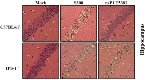
and contributes to control of nsP1 T538I………...………...…...72 Figure 2.6: Increased quantity and distribution of viral RNA and
neuronal damage in IPS-1-/- mice infected with S300 and nsP1 T538I as compared with C57BL/6 mice infected with nsP1 T538I...………..75 Supplemental Figure 2.1: TLR4 signaling is dispensable for conferring the attenuated phenotype during nsP1 T538I infection………...……80 Figure 3.1: There is no difference in CNS inflammatory infiltrate
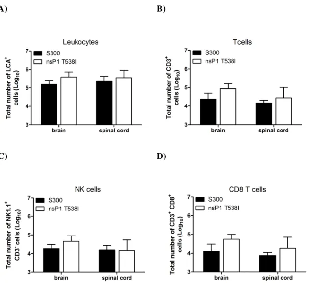
after S300 or nsP1 T538I infection……...………...……...……….100 Figure 3.2: RAG-1-/- mice succumb to nsP1 T538I infection and
fail to control nsP1 T538I replication in the CNS…...………...…..103 Figure 3.3: µMT mice are capable of recovering from nsP1 T538I
infection, yet µMT exhibit a defect in control of nsP1 T538I
xiv
Figure 3.5: IFN-γ signaling is not required for protection from
Disease or control of the nsP1 T538I virus...111 Figure 3.6: IFN-γ cytokine is pathogenic during SINV infection…….……...……...114 Figure 3.7: Expression of the IFN α/β receptor on splenocytes is not
required for control of nsP1 T538I…………...…...……...118 Figure 3.8: No differences in quantity or quality of serum antibody
responses after S300 or nsP1 T538I infection.………...…….….120 Supplemental Figure 3.1: Representative quantification of CD3, CD4,
CD8, and isotype control depleted mice...122 Figure 4.1: IFITs inhibit nsP1 T538I replication in IFN-β pretreated MEFs......134 Figure 4.2: Ifnar-/- and IPS-1-/- mice are more susceptible than C57BL/6J mice to nsP1 T538I peripheral
infection...Error! Bookmark not defined.
xv
LIST OF ABBREVIATIONS
CARD Caspase recruitment domain CHIKV Chikungunya virus
CNS Central nervous system DC Dendritic cell
dpi Days post-infection dsRNA Double-stranded RNA HS Heparan sulfate hpi Hours post-infection i.c. intracranial
IFN Interferon
IFNAR Interferon α/β receptor
IPS-1 Interferon promoter stimular-1 IRF Interferon regulatory factor ISG Interferon stimulated gene
ISGF3 Interferon-stimulated gene factor 3 Jak Janus kinase
xvi NF-κB Nuclear factor kappa B nsP nonstructural protein
NSV Neuro-adapted Sindbis virus
PAMP Pathogen-associated molecular pattern
PKR Protein kinase R/dsRNA-dependent protein kinase R POGD Pogosta virus disease
PRR Pattern recognition receptor RLR RIG-I-like receptor
RRV Ross River virus SFV Semliki Forest virus SH2 Src-homology 2 SINV Sindbis virus
SIRS Systemic inflammatory response syndrome ssRNA Single-stranded RNA
STAT Signal transducer and activator of transcription TANK TRAF family member-associated NF-κB activator TBK1 TANK-binding kinase 1
TLR Toll-like receptor
TRIF TIR-domain-containing adaptor including interferon-β Tyk2 Tyrosine kinase 2
CHAPTER ONE: INTRODUCTION
1.1 Alphavirus background
Alphaviruses are small, enveloped viruses with a positive-sense single stranded RNA genome and belong to the family Togaviridae. Alphaviruses have a nearly worldwide distribution and cycle in nature between an arthropod vector (mainly mosquito) and a vertebrate host, such as a small mammal or bird. However, these viruses can acquire mutations resulting in an increased mosquito host range and/or spillover infection of equines and humans. Thereby, alphaviruses can cause epidemics, and represent a significant public health threat. Human infection by pathogenic Old World alphaviruses can cause long-lasting arthritis and myositis, while human and equine infection by pathogenic New World alphaviruses can cause encephalitis with a threat of mortality. Currently, no specific vaccine or antiviral therapy exists to prevent or treat alphavirus infection.
2
interferon response, the immune system within the central nervous system (CNS), and known alphavirus-host interactions.
Alphavirus structure and genome organization
Alphaviruses have a single-stranded positive-sense RNA genome of approximately 11.7 Kb in length. The genome is flanked by a 5’ cap and a 3’ poly-adenylated tail, and contains two open-reading frames. Expression of the nonstructural genes is under the control of a 49S promoter, while the structural genes are regulated by an internal 26S promoter. The genomic RNA is enclosed by 240 copies of the capsid protein arranged in icosohedral symmetry. The nucleocapsid is surrounded by envelope glycoproteins, E1 and E2 that are embedded within a lipid bilayer derived from the host cell plasma membrane. E1 and E2 form 80 trimeric spikes with T = 4 quasi-symmetry (295).
Evolution of alphaviruses
Alphaviruses are arboviruses, and thus are transmitted by arthropod vectors (e.g. mosquitoes). The dual-host life cycle of alphaviruses mandates that these viruses maintain fitness in vector and reservoir hosts with markedly different microenvironments and selective pressures. Yet, the wide distribution of alphaviruses demonstrates that these viruses are very capable of usurping the distinct host machinery to replicate and spread successfully.
3
Encephalitis (WEE) complex (which includes SINV), the Venezuelan Equine Encephalitis (VEE) complex, and the Eastern Equine Encephalitis (EEE) complex (38, 81). Even the most recent comprehensive alphavirus phylogenetic analysis (81) cannot distinguish between a New World or an Old World origin of alphaviruses. However, it is clear that numerous reintroductions occurred after the initial introduction in one of the two hemispheres (81, 246). Most interestingly, these analyses suggest that alphaviruses may have emerged in the Pacific Ocean in either terrestrial or marine hosts, and likely were spread via the mosquito. This hypothesis was, in large part, derived from the sequencing and subsequent placement of the aquatic alphaviruses, salmon pancreatic disease virus and Southern elephant seal virus, at basal positions in the phylogenetic tree (81).
4 1.2 Alphavirus life-cycle
The elusive alphavirus receptor
Alphaviruses infect a broad host range and numerous cell types within each host, including dendritic cells (DCs), muscle cells, connective tissues, neurons, and fibroblasts. Alphaviruses enter the host cell via receptor-mediated endocytosis at the plasma membrane, a process that is discussed in detail below. Due to the wide array of infected cell types, mode of entry, and diverse host range, the alphavirus receptor must be evolutionarily conserved and nearly ubiquitously expressed at the plasma membrane.
Despite an immense effort, the identification of a true alphavirus receptor was not made until 2011. This delay was in part due to the confounding tendency of alphaviruses to acquire cell culture adaptations that confer the ability to interact with the glycosaminoglycan, heparan sulfate (158). Whereas heparan sulfate does not mediate entry of most wildtype alphaviruses, certain wildtype EEE strains interact with heparan sulfate (discussed in detail in section 1.4) (158).
5
than mammalian-derived RRV containing complex glycans (156, 283). Finally, the laminin receptor may serve as an alphavirus attachment factor and thus enhance infection (326).
Only recently has the SINV receptor been identified as being Natural Resistance-Associated Macrophage Protein (NRAMP) (254). NRAMPs are highly conserved proteins that have 12 transmembrane domains involved in transport of heavy metal ions such as iron across cell membranes (8, 226). Importantly, humans have 2 NRAMP genes: NRAMP1, which is expressed only on the membrane of late endosomes and lysosomes of macrophages, dendritic cells (113, 279), in granules of polymorphonuclear leukocytes (39), and in neurons (73), while NRAMP2 is ubiquitously expressed at the plasma membrane and in endosomes (226). In Drosophila cells, dNRAMP is required for SINV binding and infection, and in mammalian cells, NRAMP2 is required for SINV binding and infection. Somewhat surprisingly, Ross River virus (RRV) entry is independent of NRAMP, and whether NRAMP or other receptors mediate entry of other alphaviruses has yet to be determined. Interestingly, NRAMP2 is sensitive to the effects of iron, whereby both NRAMP2 mRNA and protein are degraded when iron levels are high (80, 191, 203). Treatment of insect or mammalian cells with iron significantly reduced SINV infection (254), thereby suggesting that high amounts of iron may affect the fitness of SINV in nature. Further studies are needed to clarify whether NRAMP1 has a role in alphavirus entry or fusion with the endosomal membrane.
Entry
6
induces a conformational change and release of E1 from the E1-E2 heterodimers. E1 homotrimers then bind the target vesicle membrane by inserting the hydrophobic fusion peptide and forming pores in the cell and viral membranes (176, 227, 274, 294, 295). While receptor-mediated, clathrin-dependent endocytosis and fusion with endosomes is likely the predominant entry route, there is evidence that alphaviruses can enter via a clathrin-independent pathway (43, 53, 125, 235, 325). Cholesterol is necessary for fusion of SINV, CHIKV, and SFV; however VEEV fusion does not require cholesterol (18, 162).
Protein translation and processing
7
295). Therefore the nsP4 protein is produced at significantly lower levels than P123. In addition, nsP4 protein levels are restricted by the presence of a destabilizing tyrosine at the N-terminus of nsP4 that initiates degradation by the N-end rule pathway of any free nsP4 that is not present within replication complexes (60).
The structural proteins are translated late during infection as a second polyprotein, and include capsid, the E1 and E2 envelope proteins, and the smaller proteins 6K and E3 (84, 295). Capsid protein cleaves itself from the structural polyprotein (233, 335). E3 contains a signal sequence that directs the remaining envelope polyprotein precursor E3-E2-6K-E1 into the lumen of the endoplasmic reticulum (ER), where host signalases process the N- and C-terminus of the 6K peptide to generate E3E2 (also referred to as PE2), 6K, and E1. Subsequently, E3E2 and E1 form heterodimers in the early Golgi, traffic to the plasma membrane, and E3E2 is cleaved by a host furin-like protease to release mature E2 and E3 (58, 266). Capsid oligomerizes and recognizes a packaging signal within either nsP1 or nsP2 to direct encapsidation of the viral genomic RNA into the virion. Capsid interacts with E2 to facilitate budding from the plasma membrane. The budded virion retains a lipid membrane from the host cell plasma membrane.
The nonstructural proteins
8
cysteine protease activity in the N-terminal region (100, 252, 319). While nsP3 is essential for replication, only recently was it shown to contain a conserved proline-rich Src homology-3 (SHhomology-3) domain that is essential for interactions with amphiphysin-1 and Bin1/amphiphysin-2, two cellular adaptor proteins involved in endocytosis and membrane trafficking (225). Mutations within the nsP3 SH3 binding site resulted in reduced viral replication in vitro and decreased pathogenicity in mice (225). The nsP4 protein serves as the RNA-dependent RNA-polymerase (RdRp) (115), and possesses adenylyltransferase activity, with both enzymatic functions attributed to a GDD amino acid motif within the C-terminus (308).
The structural proteins
To facilitate assembly of the virion, the capsid protein binds viral genomic RNA and glycoproteins (233, 234, 334). The function of E3 is not defined and only certain alphaviruses incorporate E3 into the virion, with SINV not being one of those viruses (287). The E2 protein mediates receptor binding via 30 C-terminal amino acid residues (190, 292), while the E1 protein contains a fusion peptide that facilitates fusion with the host cell (22, 230). Lastly, the palmitoylated 6K protein may be involved in transport to viral assembly sites at the plasma membrane (90, 197).
1.3 Pathogenic alphaviruses and epidemics
Symptoms
9
either encephalitis, as is the case for the New World viruses or arthralgia and arthritis, as is the case for the Old World viruses (107). Arthralgia occurs mainly in small joints and more often in adults than in children. In severe cases, joints can become swollen and arthralgia may persist for weeks or months.
The alphaviruses associated with encephalitis are WEEV, VEEV, and EEEV, and less commonly RRV and CHIKV (reviewed in (348)). WEEV and VEEV cause a febrile disease that only rarely progresses to encephalitis, however in equines encephalitis is common and the mortality rate after VEEV infection is greater than 50% (324). WEEV and VEEV have a human case fatality rate of 3-7% and 0.5-1%, respectively (348). VEEV has the capability to be transmitted by the aerosol route, and has been developed as a biological weapon in the United States and the Soviet Union.
Humans infected with the highly pathogenic North American EEEV experience fever, chills, myalgia, arthralgia, retro-ocular pain, headache, and decreased consciousness, whereas the very young often progress to neurologic symptoms in the absence of other symptoms (64). EEEV causes neurologic disease, marked by paralysis, seizures, coma and death in 30-80% of infected humans, with long-term neurologic deficits reported in 35% of surviving individuals (103).
Transmission
10
alphaviruses to enter an epizootic transmission cycle, whereby an arthropod transmits the virus to a horse, human, or other large mammal (24, 25, 328).
Alphavirus epidemics
Historically, there are multiple of examples of alphavirus crossover to human populations or spread to new regions. Although endemic to Australia, RRV caused an outbreak in 1979, when it spread to the island of Fiji, American Samoa, and the Cook islands (1, 16, 257). During this epidemic, ~500,000 humans were infected, with ~50,000 of them experiencing severe arthralgia, rash and fever (1, 16). Patients infected with RRV report symptoms of acute fever, rash, myalgia, and arthralgia that can persist for upwards of 6 months. In 1995, an outbreak of VEEV occurred in Venezuela and Columbia that resulted in 75,000-100,000 affected humans and a case fatality rate of 0.7% (44, 45, 331).
11
SINV has the distinction of being the most widely distributed of all arboviruses, and has been isolated in Europe, the Middle East, Africa, India, Asia, Australia, New Zealand, and the Philippines, with a likelihood of having been present in the Western hemisphere (159). SINVs use birds as their vertebrate hosts and the Culex mosquito species as vectors (196). Interestingly, migratory bird patterns can serve to explain the genetic relatedness of SINV genotypes, thereby supporting the idea that birds drive the spread of SINV (196). Despite its near worldwide prevalence, SINV is documented as being the etiologic agent of outbreaks of rash and arthritis only in South Africa, Sweden (Ockelbo virus), Finland (Pogosta disease), and the Karelian part of Russia (Karelian fever) (31, 195, 211). SINV does not undergo human-mosquito-human transmission, but rather outbreaks are thought to arise when strains that normally circulate in Culex or Culiseta spp. and birds gain the ability to infect human-feeding mosquitoes, such as Aedes spp. (159).
1.4 The SINV mouse-model
Isolation and selection histories
12
The outcome of SINV infection in mice is dependent upon mouse age and strain, route of inoculation, and virus strain (105, 106, 171, 316). As a group, all SINVs cause lethal disease in neonatal mice, marked by high levels of proinflammatory cytokines, and uncontrolled replication in the muscle, brain, and serum (157, 312, 313). All SINVs, with the exception of strains AR86, NSV, and SVNI, are completely avirulent in adult mice, regardless of the dose or inoculation route. However, of the three adult neurovirulent strains, SVNI is unique in that it is also neuro-invasive in adult mice. The age-dependent resistance to neuro-invasion is not specific to SINV in mice, and has been reported for numerous neurotropic virus families including rhabdoviruses, reoviruses, bunyaviruses, and flaviviruses (reviewed in (229)).
AR339
13
response syndrome (SIRS) (133). Therefore, neonatal mice infected with TR339 most likely die from SIRS (313).
To better understand the host mechanism of age-resistance to TR339, adult mice deficient in IFN-α/β receptor (Ifnar-/-), IFN-γ receptor (IFN-γ R-/-), or doubly deficient mice were infected. Ifnar-/- displayed a rapid progression to death, while IFN-γ R-/- mice showed no disease signs and were indistinguishable from wt mice. The double-knockout mice succumbed even more quickly than Ifnar-/- mice. Both mouse strains that succumbed to TR339 infection exhibited increased viral tropism for macrophage-DC lineage cells that are not normally infected in wt mice. Therefore, these results suggest that type I IFN is a critical component of age-dependent resistance, with IFN-γ having an accessory role (108). A later study compared CNS gene expression during lethal neonatal infection with TR339 and avirulent 4-week old infection with TR339 by the i.c. route of infection. Although most host inflammatory genes are up regulated in neonates infected with TR339 as compared to weanling mice, ISG12 was down regulated in neonates. However, 100% lethality was observed in neonatal mice infected with the SINV expressing ISG12, and viral replication was unchanged by ISG12 expression (172). Other studies have suggested that age-dependent resistance may be related to the resistance of adult neurons to apoptosis (reviewed in (109)).
TR339: heparan sulfate binding
14
comprehensive analysis demonstrated that HS-binding mutations are attenuated by the subcutaneous route, but are actually more virulent when delivered intracranially (i.c.). Ryman et al. postulated that this discrepancy reflected enhanced CNS-specific replication, and/or impaired peripheral replication of HS mutants (258). Furthermore, sequence data from wild type isolates of circulating strains EEEV revealed the presence of HS-binding properties (92, 347). Mutating the HS-binding site within EEEV decreased neurovirulence in vivo, but increased lymphoid tissue replication and cytokine and chemokine production (92). Therefore, HS binding may have relevance in nature, however further work is necessary to fully understand the selective pressures that drive HS binding and the resulting affects on in vivo virulence.
NSV
15
SVA/SVB and SVN/SVNI
Two pairs of SINV variants (SVA and SVB) and (SVN and SVNI) were derived by separate passages, but from a single virus isolated from a pool of culicine mosquitoes collected in southern Israel in 1983 (198). SVN was generated after 15 alternating passages in suckling and weanling mouse brains and is neurovirulent. To generate SVNI, an addition 7 passages were performed in weanling mice, and SVNI is both neurovirulent and neuro-invasive in adult mice (69, 161). The neuro-neuro-invasive determinants within SVNI were mapped to the 5’ noncoding region (NCR) and the E2 position 190 (Met-190) (69, 161). SVA and SVB are early passage isolates that are neurotropic, but not neurovirulent, and SVB is nonlethal, but neuro-invasive. The neuro-invasive determinant for SV was mapped to E2 position 55 (Gln-55) (69, 161). Of the four strains derived from the Israeli isolate, only SVNI is neurovirulent in adult rats (but not neuro-invasive), and this determinant was also mapped to the 5’ NCR (160). While these viruses have not been extensively studied, they provide excellent tools, and the exclusive opportunity to study neuro-invasion of SINVs.
AR86
16
AR86 causes lethal neurologic disease in adult mice (291). Mice exhibit signs of disease including weight loss, ruffled fur, hunched posture, hind-limb dysfunction, paralysis, and ultimately become moribund (122, 297). Even after i.c. inoculation, the closely related G100 is avirulent in adult mice, although G100 replicates in the CNS equivalently to S300 at early times post-infection. The host innate and adaptive immune pathways that modulate in vivo pathogenesis of AR86 and control of attenuated mutants of AR86 have not yet been published (with the exception of type I IFN), and are therefore the subject of this dissertation. However, extensive studies have characterized the AR86 genetic determinants of neurovirulence, and these results will be detailed in the following section.
AR86 neurovirulence determinants
17
infected animals (56), and a Thr at nsP1 position 538 is both necessary and sufficient to block Jak/STAT signaling in response to type I or II IFN (289).
Sequence analysis and comparison between S300 and G100 revealed 20 single amino acid differences, in addition to an 18 amino acid deletion within nsP3 (291, 297). Because S300 causes lethal neurologic disease and G100 is avirulent, these viruses were ideal tools to investigate the genetic determinants of neurologic disease. Chimeric S300 and Girdwood viruses were constructed, and infected by the i.c. route into adult outbred CD-1 mice to determine virulence (297). These studies revealed 4 critical genetic determinants of S300 neurovirulence, with 3 in the nonstructural genes and 1 in the structural genes. These determinants include the aforementioned nsP1 T538I (Thr is virulent), an 18 amino acid deletion within the C-terminus of nsP3 (deletion is virulent), nsP3 Opal537Cys (Cys is virulent), and E2 Leu243Ser (Ser is virulent). Initially, the mutation of these single attenuating mutations suggested their role as determinants. Furthermore, introduction of all 4 determinants into the avirulent G100 background was able to rescue nearly full virulence of S300. Similar to G100, when the 4 attenuating are introduced into the S300 backbone, this virus can establish infection within the CNS equivalent to S300, but is more efficiently controlled at late times post-infection (297).
1.5 The complexity of the immune response to invading pathogens within the CNS
18
paralysis, seizures, and cognitive impairment. Therefore, in order to protect these critical cells, the immune system employs mechanisms to orchestrate viral clearance through a highly regulated and noncytolytic process (reviewed in (110)).
Multiple mechanisms are in place as a means to protect neurons within the CNS from invasion by pathogens, and neuronal death and damage. Ironically, many of these mechanisms can be advantageous for the pathogen. Furthermore, the interconnectedness of the neurons themselves can facilitate spread of pathogens. Unlike other organs, the CNS is maintained in a relatively quiescent state in large part due to blood-brain barrier (BBB), which is composed of endothelial tight junctions and maintained by astrocytes. Although activated T cells are permitted to cross the BBB, they either die or exit when antigen is not detected (126, 135).
19
Viral clearance from the CNS
The paradigm for viral infections of the CNS is that the innate immune system and type I IFN control viral replication early during infection (37, 85, 260), however the adaptive immune response and IFN-γ and in some case perforin, are essential for clearance of the virus (17, 34, 108). Owing to the lack of MHC class II expression on neurons, astrocytes, and oligodendrocytes, CD4+ T cells likely do not possess direct antiviral activity (108). However, microglia, along with infiltrating monocytes and DCs express and can up regulate MHC class II expression, and thus cross-presentation may contribute to T cell mediated clearance (reviewed in (270)).
Clearance of virus from the CNS requires first clearance of cell-free virus, next abrogation of intracellular viral replication, and finally the elimination of viral nucleic acid (reviewed in (110). The critical protective role of IFN-γ has been extensively dissected in a SINV mouse model (strains related to TR339), and T cell production of IFN-γ is critical for clearance during other neurotropic viruses such as Borna disease virus, Theiler’s murine encephalomyelitis virus, MHV, and measles (118) (238, 239, 253). The molecular mechanism of IFN-γ-mediated clearance is not fully understood; however the efficacy of IFN-γ most likely lies in its unique ability to inhibit viral replication, while also maintaining the viability of infected neurons.
Clearance of alphaviruses from the CNS
20
infection were performed in RAG-1-/- or SCID mice which lack functional B and T cells, and support lifelong persistent infection. These studies have demonstrated a protective and synergistic role for antibody and IFN-γ (34), have been extensively discussed in literature reviews, and have served as a model of IFN-γ-mediated noncytolytic clearance from neurons.
Persistent infection of adult scid/CB17 mice with TR339 was cleared by treatment with virus-specific monoclonal antibodies to the E2 glycoprotein, but not by transfer of sensitized T cells (184). While µMT mice, which lack functional B cells, had a clearance defect in the brain, virus in the spinal cord was cleared with similar kinetics to wild type mice (21). Therefore, an antibody-independent mechanism could drive clearance in the spinal cord. Binder et al. showed that either CD8+ or CD4+ T cells could mediate clearance from the spinal cord of persistently infected RAG-1-/- mice (21). The T cell effector function that drove clearance was found to be IFN-γ. When SCID mice were infected with strain TE (TR399 expressing His at E2 position 55) expressing IFN-γ from a second subgenomic promoter, virus was cleared from the spinal cord, equivalently to µMT mice infected with TR339 expressing an irrelevant control transgene (21). The fact that neither IFN-γ R-/- nor IFN-γ-/- (GKO) displayed clearance defects was overshadowed by the phenotype observed in µMT/GKO double-knockout mice. The double deficiency resulted in a defect in clearing virus from the brain stem and spinal cord, locations from which virus was cleared in µMT mice. These studies suggested that antibody and IFN-γ may synergistically clear virus from the specific locations within the CNS (21).
21
mice deficient in CD8+ T cells showed increased survival and reduced hippocampal damage upon infection with NSV (152, 255, 336). The discrepancy between NSV phenotypes within CD8+ T cell deficient mice and β2-microglobulin deficient mice has yet to be fully understood.
The in vivo neuronal cell death and mortality that occurs with NSV infection can be diminished by blocking the α-amino-3-hydroxy-5-methyl-4-isoxazole propionic acid
(AMPA) receptor. Hyperstimulation of the AMPA receptor is known to result in excitotoxic death within neurons (49). Interestingly, blockade of AMPA receptor resulted in delayed viral clearance, thereby suggesting that NSV-induced neuronal death is not due to viral replication, but rather the host immune response. NSV infection correlates with an increase in TNF-α expression, and TNF-α was shown to down regulate the glutamate transporter (GLT-1) on astrocytes, thereby increasing excitotoxic neuronal death (41). Consequently, TNF-α deficient mice exhibit a dramatic reduction in neuronal cell death and reduced mortality.
22
immune-pathology may be responsible for disease exacerbation and death. This hypothesis is confirmed by the studies discussed earlier in this chapter that found that CD4+ T cells and IFN-γ deficient mice are protected from disease.
A recent study investigated the role of TLRs during NSV viral infection. Specifically, Esen et al. infected Unc93b13d/3d mice, which functionally ablates TLR3, TLR7, and TLR9 signaling. These mice fail to transfer TLRs from the ER to the endosome (151, 299). While Unc93b13d/3d mice were more susceptible to NSV infection, neither MyD88 -/-nor TLR3-/- mice were more susceptible, suggesting that TLRs do not regulate the pathogenesis to NSV infection (72). However, a TLR-independent effect of mutating Unc93b13d/3d may have a protective role in NSV neurovirulence.
1.6 The type I IFN system
Discovery of interferon
23
Undoubtedly the discovery of IFN, the first identified cytokine, has spawned an entire branch of scientific research.
The IFN family consists of structurally related cytokines that possess antiviral activity and are found exclusively in vertebrates (reviewed in (301)). At first, IFN was recognized for its antiviral properties, however further research demonstrated the additional immunomodulatory and antitumor effects of IFN (242). IFNs are classified into types, based upon the specific receptor through which they signal. Type I IFNs include IFN-α (that includes at least 14 subtypes), -β, -ω, and –ε, however most studies have focused on IFN-α/β (175). All type I IFNs signal through the IFN-α/β receptor, IFNAR1 and IFNAR2. Type I IFNs are induced in virally infected cells or uninfected cells with macrophages and DCs being the major type I IFN producers in vivo. Type I IFN contributes to DC maturation, NK cell activation, antibody production, and T cells responses, thereby acting as a bridge to the adaptive immune response (129).
Pattern recognition receptors
24
Activation of PRRs leads to the production of type I IFN and inflammatory cytokines, and the transcriptional induction of hundreds of ISGs. The PRR signaling cascade is initiated upon recognition of specific PAMPs, followed by the phosphorylation and translocation of latent cytoplasmic transcription factors, including IRF3, IRF7, NF-κB, and ATF2/cJun. Together, these 4 transcription factors comprise the enhanceosome, and bind the IFN-β promoter region to optimally induce its transcription (74, 132, 244).
TLR recognition of RNA virus PAMPs
All TLRs, except TLR3, signal through the MyD88 adaptor protein. TLR2 and TLR4 require the Mal/TIRAP adaptor in order to recruit MyD88 to the cell surface (52). TLR3 recruits the adaptor protein, TIR-domain-containing adapter-inducing interferon-β (TRIF). TLR4 can recruit either MyD88 or TRIF, with the latter requiring the initial recruitment of the adaptor protein TRIF-related adaptor molecule (TRAM). TLR signaling cascades result in the activation of NF-κB, mitogen-activated protein kinase (MAPK), and interferon regulatory factors (IRFs) 1, 3, 5, and 7 (130). TLR3 and TLR7 (analogous to human TLR8) localize to the endosome, often the site of viral uncoating, where they are poised to recognize viral RNA. TLR3 recognizes double-stranded RNA (dsRNA) while TLR7 recognizes ssRNA (reviewed in (28)). TLR2 and TLR4 localize to the cell surface, where they recognize viral envelope proteins.
RLR recognition of RNA virus PAMPs
25
and MDA5 also contain an N-terminal caspase activation and recruitment (CARD) domain, and RIG-I and LGP2 possess a C-terminal repressor domain. RIG-I recognizes free 5’ triphosphates on viral RNA often in combination with polyUridine-rich motifs, and RIG-I has been shown to recognize hepatitis C virus, Sendai virus, influenza virus, vesicular stomatitis virus, rabies virus, and JEV (97, 132, 145, 244). Efficient activation of RIG-I may require not only RNA-motif recognition, but also Lys-63-linked ubiquitination by tripartite motif 25 (TRIM 25) (89). MDA5 recognizes long double-stranded RNA, and has been shown to recognize picornoviruses and noroviruses (144, 145, 208). Conflicting evidence exists as to the role of LGP2, with original reports suggesting it was a negative regulator of RLR signaling, and recent reports suggesting the opposite (269, 320). One possible mechanism of LGP2 regulation is that LGP2 acts upstream of RIG-I and MDA5 by unwinding or removing nucleoproteins from viral RNA (89).
Once activated, both RIG-I and MDA5 interact with IPS-1(also known as MAVS, Cardif, or Visa) via CARD-CARD interactions (148, 212, 281, 342). IPS-1 localizes to mitochondrial associated membranes (MAMs) and peroxisomes (131). The activated RIG-I-IPS-1 or MDA5-IPS1 complex requires an additional interaction with tumor necrosis factor (TNF) receptor-associated factor (TRAF)3 to mediate IRF3/IRF7 activation and downstream type I IFN induction (264). IRF3 and IRF7 can either form homodimers or heterodimers, whereby IRF3 homodimers strongly induces IFN-β over IFN-α, and IRF7 activates both type I IFNs (268). NF-κB activation requires interaction between IPS-1 and TRAF2 and TRAF6 (342).
26
kinase R (PKR) that recognizes dsRNA. PKR is involved in the host response to stress, and has recently been shown to enhance type I IFN by a distinct mechanism (13, 42, 96). PKR stabilizes and preserves IFN-α/β mRNA, thus allowing for ongoing translation (278). In addition, PKR phosphorylates eIF-2α, which inhibits host and certain viral protein synthesis, and activates the cellular stress response (340, 341). Interestingly, translation of SINV 26S RNA, but not genomic RNA persists, despite the fact that nearly all of eIF2α is activated by
phosphorylation during SINV infection (321). The translation of 26 RNA in the presence of phosphorylated eIF2α was dependent upon a hairpin loop RNA (DLP) structure downstream
of the AUG codon. Ventoso et al. hypothesize that the DLP may allow the ribosome to pause at the first AUG and deliver the Met-tRNAi to the ribosome (321). Thus SINV is able to overcome translation inhibition despite abundant eIF2α phosphorylation. Even more
surprisingly, shut-off of host translation during SINV infection is IFN-dependent but independent of PKR (262). PKR deficient mice are not more susceptible to SINV infection (262). However, PKR-/- DCs support greater levels of SINV infection (262, 263).
PRRs that mediate type I IFN induction in response to alphavirus infection
27
require active viral replication. Alphaviruses possess dramatically different potencies of antagonism of and sensitivity to type I IFN, and therefore careful evaluation and comparison among virus strains is critical.
In myeloid DCs (mDCs) infected with SFV, IRF3 but not MyD88 was required for IFN-α/β production, and this effect did not require replication (127). However, viral replication was required for IRF3 activation in fibroblasts (14). Primary DCs and macrophages infected with SINV showed a minor contribution of MDA5 to IFN-α/β production, but no role for PKR (97, 262). Induction of type I IFN in MEFs infected with a noncytopathic SINV that does not shut-off host macromolecular synthesis required MDA5 and PKR, but not RIG-I. However, wild type SINV (TR339) did not induce detectable type I IFN in MEFs (35). In human foreskin fibroblasts, CHIKV induction of IFN-β and ISGs mRNAs is IRF3 and IPS-1-dependent, however CHIKV was so efficient at blocking cellular protein synthesis that no corresponding IFN-β protein could be detected, even at high MOIs (337). Finally, in MEFs, Schilte et al. demonstrate that RIG-I, but not MDA5 is required for IFN-β transcription during CHIKV infection (273).
The aforementioned studies illustrate the complexity of alphavirus induction of type I IFN, which depends upon cell type, virus strain, and may or may not require replication. Therefore future studies should assess the PRRs required for type I IFN induction within multiple cell types upon infection with multiple alphaviruses.
Type I IFN signaling/amplification
28
heterodimeric IFNAR1 and IFNAR2 receptor subunits on either infected or uninfected surrounding cells. The IFNAR1 and IFNAR2 receptors are constitutively associated at their cytoplasmic domains with the Janus kinases (Jaks), Tyk2 and Jak1, respectively (339). Once IFNAR recognizes its ligand, Tyk2 and Jak1 undergo trans or auto-phosphorylation, to allow docking sites for STAT1 and STAT2 proteins via SH2 domain interactions (51, 93, 220, 344). STAT1 and STAT2, themselves become phosphorylated by Jak1 and Tyk2, and subsequently heterodimerize and disassociate from the receptor complex. The STAT heterodimer associates with IRF9 in the cytoplasm, and this complex is named the IFN-stimulated gene factor 3 (ISGF3) complex (88, 185). The ISGF3 complex translocates to the nucleus, where it binds to promoters with interferon-stimulated response elements (ISREs) elements and activates the transcription of hundreds of genes (245).
Evidence for the contribution of type I IFN to the adaptive immune response
29
effect of type I IFN, the unique ability of activated T cells to maintain low STAT1 expression may explain part of the proliferative effect.
Interestingly, a recent study showed that B cells contributed to protection against VSV, and that this effect was independent of humoral or cell-mediated adaptive immunity. Instead, B cells were the source of lymphotoxin (LT) α1β2, which directed subcapsular sinus macrophage differentiation, a prerequisite for VSV permissiveness and subsequent type I IFN production (218).
Type II IFN
IFN-γ was discovered in 1965, as a protein secreted by activated lymphocytes and possessing antiviral activity (241). IFN-γ is the only type II IFN, and unlike type I IFN, IFN-γ is not secreted by virally infected cells (11). IFN-γ is produced by NK cells, CD4+ and
30
recognized by STAT1, 3, 4, 5, or 6 homodimers, and multiple combinations of heterodimers (245). Subsequently IRF1 binds to ISREs to induce a multitude of ISGs. Since prolonged IFN-γ expression is harmful, IFN-γ induces the expression of suppressor of cytokine signaling (SOCS) genes that serve to dampen IFN-γ signaling (245).
Type III IFN
Type III IFNs include IFN-λs, also referred to as IL-28/29 (285, 165). Type III IFNs bind to the IL-28Rα/IL-10Rβ heterodimeric receptor, and also signal through the ISGF3 complex (9). This family of antiviral cytokines is not as well-studied as type I or type II IFN, and will not be discussed further in this dissertation.
Type I and type II IFN crosstalk
The type I and II IFN induction pathways do not always follow the canonical pathways as described above. For example, type I IFNs can promote STAT1 homodimer formation and thereby stimulate expression of GAS-containing genes, and conversely IFN-γ can induce a modified ISGF3 complex that can bind ISREs (187, 217)
Recent studies have demonstrated a critical role for the constitutive, albeit low level expression of IFN-α/β. Specifically, constitutive IFN-α/β is required for robust responses to IFN-γ and IL-6 (215, 300). However, IFN-α/β is maintained at basally low levels by the negative regulation by IRF-2 (304).
31
molecules, Mx and RNaseL do not possess in vivo anti-SINV capacity (263), 2’-5’ OAS inhibits TR339 replication in vitro (27), PKR does not affect SINV replication in vivo but has a minor role in vitro, and inhibition of NOS increases NSV mortality in vivo (189, 262, 314). NO can be produced in the CNS by any of the 3 isoforms of nitric oxide synthase (NOS): by neuronal NOS (nNOS or type I NOS), by glial or macrophage inducible NOS (iNOS or type II NOS), and by neuronal and endothelial type III NOS (12, 224). IFN-γ inhibits replication of Vesicular Stomatitis virus (VSV), JEV, polio virus, HSV-1, influenza, and AR339 in a neuroblastoma cell line, an effect that was mediated by nNOS for all viruses except AR339 and influenza (19, 164, 189). Inhibition of NOS increased mortality in vivo following NSV, JEV, and VSV infection (121, 123, 189, 314).
While the protective role of IFN-γ has been extensively discussed, there is also increasing, albeit controversial data suggesting that IFN-γ is involved in autoimmune disorders such as allergic encephalomyelitis, and insulin-dependent diabetes mellitus (77, 322). Moreover, prolonged exposure of neurons to IFN-γ results in dendritic retraction (204).
1.7 Alphavirus interactions with the host innate immune system
Alphavirus evasion of innate immunity
shut-32
off host synthesis, while New World viruses carry out this function with capsid (3, 26, 85). The precise mechanism of alphavirus shut off of host translation remains unclear. Mechanistic understanding of host shut off has been complicated by the fact that alphavirus infection results in PKR-dependent phosphorylation of nearly all of the available eIF2α (321). eIF2 contains three subunits (α, β, and γ), that are required for the delivery of Met -tRNAi to the ribosome, and thereby initiate protein synthesis at the corresponding AUG codon. Surprisingly, shut off of host translation occurred equivalently after SINV infection of wild-type or PKR-/- NIH 3T3 cells, thereby suggesting that PKR does not mediate inhibition of host macromolecular synthesis (101).
Recent evidence suggests that Old World viruses block cellular transcription by an nsP2-dependent degradation of Rpb1, a catalytic component of RNA polymerase II (5). New World alphaviruses encode a nuclear localization signal within capsid protein, which blocks nuclear pores and nuclear import of cellular proteins with nuclear localization signals (10). This mechanism is thought to result in the downregulation of cellular transcription.
Until recently, global, nonspecific shut off was proposed to be the sole mechanism of alphavirus antagonism of the type I IFN response. Simmons et al. described a specific abrogation of Jak/STAT signaling upon infection with VEEV or SINV that occurred prior to shut off, and further demonstrated that in the case of SINV, this activity was dependent on the nsP1 position 538 Thr virulence determinant (289).
ISGs
33
identified host molecules with anti-SINV properties both in vitro and in vivo. Of note, the studies described below were performed with TR339 or a derivative thereof, using the neonatal model of a SIRS-like infection. Therefore the role of these molecules in protection from AR86 or NSV may or may not be similar. The SINV genome is highly sensitive to the addition of nucleotides, as marked by its small genome. Therefore, nearly all double-subgenomic viruses are attenuated in vivo, and difficulty arises in the interpretation of pathogenesis studies performed with such viruses. However, these studies have begun to describe the complex interaction between virus and host, with a focus on the type I IFN response.
Zhang et al. expressed several candidate ISGs off of a double-subgenomic SINV, and demonstrated that zinc finger antiviral protein (ZAP), and viperin expressing viruses were attenuated in vivo (350). In vitro siRNA and overexpression experiments suggested that ISG20 and ZAP were strong inhibitors of SINV replication, while viperin, p56 (ISG56), and ISG15 moderately inhibited SINV replication (350). In addition, ZAP was shown to inhibit in vitro replication of other alphaviruses, including RRV, SFV, and VEEV, along with other viruses (20).
ISG15
34
subgenomic promoter confers protection in the highly susceptible adult Ifnar-/- mice (181). In this case, utilizing a double-subgenomic virus for a gain-of-function readout does not have the same caveats as mentioned above.
ISG15 possesses antiviral activity against a diverse range of viruses, and this activity is sometimes, but not always linked to the conjugation of ISG15 to proteins (182). In the case of HIV-1, Ebola, and VSV, ISG15 blocks budding from the infected cell by antagonizing the pro-viral ubiquitination modification of viral proteins. Further confirmation of ISG15’s antiviral role is that viruses have evolved strategies to directly antagonize ISG15.
Viperin
Viperin is a highly conserved protein that localizes to the ER, lipid droplets, and in the case of human cytomegalovirus (HCMV) infection, to the mitochondria (reviewed in (280)). Despite being identified as having antiviral activity towards numerous viruses, the mechanism by which viperin exerts antiviral activity is largely unknown. Viperin is induced by IFN-γ in macrophages, and by type I IFN in most cell types. Viperin is also induced an IRF-3 and IRF7-dependent manner by dsRNA, poly I:C, and SINV, along with other viruses. Upon infection with vesicular stomatitis (VSV), viperin is induced early via a peroxisome-localized IPS-1 pathway, and late via a mitochondrial-peroxisome-localized IPS-1 pathway (67).
ZAP
35
cytoplasm (20, 91, 114). The mechanism of anti-alphavirus activity by ZAP has not been confirmed, however ZAP was shown to block an early stage of SINV replication, prior to viral polyprotein synthesis (20).
The mechanism by which ZAP exerts its antiviral activity during Moloney murine leukemia virus involves the binding of ZAP to viral RNA and subsequent shuttling of the RNA to the exosome. Cytoplasmic exosomes mediate degradation of certain unused or erroneous RNAs, however the details of exosomal RNA degradation via ZAP is poorly understood.
Alternative splicing results in production of two ZAP isoforms, the longer of which possesses greater anti-alphavirus activity (149). Interestingly, ZAP was found to synergize with multiple ISGs during SINV infection, including some members of the RNA sensing and induction pathways (IRF7, MYD88, MDA5, RIG-I, and IRF2) (143). In addition, the short isoform of ZAP (ZAPS) interacts with RIG-I to increase the type I IFN response, an effect which may have implications during SINV infection (120).
36
nuclear. Remarkably, during SINV infection, HuR is translocated from the nucleus to the cytoplasm (293).
Potential anti-SINV role for IFIT family members
An initial study suggested that ISG54 and ISG56 (hereafter referred to as interferon-induced protein with tetratricopeptide repeats, IFIT2 and IFIT1, respectively) did not play major roles in modulation of SINV in vitro or in vivo (349). However, because studies from our lab suggest that attenuated RRV (Cruz, C, unpublished results), and possibly SINV mutants activate the type I IFN response by exposing free 5’ RNA triphosphates, these molecules may have relevance during the attenuated alphavirus infection. Furthermore, initial studies in our laboratory suggest that IFIT1 and IFIT2 may inhibit SINV replication in IFN-β primed cells.
IFITs are induced by type I IFN, and reach extremely high expression levels. By using a proteomics approach, IFIT1 was shown to bind uncapped RNA, and IFIT1, IFIT2 and IFIT3 all form a complex (243). Mice lacking Ifit1 were more susceptible and supported higher levels of infection when infected with viruses that express uncapped RNA, but not with those that do not (243). Interestingly, a WNV mutant which lacks 2’O-methylase activity, a modification that occurs on cellular and viral RNA CAP structures, was more sensitive than wt WNV to the antiviral effects of IFITs (57).
Autophagy
37
protective during SINV infection of mice. Overexpression of Beclin 1 (the mammalian Atg6 ortholog) in neurons protects neonatal mice against lethal SINV infection (188). A recent study demonstrated that Atg5 deficient mice are also more susceptible to lethal SINV infection, which is characterized by increased neuronal death (231). Furthermore, the cellular autophagy protein, p62, binds to and targets the SINV capsid protein to the autophagosome (231). Autophagy involves the formation of intracellular double membrane-bound autophagosomes, which, in general mediate digestion of cellular material. Interestingly, autophagy pathways interact with PRRs, and autophagy has been shown to be a negative regulator of RLR signaling (reviewed in (265)). Future studies should be undertaken to determine the potential cross-talk between RLR pathways and autophagy during SINV infection.
1.8 Dissertation objectives
38
IFN-γ is indeed pathogenic during infection with neurovirulent strains of SINV, then further studies are necessary to understand this mechanism.
The first goal of this dissertation was to determine which host sensing pathway was required for the attenuation of nsP1 T538I in vivo. To address this aim, mice deficient in each of the adaptor molecules essential for PRR signaling were infected with S300 or nsP1 T538I and intensively analyzed for disease correlates. These studies demonstrated that the RLR-dependent IPS-1 pathway is essential for the attenuation of nsp1 T538I, while the TLR3-dependent TRIF pathway is modestly protective against SINV in general.
A second major aim of these studies was to understand the components of adaptive immunity that mediated the attenuation of nsP1 T538I. These studies demonstrated additive contributions of T and B cells to mediating protection and control of the mutant nsP1 T538I virus. Furthermore, expression of the IFN-α/β receptor on lymphocytes was dispensable for mediating control of nsP1 T538I replication. While the IFN-γ receptor did not regulate SINV pathogenesis, mice lacking IFN-γ cytokine showed ameliorated disease with both S300 and nsP1 T538I.
These studies were designed to begin to understand how a mutation within the nsPs is detected, eliminated, and cleared in vivo. The following aims were addressed:
Aim 1: To determine the roles of the Toll-like receptor (TLR) and the RIG-I-like receptor (RLR) host sensing pathways in promoting viral control and protection during AR86 infection.
Submitted to Virology, April 20, 2012
Amy C. Wollish,1,2,3 Martin Ferris,1,2,3 Lance Blevins,1,2,3 Yueh-Ming Loo, 4 Michael Gale Jr,4 Mark T. Heise1,2,3
1,2,3
Department of Genetics, Department of Microbiology and Immunology, Carolina Vaccine Institute, University of North Carolina at Chapel Hill, North Carolina 27599.
4
Department of Immunology, University of Washington School of Medicine, Seattle, WA 98195.
CHAPTER TWO:
AN ATTENUATING MUTATION IN A NEUROVIRULENT SINDBIS VIRUS STRAIN INTERACTS WITH THE IPS-1 SIGNALING PATHWAY IN VIVO
2.1 Introduction
Encephalitic viruses represent an important public health threat due to their high rate of mortality and worldwide distribution. Sindbis virus (SINV), the prototypic alphavirus, provides an excellent model of viral-induced encephalomyelitis, and serves as a model of the complex process of viral clearance from neurons (reviewed in (110)). Survival of mice infected with SINV is dependent on a number of factors including viral genetics, host age, and interactions with the host innate and adaptive immune pathways (68, 112, 122, 142, 160, 170, 236, 260, 315, 316).
40
proteins (297). One of these determinants, a threonine (Thr) codon at nsP1 position 538, is essential for S300 neurovirulence, where an attenuating isoleucine (Ile, consensus among non-neurovirulent SINV viruses) at this position accelerates polyprotein processing and expression from the 26S RNA promoter (123). S300 expressing this Ile (referred to as nsP1T538I hereafter), has no defect in its ability to establish infection within the CNS, even exhibiting increased early replication as compared to S300. However, in CD-1 mice, nsP1 T538I viral RNA within the brain was nearly undetectable at late times pi (122).
Type I interferon (interferon alpha/beta [IFN-α/β]) is critical for host survival upon infections with alphaviruses (260, 338). Indeed, SINV is highly sensitive to the antiviral effects of IFN-α/β (345), and in vitro data suggest that SINV antagonizes IFN-α/β induction via global shutoff of host macromolecular synthesis (35, 85). However, the nsP1 T538I virus shuts off host macromolecular synthesis equivalently to S300, but nsP1 T538I induces significantly more type I IFN both in vitro and vivo, and nsP1 T538I fails to antagonize early type I or II IFN-dependent signaling at or before the level of STAT phosphorylation (56, 289), suggesting that this determinant affects neurovirulence via specific interactions with the type I IFN pathway.
41
detect PAMPs, and a cytoplasmic Toll/interleukin-1 receptor (TIR) domain, required for activation of downstream signaling pathways. TLR3 and TLR7/8 are localized to intracellular vesicles, where they recognize viral double-stranded RNA and single-stranded RNA, respectively (66, 194, 206). TLR2 and TLR4 are localized to the cell surface, where they recognize viral structural proteins, along with bacterial PAMPs (reviewed in (146)). MyD88 is the essential adaptor protein required for TLR signaling through all of the TLRs, except for TLR3 and the MyD88-independent pathway of TLR4 signaling, both of which can signal via the adaptor Toll/IL-1R domain-containing adaptor inducing IFN-β (TRIF, also known as Ticam-1) (52, 146, 147, 207, 232, 267, 343).
The RLRs: melanoma differentiation-associated gene (Mda)5 and retinoic acid-inducible gene (RIG)-I, are localized to the cytoplasm, and recognize specific RNA motifs within viral RNA such as 5’ triphosphates on uncapped RNA and dsRNA (132, 144, 145, 193, 244). Once activated, the RLRs undergo a conformational change that allows them to bind via a caspase recruitment domain (CARD) to Interferon promoter stimulator (IPS)-1 (also known as Cardif, Visa, and MAVS) which is located on mitochondria-associated membranes (131, 167).
42
manner, and upon activation, PKR activates IRF-3 and induces the phosphorylation of eIF2α, which inhibits host and certain viral translation and activates the host stress response (223).
Despite the wealth of known host factors that inhibit SINV, the precise alphavirus PAMPs and cognate host PRRs required for IFN-α/β production in vitro and in vivo have not been fully elucidated, and the role of these molecules in the pathogenesis of neurovirulent SINVs has not been determined. Type I IFN induction by SINV in primary murine fibroblast cells was shown to be largely dependent on PKR and Mda5 (35), however Mda5 was not required for SINV-induced IFN-α production in primary bone-marrow derived macrophages (97). Recently, IPS-1 was shown to be critical for activation of IRF3 and IFN-α/β production in fibroblasts in a related alphavirus, chikungunya virus (CHIKV) (337). However, IPS-1 deficient mice were only slightly more susceptible to CHIKV peripheral infection, and exhibited increased CHIKV replication in the serum at 48 hours pi, but not in other organs or at other time-points (273). A study with another neurotropic virus, West Nile virus, demonstrated that IPS-1 is essential for the regulated activation of the innate and development of the adaptive immune response (296).
43
IPS-1 signaling pathway plays a major role in controlling the attenuated nsP1 T538I virus and limiting virus-induced morbidity and mortality. Furthermore, we demonstrate that while the TLR pathways are dispensable for nsP1 T538I replication control, TLR signaling via the TRIF adaptor molecule and not MyD88 may modulate the neurovirulence of SINV.
2.2 Materials and Methods
Mice. Female C57BL/6J, TRIF/Ticam-1 deficient (stock 005037), and MyD88 deficient (stock 009088) mice were obtained commercially from Jackson Labs. IFN-α/βR -/-mice on the C57BL/6J background were provided by Jason Whitmire (UNC-Chapel Hill), and were originally made by Jonathan Sprent (The Scripps Research Institute) (163). IPS-1 -/-(STI) mice were generated in the Gale laboratory as previously described (65). Mice were genotyped and bred in-house under specific-pathogen-free conditions. Adult 9-12-week-old female mice were anesthetized with a ketamine-xylazine mixture prior to intracranial (i.c.) inoculation with 103 PFU of virus in diluent (Dulbecco’s Phosphate Buffered Saline [1X DPBS; GIBCO] supplemented with 1% donor calf serum (DCS), 0.122 mg/mL CaCl2, and
44
Viral burden analysis. For in vivo growth studies, mice were inoculated as above and sacrificed by exsanguination followed by perfusion with 15 ml of PBS. Indicated tissues were removed, weighed, homogenized using glass-beads in a tissue homogenizer and stored at -80˚C until viral load was assessed by a standard plaque assay on baby hamster kidney (BHK-21) cells as previously described (122).
Virus production. The wild-type AR86 infectious clone is denoted as S300, and the mutant virus with a single amino acid change (nsP1 T538I) is denoted as S340. Both pS300 and pS55 plasmids, which differ only in the linearization site used, each encode wild-type AR86; thus, S300 and the previously designated S55 (122, 123) are synonymous. Similarly, S51 is synonymous with S340/nsP1 T538I (122, 297). Virus stocks were made as described previously (122). Briefly, viral cDNA plasmids were linearized with PmeI and used as templates for the synthesis of full-length transcripts by using SP6-specific mMessage Machine in vitro transcription kits (Ambion). Capped, poly-adenylated transcripts were then electroporated into BHK-21 cells, and after 24 hours, supernatants were harvested, and clarified at 3,000 RPM for 15 minutes. All virus stocks were titrated by plaque assay on BHK-21 cells.
In situ hybridization. Hybridizations were performed with a 35S-UTP-labeled
45
according to the method of Charles et al., (47) by using 25 µL of probe/slide at 5 x 10⁴ cpm/µL.
Histological analysis. Mock-infected or SINV-infected mice were sacrificed by exsanguination followed by perfusion with 15 ml of PBS, 4% paraformaldehyde (PFA), pH 7.3. Brains were embedded in paraffin and 10µm sections were prepared and stained with hematoxylin and eosin (H&E) by the UNC histopathology core facility. Sections were analyzed using an Olympus BX61 microscope fitted with a QImaging RETIGA 4000R color camera for digital imaging.
Real-time PCR. For SINV genome analysis, a TaqMan primer-probe set (designed
with the Primer Express software) specific for the nsP3 and the E2 region of SINV were used. The primer and probe sequences for the TaqMan primer-probe set were as follows: nsP3: forward primer, 5′ - ATATCGCCTCGTTCGACAAAA-3′; reverse primer, 5′- ACACCCAGGTCTTCCAAGATCA-3′; and probe, 5′- TATGGCGTTAACCGGCCT-3′, E2: forward primer, 5′ - CGCGGCCTGGTGTACAAC-3′; reverse primer, 5′- CCAAACGCTCCTGGTTTCA-3′; and probe, 5′- TGACTTTCCGGAATACGGA-3′. Brain RNA was isolated with an RNeasy Lipid Tissue Mini kit (Qiagen). cDNAs were synthesized from mRNA by reverse transcription with the respective tagged primer. RT-PCR (qRT-PCR) was then performed with Prism 7000 real-time PCR system. cDNA standard curves for the SINV genome and 26S RNA were generated to ensure optimal primer-probe efficiency and to assign relative genome and 26S RNA copy numbers to directly compare each sample.
46
If overall differences were observed across weight loss curves, ANOVA and Tukey’s Honestly Significant Difference test were used to determine factors influencing weight-loss within a given day. Survival curves and weight loss data were analyzed using the R statistical language (249), with the survival package for survival analysis. For in vivo viral burden analysis, a two-way ANOVA, with Bonferonni correction was used to determine statistical differences. Viral burden data were analyzed using Prism software (GraphPad Prism5, San Diego, CA).
2.3 Results
47
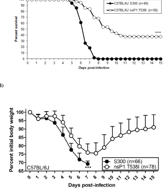
2.1). Moreover, S300 infected C57BL/6J mice displayed enhanced weight loss from 4-7 dpi as compared to nsP1 T538I infected mice. C57BL/6J mice infected with nsP1 T538I began to regain weight on day 9 dpi, while S300 infected mice never showed signs of weight gain/recovery (Fig. 2.1B). Therefore, although the attenuated mutant caused more severe disease in the C57BL/6J background than was previously reported in outbred CD-1 mice, the mutant virus is still significantly attenuated in C57BL/6J mice when compared to S300 virus (Fig. 2.1). Further analysis indicated that the nsP1 T538I virus replicated equivalently to S300 virus at 1 and 3 dpi within the CNS, however nsP1 T538I titers were significantly reduced as compared to S300 in the brain on 5 and 6 dpi, and in the spinal cord on day 6 pi (Fig. 2.1C and D). These results suggest that the nsP1 538 virus is able to efficiently establish infection within the CNS, but that the virus is likely cleared more efficiently than S300.
48
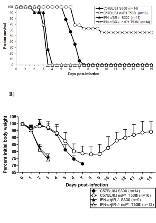
brain and spinal cord of IFN-α/βR-/- mice as compared to C57BL/6J mice on day 3 pi (Fig. 2.2C and D). These data demonstrate that the mutant virus displays virulence comparable to S300 in the absence of type I IFN signaling. When combined with our previous in vitro and in vivo studies (56), this observation suggests that interactions with the type I IFN system contribute to the attenuation of the nsP1 T538I mutant in vivo, and contribute to overall protection from S300 and nsP1 T538I induced disease.
MyD88-dependent TLR signaling is dispensable for protection and control of nsP1 T538I. Given that the nsP1 T538I mutant exhibited similar levels of virulence as S300 in IFN-α/βR-/- mice, we sought to determine which type I IFN induction pathway(s) was responsible for regulating the attenuation of the nsP1 T538I mutant. Therefore, we assessed the role of the TLR and RLR-mediated type I IFN induction pathways in the virulence of S300 and nsP1 T538I.