The handle
http://hdl.handle.net/1887/23937 holds various files of this Leiden University
dissertation.
Author
: Liu, Zhen
Title
: Exploring the interplay between TGF-β and VEGF signalling in endothelial cell
function
TGF-β and VEGF signalling
in endothelial cell function
Zhen Liu, University of Leiden, The Netherlands, 2014. Thesis ISBN: 978-90-8891-807-0
Cover: QiaoQiao Li
Lay-out and Print: Uitgeverij BOXPress || Proefschriftmaken.nl
TGF-β and VEGF signalling in
endothelial cell function
PROEFSCHRIFT ter verkrijging van
de graad van Doctor aan de Universiteit Leiden, op gezag van Rector Magnificus prof. mr. C.J.J.M. Stolker,
volgens besluit van het College voor Promoties te verdedigen op dinsdag 18 februari 2014
om 15.00 uur
door
Zhen Liu
Prof. dr. M. J. T. H. Goumans Co-promoter: Dr. E. Pardali
Overige Leden: Prof. dr. A. Sonnenberg1
Prof. dr. A. J. van Zonneveld Prof. dr. P. H. A. Quax
1 Nederlands Kanker Instituut, Amsterdam
The research described in this thesis was performed at the department of Molecular Cell Biology, Leiden University Medical Center, Leiden, The Netherlands. This research is supported by the Centre for Biomedical Genetics, Dutch Cancer Society (RUL 2005-3371), FP6 EC Integrated Project Angiotargeting 504743 and the LeDucq foundation.
Scope of the investigation ... 9
Thesis Outline ... 10
Chapter 1 ... 11
TGF-β signalling in vascular biology and dysfunction Chapter 2 ... 33
VEGF and inhibitors of TGF-β type-I receptor kinase synergistically promote blood-vessel formation by inducing α5-integrin expression Chapter 3 ... 57
BMP-9 signals via ALK1 and inhibits bFGF-induced endothelial cell proliferation and VEGF-stimulated angiogenesis Chapter 4 ... 79
Endoglin is dispensable for vasculogenesis, but required for vascular endothelial growth factor-induced angiogenesis Chapter 5 ... 103
Matrix Metalloproteinase-14 (MT1-MMP)-mediated endoglin shedding inhibits tumour angiogenesis Chapter 6 ... 127
Soluble fms-like tyrosine kinase 1 and soluble endoglin are elevated circulating anti-angiogenic factors in pre-eclampsia Chapter 7 ... 147
General Discussion Abbreviations ... 165
Summary ... 167
Nederlandse samenvatting ... 169
Curriculum Vitae ... 173
Scope of the investigation
Endothelial cell sprouting is a multi-step process, tightly regulated by diverse signalling pathways. Vascular endothelial growth factor (VEGF) is an essential inducer for angiogenesis as evidenced by in vivo and in vitro studies. Transforming growth factor (TGF)-β remodels the vascular morphogenesis in vivo and regulates the expression of VEGF and VEGF receptors in vitro. However, little is known about how these two factors orchestrate the modulation of endothelial cell function. The scope of the research presented in this thesis is to study the interplay between TGF-β and VEGF signalling on endothelial cell function, with the focus on the effect of TGF-β signalling on VEGF- induced endothelial cell function.
TGF-β transduces its effect by binding to two distinct TGF-β type I receptors, ALK1 and ALK5 on endothelial cells. In addition, the concentration of TGF-β affects the degree of activation of these two receptors. In Part I, to investigate the influence of ALK5 activity in response to VEGF stimulation, a selective inhibitor of ALK5 (SB-431542) was applied to investigate the role of ALK5 in VEGF-induced vascular network formation; to address the role of ALK1 in VEGF-induced endothelial cell behavior, BMP9 as a potent ALK1 ligand was used in the presence of VEGF.
Endoglin is a co-receptor for TGF-β and functions as a modulator for ALK1 and ALK5 in endothelial cells. The involvement of endoglin in VEGF-induced endothelial cell function remains unclear. In Part II, the studies were designed to elaborate the role of endoglin and its soluble form on VEGF-stimulated endothelial cell sprouting. Of note, Chapter 6 discusses the pathological contribution of elevated soluble endoglin levels to pre-eclampsia.
Thesis Outline
The studies presented in this thesis have focused on the crosstalk between the TGF-β, BMP9 and VEGF signalling pathways and their roles on EC function, and how endoglin, a co-receptor of TGF-β signalling influences VEGF-induced endothelial sprouting.
Chapter 1 provides a general overview of the role of TGF-β family signalling in vascular development
concerning its role in endothelial cell and mural cells as well as the impact of TGF-β signalling in pathological conditions.
Part I: The effect TGF-β signalling in endothelial cell function in response to VEGF
Chapter 2 demonstrates that inhibition of the TGF-β signalling pathway using the TGF-β receptor I
inhibitor SB-431542 enhances VEGF-induced endothelial cell function. Sub-optimal doses of VEGF and SB-431542 synergistically induce endothelial cell migration and sprouting.
Chapter 3 reports the new ligand BMP9 for ALK1 signalling and its inhibitory role in endothelial
cell function.
Part II: The role of endoglin in endothelial cell function in response to VEGF
Chapter 4 describes the new observation that endoglin is required for efficient VEGF-induced
endothelial cell sprouting.
Chapter 5 demonstrates that endoglin is cleaved by MMP14, and that the derived soluble form of
endoglin exerts inhibitory effect on VEGF-induced endothelial cell sprouting
Chapter 6 gives an overview of the role of soluble endoglin and soluble Flt1 in pre-eclampsia.
Part III: General Discussion
Chapter 7 discusses the perspectives of the main focuses of this thesis reflected towards the
Ch
ap
te
r 1
Chapter 1
TGF-β signalling in vascular biology and dysfunction
Marie-José Goumans, Zhen Liu, Peter ten Dijke
Department of Molecular Cell Biology and Centre for Biomedical Genetics, Leiden University Medical Center, PO Box 9600, 2300 RC Leiden, The Netherlands
Abstract
Transforming growth factor (TGF)-β family members are multifunctional cytokines that elicit their effects on cells, including endothelial and mural cells, via specific type I and type II serine/threonine kinase receptors and intracellular Smad transcription factors. Knock-out mouse models for TGF-β family signalling pathway components have revealed their critical importance in proper yolk sac angiogenesis. Genetic studies in humans have linked mutations in these signalling components to specific cardiovascular syndromes such as hereditary hemorrhagic telangiectasia, primary pulmonary hypertension and Marfan syndrome. In this review, we present recent advances in our understanding of the role of TGF-β receptor signalling in vascular biology and disease, and discuss how this may be applied for therapy.
Keywords: angiogenesis, BMP, Marfan syndrome, Smad, pre-eclampsia, pulmonary
Ch
ap
te
r 1
Introduction
Transforming growth factor (TGF)-β is the prototypic member of a large family of evolutionarily conserved pleiotropic cytokines. Thirty three members are present in mammals, including three TGF-β isoforms, activins, and bone morphogenetic proteins (BMPs) [1-3]. TGF-β family members have critical and specific roles during embryogenesis and in maintaining the homeostasis of adult tissues. Perturbations in their signalling pathways have been linked to a diverse set of developmental disorders and diseases, including cancer, fibrosis, auto-immune and cardiovascular diseases.
All TGF-β family ligands are generated as dimeric precursor proteins and subsequently cleaved by proteases and secreted [4]. Members of the TGF-β family elicit their cellular effects by binding to a complex of type II and type I serine/threonine kinase transmembrane receptors (Figure 1). Five type II receptors and seven type I receptors, also termed activin receptor-like kinases (ALKs) are present in mammals [1-3]. Within the ligand-induced heteromeric receptor complex, the constitutively active type II receptor phosphorylates the type I receptor on specific serine and threonine residues in the intracellular juxtamembrane region. TGF-β signals in most cells via TGF-β type II receptor (TβRII) and ALK5, activins via activin receptor type IIA (ActRIIA) and IIB and ALK4, and BMPs via BMP type II receptor (BMPRII), ActRIIs and ALK1, 2, 3 and 6. In endothelial cells (ECs) TGF-β can, in addition to ALK5, also signal via ALK1 [5, 6]. Upon type I receptor activation, receptor-regulated Smads (R-Smads) are recruited to, and phosphorylated by the type I receptor at the two serine residues in their extreme carboxyl termini. ALK4 and 5 (and 7) induce R-Smad2 and 3 phosphorylation, while ALK1, 2, 3 and 6 mediate phosphorylation of R-Smad1, 5 and 8. Activated R-Smads form complexes with the common mediator Smad4, and translocate into the nucleus, where they can regulate, together with other partner proteins, the transcription of specific target genes. Inhibitory (I)-Smads, i.e. Smad6 and -7, can inhibit the activation of R-Smads by competing with R-Smads for type I receptor interaction and by recruiting specific ubiquitin ligases or phosphatases to the activated receptor complex thereby targeting it for proteosomal degradation or de-phosphorylation, respectively [1-3].
In contrast to factors such as vascular endothelial growth factors (VEGFs) and angiopoietins that have prominent effects on EC behavior [16], TGF-β was initially discovered through its effects on fibroblasts [17] and subsequently shown to affect among other cell types, epithelial-, immune-, stem-, endothelial- and mural cells. This together with its highly cellular context dependent properties, frequently having opposite effects depending on the cellular differentiation state or the presence of other specific extracellular cues, has left the elucidation of the complex role of TGF-β family members in the cardiovascular system somewhat under-investigated. However, phenotypic and molecular characterizations of knock-out mice for TGF-β signalling components have demonstrated their critical role in angiogenesis, and importantly several cardiovascular syndromes were directly linked to mutations in their genes [4]. TGF-β family members have now gained a prominent spot among other key cytokines that control vascular function. Our understanding of their complex role in cardiovascular biology and interplay with VEGF and other angiogenesis regulators is proceeding at a rapid pace. In this review we focus on recent insights into the function of TGF-β family members in the cardiovascular system and discuss how dys-regulation of their signalling pathways contributes to vascular pathologies.
Fig.1 Signal transduction by TGF-β family members is mediated via specific heteromeric complexes of type I and type
Ch
ap
te
r 1
Vascular morphogenesis
Neovascularization, the formation of new functional microvascular networks, is a tightly controlled process regulated by several converging signalling pathways that are tightly coordinated in time and space. The formation of new blood vessels can occur via two processes, vasculogenesis and angiogenesis, both of which result in the formation of endothelial-lined tubes [16]. During vasculogenesis new vessels arise de novo from a mass of proliferating cells, classically forming an inner core of hematopoietic precursor cells and an outer layer of ECs termed a blood island, followed by their subsequent migration, fusion and organization into a primary capillary plexus [18]. Angiogenesis refers to the formation of new capillary networks by sprouting from pre-existing vessels [18, 19]. Neovascularization is involved in growth and development, wound healing and several pathological situations such as tumor growth and metastasis, and cardiovascular disorders [20]. While angiogenesis occurs during embryogenesis and in adult life, vasculogenesis was initially thought to occur only in the embryo, but some studies have suggested that circulating endothelial progenitor cells may also contribute to vessel formation in the adult [21]. The formation of new capillaries involves EC activation, migration, alignment, proliferation, tube formation, branching and maturation of intercellular junctions and the surrounding basement membrane. All new blood vessels begin as simple EC-lined capillaries, but during vessel maturation, some vessels remain as capillaries covered by pericytes, and others develop into large vessels with support from a layer of smooth muscle cells (SMCs), forming a strong vessel wall.
Angiogenesis is a carefully balanced process, under the control of and fine tuned by a multitude of factors, including stimulators like VEGF and inhibitors such as thrombospondin [20]. Considering the context-dependent effects of TGF-β in other cell systems, it is not surprising that its effect on blood vessel formation is biphasic [4]. It can act as both a promoting and an inhibitory factor of angiogenesis for which the underlying mechanisms are starting to be uncovered.
Insights from knock-out mice
In vivo studies show that loss of TGF-β signalling components leads to abnormal formation
not only affects the ECs, but is also important for proper differentiation and function of SMCs and pericytes. Mice lacking TβRII specifically in vascular SMCs also showed vascular defects in the yolk sac but at later stages of development, allowing the embryo to survive to E12.5 [23].
Fig.2 TβRI/ALK5-deficient embryos exhibit severe defects in vascular development. Gross morphology of whole-mount yolk sacs in mutant embryos is compared with wild type littermates. Arrow indicates the pericardial effusion in the mutant embryo probably caused by a circulation defect [116].
At the intracellular level Smad1-, Smad2- or Smad4- deficient mice demonstrate pre-angiogenesis lethality in embryos [22]. In Smad1-deficient embryos this is accompanied with defects in chorion-allantoic circulation [24, 25]. Smad5 knock-out and endothelial specific Smad4 knock-out have phenotypes reminiscent of TGF-β receptor knock-out mice [26, 27]. Smad3-lacking mice are viable but die of impaired immunity and colon cancer [22].
Ch
ap
te
r 1
Role of TGF-β signalling in ECs
TGF-β has been proposed to regulate the activation state of ECs by differentially activating two type I receptors, ALK5 and ALK1 [5, 6]. ALK5 is widely expressed in almost all tissues, but the expression of ALK1 is restricted to ECs and during embryogenesis at sites of angiogenesis [32].
TGF-β/ALK5 signalling induces Smad2/3 phosphorylation and blocks angiogenesis by inhibiting EC proliferation, tube formation and migration [6, 33]. ALK5 induces the expression of fibronectin and plasminogen activator inhibitor type 1 (PAI-1), a negative regulator of EC migration [34]. Interestingly, in mouse embryonic stem cell-derived ECs, the ALK5 kinase inhibitor SB-431542 enhances EC growth and integrity via up-regulation of the tight junction component Claudin-5, suggesting a role for ALK5 signalling in regulating vascular permeability [35]. ALK5 indeed has been reported to increase TGF-β-induced EC permeability and actin cytoskeleton remodeling [36]. By enhancing TβRII/ALK5 assembly clustered VE-cadherin promotes persistent and elevated TGF-β-induced Smad2/3 activation, indicating a positive role for VE-cadherin in TGF-β/ALK5-induced vessel stabilization [37]. Taken together, TGF-β/ALK5 signalling plays an important role in keeping the endothelium quiescent.
In contrast to TGF-β/ALK5, TGF-β/ALK1 signalling induces Smad1/5 activation and has been shown to stimulate EC migration, proliferation and tube formation [33]. Caveolin1 was shown to associate with ALK1 and to promote TGF-β/ALK1-induced responses [38]. An important intracellular effector of ALK1 is Id1; its upregulation was shown to be required for TGF-β/ALK1- induced EC migration and tube formation [6]. However, an inhibitory effect of ALK1 signalling on EC proliferation, migration and spouting has also been reported [39, 40]. BMP9, identified as a ligand for the ALK1 and BMPR-II complex in ECs, was shown to inhibit EC migration and VEGF-induced angiogenesis [13, 14]. These observations suggest that the effect of ALK1 signalling on angiogenesis is dependent on the context and specific ligand by which it is activated.
ALK1 and ALK5 signalling not only elicit opposite responses, but also physically interact with each other in ECs. ALK5-deficient ECs are not only defective in TGF-β/ALK5 signalling but also exhibit impaired TGF-β/ ALK1 responses; ALK5 was found to be necessary for recruitment of ALK1 into a TGF-β receptor complex, and the kinase activity of ALK5 is essential for maximal ALK1 activation [33]. Furthermore, ALK1 can directly antagonize ALK5/Smad2/3 signalling at the level of Smads [5, 6]. The cross-talk between ALK1 and ALK5 signalling provides ECs with a TGF-β-dependent switch to fine tune EC function.
to induce tumor angiogenesis [43]. BMP endothelial cell precursor-derived regulator (BMPER) interacts with BMP4, and regulates BMP4-mediated angiogenesis [44]. Interestingly, inhibition of BMPRII using specific siRNA in human pulmonary arterial ECs was found to induce EC apoptosis via an increase in activated caspase-3 [45]. Upon exposure to hypoxia, BMPRII, phosphorylated Smad1/5/8 and Id1 expression were strongly reduced in these ECs, which may be of relevance to the pathogenesis of hypoxia-induced pulmonary hypertension [46]. BMP9, which interacts with ALK1, is reported to be a circulating vascular quiescence factor [15].
Interestingly, there are ECs that express betaglycan and those that express endoglin. ECs expressing betaglycan respond to all three isoforms of TGF-β, whereas ECs that express endoglin (and not betaglycan) respond with high potency to TGF-β1 and -β3, but not -β2 [47]. In ECs that co-express betaglycan and endoglin, both proteins were shown to be part of a common TGF-β receptor complex [48]. The co-receptor endoglin is predominantly expressed in highly proliferating vascular ECs, but is also reported to be detected on hematopoietic cells, syncytiotrophoblasts of term placenta, stromal cells and mesenchymal cells. Ectopic expression of endoglin inhibits TGF-β-induced growth inhibition in ECs, monocytes and myoblasts [12, 49] and extracellular matrix deposition [50]. Endoglin regulates the fine-tuning between the ALK1 and ALK5 signalling pathways. It is required for TGF-β/ALK1 signalling and indirectly inhibits TGF-β/ ALK5 signalling [49, 51]. Suppression of endoglin gene expression using siRNA resulted in impaired TGF-β/ ALK1 signalling responses [49]. ECs derived from Eng–/– embryos were unable to proliferate in culture, displayed reduced migration, VEGF secretion and eNOS expression [49, 52]. Interestingly, up-regulation of endoglin protects ECs from the apoptotic action of TGF-β1 [53]. Endoglin was recently shown to interact via its C- terminal PDZ binding motif with the scaffolding protein GIPC, which promotes endoglin cell surface retention [54]. Elevated endoglin expression levels are used as a marker of tumor angiogenesis and endoglin antibodies coupled with toxin or radioactivity have been successfully used to target ECs in anti-angiogenic therapy [55, 56]. Two endoglin splice forms have been reported, termed long (L) and short (S)-endoglin with pro- and anti-angiogenic activity, respectively [57]. While the L-form is most abundantly expressed and promotes TGF-β/ALK1 signalling, S-endoglin expression was recently shown to be specifically high in senescent ECs and it preferentially promotes TGF-β/ALK5 signalling [58,59]. Transgenic mice overexpressing S-endoglin in ECs showed hypertension. Taken together, whereas L-endoglin contributes to the activation phase of angiogenesis and contributes to pathological neovascularization, S-endoglin is induced during EC senescence and may lead to age-associated pathologies such as hypertension [59].
Role of TGF-β signalling in vascular SMCs
Ch
ap
te
r 1
includes a large set of SMC differentiation marker genes [61]. Myocardin, an important coactivator of serum response factor, was shown to potently enhance TGF-β/Smad3-mediated activation of SM22α actin transcription [62]. The zinc finger E-box binding transcription factor DeltaEF1 was also shown to have an important effector role in this respect by forming a complex with serum response factor and Smad3 [63]. TGF-β-induced growth inhibition of vascular SMCs was found to be ALK5-mediated via both Smad3-dependent and p38 MAP kinase-dependent signalling pathways [64]. TGF-β was also shown to inhibit SMC migration in a non-Smad3-dependent pathway via up-regulation of cysteine rich protein 2 expression [65, 66]. Interestingly, Smad3-deficient vascular SMCs demonstrated reduced growth inhibition by TGF-β, but did not show any attenuated TGF-β-mediated migratory response [64, 67].
Endoglin is also important for the formation of vascular SMCs. Ectopic endoglin expression in neural crest stem cells causes pericardial hemorrhage associated with altered vascular SMC investment in the walls of major vessels, suggesting a direct role of endoglin in myogenic differentiation [68].
Vascular SMC differentiation and function are also influenced by BMPs. The effect of BMPs depends on the source of SMCs studied as well as the local environment. While BMP2 stimulates vascular SMC migration, it prevents platelet-derived growth factor induced vascular SMC proliferation via induction of the PPARγ/apoE axis [69, 70]. BMP7 also inhibits the growth of vascular SMCs and helps maintain the vascular SMC phenotype in culture [71]. In pulmonary SMCs both BMP7 and BMP4 induce apoptosis via a caspase 8- and 9-dependent mechanism [72]. Interestingly, BMP4 induces micoRNA- 21, leading to the repression of PDCD4, an inhibitor of smooth muscle cell gene expression; SMC differentiation is thereby stimulated [73].
Interaction between ECs and vascular SMCs
Tight regulation and close coordination between ECs and vascular SMCs are needed to form a mature vascular network. Vascular SMCs form abundant gap junctions with ECs and upon receiving signals from SMCs, ECs line up and recruit more SMCs [74].
TGF-β can also stimulate SMCs to produce VEGF partly in a p38 MAP kinase [77], and a reactive oxygen species generation-dependent manner [78]. The produced VEGF can influence both the ECs as well as SMCs, by inducing their growth and differentiation. Interestingly, VEGF was found to inhibit TGF-β release by ECs [79].
Besides influencing their growth, TGF-β and BMP are also potent SMC differentiation factors and crucial regulators to switch SMCs from an undifferentiated phenotype to a contractile phenotype [61]. Furthermore, TGF-β can also induce trans-differentiation of ECs (Endo-MT) into smooth muscle-like cells. TGF-β induces the expression of α-smooth muscle actin and smooth muscle myosin in ECs in a Snail-dependent manner [80] and blocking TGF-β signalling with neutralizing antibodies abrogates the induction of smooth muscle markers in ECs [75].
Vascular disorders and TGF-β signalling
In recent years multiple cardiovascular disorders have been linked to alterations in TGF-β/ BMP signalling pathways, several of which will be discussed below. Increased understanding of the molecular mechanisms has been achieved by studying mouse models that mimic these human diseases. Importantly, these mouse models also allow for testing of therapeutic strategies that aim to normalize the perturbed signalling balance.
Hereditary hemorrhagic telangiectasia (HHT)
Hereditary hemorrhagic telangiectasia (HHT), or Osler-Rendu-Weber syndrome, is an autosomal dominant vascular dysplasia, characterized by the development of mucocutaneous telangiectasias and arteriovenous malformations in the brain, lungs, liver, and gastrointestinal tract (Figure 3). There is variability in the organs affected, even between individuals within families [81]. Three genes are causally related to HHT, i.e. endoglin mutated in HHT type 1 (HHT-1) [82]; ALK1 mutated in HHT-2 [83] and mutation in Smad4 causing a syndrome consist- ing of both juvenile polyposis and an HHT phenotype [84]. In vivo, EC-specific deletion of ALK1 caused vascular malformations mimicking all pathologic features of HHT-2 vascular lesions [85], while endoglin heterozygous mice have dilated and fragile blood vessels which resemble clinical manifestations of HHT-1 patients [76, 86].
Ch
ap
te
r 1
against VEGF, thalidomide and interferon α-2b to inhibit bleedings and other vascular malformations associated with HHT (http://www.hht.org/).
Marfan syndrome and Loeys-Dietz syndrome (MFS and LDS)
Marfan syndrome (MFS) is a genetic connective tissue disorder caused by mutations in the fibrillin gene [87]. Typical MFS can affect the skeletal system, ocular system, cardiovascular system, pulmonary system as well as other organs, with defects in aorta and heart valves giving the most severe complications. Fibrillin contains several motifs homologous to latent TGF-β binding protein (LTBP), and has been shown to interact with LTBP and control TGF-β bioavailability [4]. TGF-β is bound and kept inactive by the LTBP-fibrillin complex. In fibrillin-1-deficent mice, a model of MFS, fibrillin-1 deficiency was found to diminish the sequestration of latent TGF-β to the extracellular matrix, thereby leading to increased TGF-β activation [88]. Marfan syndrome type 2 is caused by mutation in the TβRII gene locus. Three prominent features of Marfan’s syndrome in humans, i.e. dilatation of the aortic root, air-space widening and skeletal myopathy, can be prevented and even reversed in the mouse model by treatment with losartan, which blocks the angiotensin II type 1 receptor and antagonizes TGF-β signalling [89, 90].
The Loeys-Dietz syndrome is a recently described autosomal dominant aortic-aneurysm syndrome presenting with cardiovascular and skeletal manifestations consistent with those seen in MFS, along with other features not present in MFS. Typical Loeys-Dietz syndrome is characterized by a mutation in either TβRI or TβRII [91]. The molecular mechanism of Loeys-Dietz syndrome is complex and poorly understood; although mutations in TβR are inactivating its function, they lead paradoxically to overactive TGF-β signalling, as measured by accumulation of phosphorylated
Fig. 3 Clinical symptoms of hereditary
Smad2 in the nucleus and expression levels of connective-tissue growth factor [92]. Thus TGF-β antagonists may also alleviate manifestations of Loeys-Dietz syndrome. In a recent clinical study with 18 MFS patients, losartan decreased the rate of progressive aortic-root dilation [93] (http://www.marfan.org/ nmf/index.jsp).
Pre-eclampsia
Pre-eclampsia, which involves a raise in blood pressure, is a major cause of maternal, fetal, and neonatal mortality. The clinical manifestations of pre-eclampsia reflect widespread EC dysfunction, resulting in vasoconstriction, endo-organ ischemia and increased vascular permeability [94]. Soluble endoglin (solEng), a placenta- derived 65 kD soluble form of endoglin and soluble fms-like tyrosine kinase 1 (sFlt1, an inhibitor of VEGF) are both considered to be clinical indicators for pre-eclampsia [95]. Just before the onset of pre-eclampsia circulating levels of solEng are found to be markedly el- evated and it cooperates with sFlt in the pathogenesis of pre-eclampsia in rats [96]. SolEng was found to inhibit TGF-β-mediated activation of eNOS, thereby affecting vascular tone. SolEng disrupts EC function, inhibiting EC tube formation and enhancing capillary permeability. SolEng is formed by proteolytic cleavage, but the protease involved remains to be identified. Upon identification of this protease involved in endoglin shedding, it will be interesting to explore whether its targeting could be beneficial for treatment of pre-eclampsia. Circulating solEng levels may be a useful diagnostic marker in pre-eclampsia [4].
Pulmonary arterial hypertension
Ch
ap
te
r 1
Fig.4 Photomicrographs of lung sections from patients with primary pulmonary hypertension (PPH). The occlusion of
small pulmonary arteries that is typical of PPH is shown. Stainings of lung sections from PPH patients are shown using anti- bodies against (A) CD31 (also known as platelet endothelial cell adhesion molecule-1), an endothelium-specific marker (arrow is pointing to single layer of ECs); and (B) SMC α-actin (arrow is pointing to concentric layers of SMCs). Figure was repro- duced from [97], with permission from Dr N Morrell and Lippincott, Williams and Wilkins.
Cardiac remodeling and hypertrophy
Cardiac remodeling describes the alteration in size, shape and function of the left ventricle in response to changes in hemodynamic loading conditions, neurohormonal activation, or induction of local mediators that alter the structural characteristics of the myocardium. TGF-β is a crucial regulator of cardiac remodeling through its direct and potent actions in mediating cardiomyocyte growth, fibroblast activation and extracellular matrix deposition [106].
myocytes [114]. The local production of TGF-β in hypertrophic myocardium and the link between the RAS system and TGF-β strongly implicate the role of TGF-β in hypertrophic response. All these results demonstrate the importance of TGF-β in mediating hypertrophic cardiac remodeling, however, limited knowledge is available on the signalling pathways responsible for TGF-β action in hypertrophy.
Concluding remarks
The pivotal importance of TGF-β family members in angiogenesis is underscored by the observations that nearly all knock-out mice for specific TGF-β family signalling components die during midgestation due to a yolk sac angiogenesis defect and that mutations in the genes encoding TGF-β pathway components are linked to an increasing number of human pathologies with vascular dysfunction. In concordance with these findings in vitro studies with different TGF-β family members have revealed potent effects on the function of ECs and vascular SMCs, affecting e.g., proliferation, differentiation, migration and extracellular matrix production. However, results obtained by different laboratories are sometimes in apparent conflict, but this can largely be attributed to context-dependent functions of TGF-β family members. Ligand concentration, cell density, cell type, media and matrix coating may determine specific cellular responses to TGF-β family members. TGF-β also plays an important role in the interplay between EdCs and vascular SMCs. Recent data indicate that TGF-β is capable of mediating EndoMT, a transdifferentiation of ECs into SMC- like cells [115].
With misregulation of TGF-β signalling at the heart of cardiovascular disorders, the cognate signalling components represent interesting targets for therapy. The first large-scale clinical trials with losartan to treat MFS are underway and results are eagerly awaited. Results from one vascular disorder may open opportunities for development of therapies in other vascular pathologies. Whereas high levels of circulating solEng may lead to pre-eclampsia by causing an EC dysfunction in multiple organs, solEng may be explored for anti-angiogenic activity in curbing tumor angiogenesis.
Acknowledgments
Ch
ap
te
r 1
References
1. Heldin C-H, Miyazono K, ten Dijke P. TGF-β signalling from cell membrane to nucleus through SMAD proteins. Nature 1997; 390:465-471.
2. Shi Y, Massagué J. Mechanisms of TGF-β signalling from cell membrane to the nucleus. Cell 2003; 113:685-700. 3. Schmierer B, Hill CS. TGFβ-SMAD signal transduction: molecular specificity and functional flexibility. Nat Rev Mol
Cell Biol 2007; 8:970-982.
4. ten Dijke P, Arthur HM. Extracellular control of TGFβ signal- ling in vascular development and disease. Nat Rev Mol
Cell Biol 2007; 8:857-869.
5. Oh SP, Seki T, Goss KA, et al. Activin receptor-like kinase 1 modulates transforming growth factor-β1 signalling in the regulation of angiogenesis. Proc Natl Acad Sci USA 2000; 97:2626-2631.
6. Goumans MJ, Valdimarsdottir G, Itoh S, Rosendahl A, Sideras P, ten Dijke P. Balancing the activation state of the endothe- lium via two distinct TGF-β type I receptors. EMBO J 2002; 21:1743-1753.
7. ten Dijke P, Goumans MJ, Pardali E. Endoglin in angiogenesis and vascular diseases. Angiogenesis 2008; 11:79-89. 8. López-Casillas F, Wrana JL, Massagué J. Betaglycan presents ligand to the TGFβ signalling receptor. Cell 1993;
73:1435- 1444.
9. Del Re E, Babitt JL, Pirani A, Schneyer AL, Lin HY. In the absence of type III receptor, the transforming growth factor (TGF)-β type II-B receptor requires the type I receptor to bind TGF-β2. J Biol Chem 2004; 279:22765-2272.
10. Sankar S, Mahooti-Brooks N, Centrella M, McCarthy TL, Madri JA. Expression of transforming growth factor type III receptor in vascular endothelial cells increases their responsiveness to transforming growth factor β2. J Biol
Chem 1995; 270:13567-13572.
11. Cheifetz S, Bellón T, Calés C, et al. Endoglin is a component of the transforming growth factor-β receptor system in human endothelial cells. J Biol Chem 1992; 267:19027-19030.
12. Lastres P, Letamendía A, Zhang H, et al. Endoglin modulates cellular responses to TGF-β1. J Cell Biol 1996; 133:1109- 1121.
13. Scharpfenecker M, van Dinther M, Liu Z, et al. BMP-9 signals via ALK1 and inhibits bFGF-induced endothelial cell proliferation and VEGF-stimulated angiogenesis. J Cell Sci 2007; 120:964-972.
14. David L, Mallet C, Mazerbourg S, Feige JJ, Bailly S. Identification of BMP9 and BMP10 as functional activators of the orphan activin receptor-like kinase 1 (ALK1) in endothelial cells. Blood 2007; 109:1953-1961.
15. David L, Mallet C, Keramidas M, et al. Bone morphogenetic protein-9 is a circulating vascular quiescence factor.
Circ Res 2008; 102:914-922.
16. Adams RH, Alitalo K. Molecular regulation of angiogenesis and lymphangiogenesis. Nat Rev Mol Cell Biol 2007; 8:464- 478.
17. Roberts AB, Anzano MA, Meyers CA, et al. Purification and properties of a type beta transforming growth factor from bo- vine kidney. Biochemistry 1983; 22:5692-5698.
18. Risau W. Mechanisms of angiogenesis. Nature 1997; 386:671-674.
21. Eguchi M, Masuda H, Asahara T. Endothelial progenitor cells for postnatal vasculogenesis. Clin Exp Nephrol 2007; 11:18- 25.
22. Goumans MJ, Mummery C. Functional analysis of the TGFβ receptor/Smad pathway through gene ablation in mice.
Int J Dev Biol 2000; 44:253-265.
23. Carvalho RL, Itoh F, Goumans MJ, et al. Compensatory signalling induced in the yolk sac vasculature by deletion of TGFβ receptors in mice. J Cell Sci 2007; 120:4269-4277.
24. Lechleider RJ, Ryan JL, Garrett L, et al. Targeted mutagenesis of Smad1 reveals an essential role in chorioallantoic fusion. Dev Biol 2001 240:157-167.
25. Tremblay KD, Dunn NR, Robertson EJ. Mouse embryos lacking Smad1 signals display defects in extra-embryonic tissues and germ cell formation. Development 2001; 128:3609-3621.
26. Lan Y, Liu B, Yao H, et al. Essential role of endothelial Smad4 in vascular remodeling and integrity. Mol Cell Biol 2007; 27:7683-7692.
27. Chang H, Zwijsen A, Vogel H, Huylebroeck D, Matzuk MM. Smad5 is essential for left-right asymmetry in mice. Dev
Biol 2000; 219:71-78.
28. Topper JN, Cai J, Qiu Y, et al. Vascular MADs: two novel MAD-related genes selectively inducible by flow in human vascular endothelium. Proc Natl Acad Sci USA 1997; 94:9314- 9319.
29. Zwijsen A, Goumans MJ, Lawson KA, Van Rooijen MA, Mummery CL. Ectopic expression of the transforming growth factor beta type II receptor disrupts mesoderm organisation during mouse gastrulation. Dev Dyn 1999; 214:141-151.
30. Galvin KM, Donovan MJ, Lynch CA, et al. A role for smad6 in development and homeostasis of the cardiovascular system. Nat Genet 2000; 24:171-174.
31. Chen Q, Chen H, Zheng D, et al. Smad7 is required for the development and function of heart. J Biol Chem 2008 Oct 24. doi:10.1074/jbc.M807233200.
32. Roelen BA, van Rooijen MA, Mummery CL. Expression of ALK-1, a type 1 serine/threonine kinase receptor, coincides with sites of vasculogenesis and angiogenesis in early mouse development. Dev Dyn 1997; 4:418-430.
33. Goumans MJ, Valdimarsdottir G, Itoh S, et al. Activin receptor-like kinase (ALK)1 is an antagonistic mediator of lateral TGFβ/ALK5 signalling. Mol Cell 2003; 12:817-828.
34. Deng G, Curriden SA, Wang S, Rosenberg S, Loskutoff DJ. Is plasminogen activator inhibitor-1 the molecular switch that governs urokinase receptor-mediated cell adhesion and release? J Cell Biol. 1996; 134:1563-1571.
35. Watabe T, Nishihara A, Mishima K, et al. TGF-β receptor kinase inhibitor enhances growth and integrity of embryonic stem cell-derived endothelial cells. J Cell Biol 2003; 163:1303- 1311.
36. Birukova AA, Adyshev D, Gorshkov B, Birukov KG, Verin AD. ALK5 and Smad4 are involved in TGF-β1-induced pulmonary endothelial permeability. FEBS Lett 2005; 579:4031- 4037.
37. Rudini N, Felici A, Giampietro C, et al.VE-cadherin is a criti- cal endothelial regulator of TGF-β signalling. EMBO J 2008; 27:993-1004.
38. Santibanez JF, Blanco FJ, Garrido-Martin EM, Sanz-Rodri- guez F, del Pozo MA, Bernabeu C. Caveolin-1 interacts and cooperates with the transforming growth factor-β type I receptor ALK1 in endothelial caveolae. Cardiovasc
Res 2008; 77:791-799.
Ch
ap
te
r 1
40. Mallet C, Vittet D, Feige JJ, Bailly S. TGFβ1 induces vasculogenesis and inhibits angiogenic sprouting in an embryonic stem cell differentiation model: respective contribution of ALK1 and ALK5. Stem Cells 2006; 24:2420-2427.
41. Valdimarsdottir G, Goumans MJ, Rosendahl A, et al. Stimulation of Id1 expression by bone morphogenetic protein is sufficient and necessary for bone morphogenetic protein-induced activation of endothelial cells. Circulation 2002; 106:2263- 2270.
42. Suzuki Y, Montagne K, Nishihara A, Watabe T, Miyazono K. BMPs promote proliferation and migration of endothelial cells via stimulation of VEGF-A/VEGFR2 and angiopoietin-1/Tie2 signalling. J Biochem 2008; 143:199-206.
43. Langenfeld EM, Langenfeld J. Bone morphogenetic protein-2 stimulates angiogenesis in developing tumors. Mol
Cancer Res 2004; 2:141-149.
44. Heinke J, Wehofsits L, Zhou Q, et al. BMPER is an endothelial cell regulator and controls bone morphogenetic protein-4- dependent angiogenesis. Circ Res 2008; 103:804-812.
45. Teichert-Kuliszewska K, Kutryk MJ, Kuliszewski MA, et al. Bone morphogenetic protein receptor-2 signalling promotes pulmonary arterial endothelial cell survival: implications for loss-of-function mutations in the pathogenesis of pulmonary hypertension. Circ Res 2006; 98:209-217.
46. Takahashi K, Kogaki S, Matsushita T, Nasuno S, Kurotobi S, Ozono K. Hypoxia induces alteration of bone morphogenetic protein receptor signalling in pulmonary artery endothelial cell. Pediatr Res 2007; 61:392-397.
47. Cheifetz S, Hernandez H, Laiho M, ten Dijke P, Iwata KK, Massagué J. Distinct transforming growth factor-β (TGF-β) receptor subsets as determinants of cellular responsiveness to three TGF-β isoforms. J Biol Chem 1990; 265:20533-20538.
48. Wong SH, Hamel L, Chevalier S, Philip A. Endoglin expression on human microvascular endothelial cells association with betaglycan and formation of higher order complexes with TGF-β signalling receptors. Eur J Biochem 2000; 267:5550- 5560.
49. Lebrin F, Goumans MJ, Jonker L, et al. Endoglin promotes endothelial cell proliferation and TGF-β/ALK1 signal transduc- tion. EMBO J 2004; 23:4018-4028.
50. Obreo J, Díez-Marques L, Lamas S, et al. Endoglin expression regulates basal and TGF-β1-induced extracellular matrix synthesis in cultured L6E9 myoblasts. Cell Physiol Biochem 2004; 14:301-310.
51. Blanco FJ, Santibanez JF, Guerrero-Esteo M, Langa C, Vary CP, Bernabeu C. Interaction and functional interplay between endoglin and ALK-1, two components of the endothelial transforming growth factor-β receptor complex. J Cell Physiol 2005; 204:574-584.
52. Jerkic M, Rodríguez-Barbero A, Prieto M, et al. Reduced an- giogenic responses in adult Endoglin heterozygous mice. carduivasc Res 2006; 69:845-854.
53. Li C, Issa R, Kumar P, et al. CD105 prevents apoptosis in hypoxic endothelial cells. J Cell Sci 2003; 116:2677-2685. 54. Lee NY, Ray BN, How T, Blobe GC. Endoglin promotes trans- forming growth factor-β-mediated smad 1/5/8 signalling
and inhibits endothelial cell migration through its association with GIPC. J Biol Chem 2008; 283: 32527-32533 .
55. Fonsatti E, Altomonte M, Nicotra MR, Natali PG, Maio M. Endoglin (CD105): a powerful therapeutic target on tumor-associated angiogenetic blood vessels. Oncogene 2003; 22:6557- 6563.
57. Pérez-Gómez E, Eleno N, López-Novoa JM, et al. Character- ization of murine S-endoglin isoform and its effects on tumor development. Oncogene 2005; 24:4450-4461.
58. Velasco S, Alvarez-Muñoz P, Pericacho M, et al. L- and S-endoglin differentially modulate TGFβ1 signalling mediated by ALK1 and ALK5 in L6E9 myoblasts. J Cell Sci 2008; 121:913-919.
59. Blanco FJ, Grande MT, Langa C, et al. S-Endoglin expression is induced in senescent endothelial cells and contributes to vascular pathology. Circ Res 2008; 103:1383-1392.
60. Bobik A.Transforming growth factor-βs and vascular disor- ders. Arterioscler Thromb Vasc Biol 2006; 26:1712-1720. 61. Owens GK. Regulation of differentiation of vascular smooth muscle cells. Physiol Rev 1995; 75:487-517.
62. Qiu P, Ritchie RP, Fu Z, et al. Myocardin enhances Smad3- mediated transforming growth factor-β1 signalling in a CArG box-independent manner: Smad-binding element is an important cis-element for SM22a transcription in
vivo. Circ Res 2005; 97:983-991.
63. Nishimura G, Manabe I, Tsushima K, et al. DeltaEF1 mediates TGF-β signalling in vascular smooth muscle cell differentiation. Dev Cell 2006; 11:93-104.
64. Feinberg MW, Watanabe M, Lebedeva MA, et al. Transforming growth factor-β1 inhibition of vascular smooth muscle cell activation is mediated via Smad3. J Biol Chem 2004; 279:16388-16393.
65. Wei J, Gorman TE, Liu X, et al. Increased neointima formation in cysteine-rich protein 2-deficient mice in response to vascular injury. Circ Res 2005; 97:1323-1331.
66. Lin DW, Chang IC, Tseng A, et al. Transforming growth fac- tor β up-regulates cysteine-rich protein 2 in vascular smooth muscle cells via activating transcription factor 2. J Biol Chem 2008; 283:15003-15014.
67. Kobayashi K, Yokote K, Fujimoto M, et al. Targeted disruption of TGF-β-Smad3 signalling leads to enhanced neointimal hyperplasia with diminished matrix deposition in response to vascular injury. Circ Res 2005; 96:904-912.
68. Mancini ML, Verdi JM, Conley BA, et al. Endoglin is required for myogenic differentiation potential of neural crest stem cells. Dev Biol 2007; 308: 520-533.
69. Willette RN, Gu JL, Lysko PG, Anderson KM, Minehart H, Yue T. BMP-2 gene expression and effects on human vascular smooth muscle cells. J Vasc Res 1999; 36:120-125.
70. Hansmann G, de Jesus Perez VA, Alastalo TP, et al. An anti- proliferative BMP-2/PPARgamma/apoE axis in human and murine SMCs and its role in pulmonary hypertension. J Clin Invest 2008; 118:1846-1857.
71. Dorai H, Vukicevic S, Sampath TK. Bone morphogenetic protein-7 (osteogenic protein-1) inhibits smooth muscle cell proliferation and stimulates the expression of markers that are characteristic of SMC phenotype in vitro. J Cell
Physiol 2000; 184:37-45.
72. Lagna G, Nguyen PH, Ni W, Hata A. BMP-dependent activation of caspase-9 and caspase-8 mediates apoptosis in pulmonary artery smooth muscle cells. Am J Physiol Lung Cell Mol Physiol 2006; 291:L1059-1067
73. Davis BN, Hilyard AC, Lagna G, Hata A. SMAD proteins control DROSHA-mediated microRNA maturation. Nature 2008; 454:56-61.
74. Hungerford JE, Owens GK, Argraves WS, Little CD. Development of the aortic vessel wall as defined by vascular smooth muscle and extracellular matrix markers. Dev Biol 1996; 178:375-392.
Ch
ap
te
r 1
76. Arthur HM, Ure J, Smith AJ, et al. Endoglin, an ancillary TGFβ receptor, is required for extraembryonic angiogenesis and plays a key role in heart development. Dev Biol 2000; 217: 42-53.
77. Yamamoto T, Kozawa O, Tanabe K, et al. Involvement of p38 MAP kinase in TGF-β-stimulated VEGF synthesis in aortic smooth muscle cells. J Cell Biochem 2001; 82:591-598.
78. Black SM, Grobe A, Mata-Greenwood E, Noskina Y. Cyclic stretch increases VEGF expression in pulmonary arterial smooth muscle cells via TGF-β1 and reactive oxygen species: a requirement for NAD(P)H oxidase. Conf Proc IEEE
Eng Med Biol Soc 2004; 7:5053-5056.
79. Dongmei L, Cuili Z, Song F, Lubenec I, Tian Y, Song QH. VEGF regulates FGF-2 and TGF-β1 expression in injury endothelial cells and mediates smooth muscle cells prolifera- tion and migration. Microvasc Res 2008 Oct 2. doi: 10.1016/ j.mvr.2008.09.007.
80. Kokudo T, Suzuki Y, Yoshimatsu Y, Yamazaki T, Watabe T, Miyazono K. Snail is required for TGFβ-induced endothelial-mesenchymal transition of embryonic stem cell-derived endothelial cells. J Cell Sci 2008; 121:3317-3324.
81. Abdalla SA, Letarte M. Hereditary haemorrhagic telangiectasia: current views on genetics and mechanisms of disease. J Med Genet 2006; 43:97-110.
82. McAllister KA, Grogg KM, Johnson DW, et al. Endoglin, a TGF-β binding protein of endothelial cells, is the gene for hereditary haemorrhagic telangiectasia type 1. Nat Genet 1994; 8:345-351.
83. Johnson DW, Berg JN, Baldwin MA, et al. Mutations in the activin receptor-like kinase 1 gene in hereditary haemorrhagic telangiectasia type 2. Nat Genet 1996; 13:189-195.
84. Gallione CJ, Richards JA, Letteboer TG, et al. SMAD4 mutations found in unselected HHT patients. J Med Genet 2006; 43:793-797.
85. Park SO, Lee YJ, Seki T, et al. ALK5- and TGFBR2-indepen- dent role of ALK1 in the pathogenesis of hereditary hemorrhagic telangiectasia type 2. Blood 2008; 111:633-642.
86. Bourdeau A, Faughnan ME, McDonald ML, Paterson AD, Wanless IR, Letarte M. Potential role of modifier genes influencing transforming growth factor-β1 levels in the development of vascular defects in endoglin heterozygous mice with hereditary hemorrhagic telangiectasia. Am J Pathol 2001; 158:2011-2020.
87. Robinson PN, Arteaga-Solis E, Baldock C, et al. The molecular genetics of Marfan syndrome and related disorders.
J Med Genet 2006; 43:769-787.
88. Neptune ER, Frischmeyer PA, Arking DE, et al. Dysregulation of TGF-β activation contributes to pathogenesis in Marfan syndrome. Nat Genet 2003; 33:407-411.
89. Cohn RD van Erp C, Habashi JP, et al. Angiotensin II type 1 receptor blockade attenuates TGF-β-induced failure of muscle regeneration in multiple myopathic states. Nat Med 2007; 13:204-210.
90. Habashi JP, Judge DP, Holm TM, et al. Losartan, an AT1 antagonist, prevents aortic aneurysm in a mouse model of Mar- fan syndrome. Science 2006; 312:117-121.
91. Loeys BL, Schwarze U, Holm T, et al. Aneurysm syndromes caused by mutations in the TGF-β receptor. N Engl J
Med 2006; 355:788-798.
92. Loeys BL, Chen J, Neptune ER, et al. A syndrome of altered cardiovascular, craniofacial, neurocognitive and skeletal development caused by mutations in TGFBR1 or TGFBR2. Nat Genet 2003; 37:275-281.
94. Sibai BM. Preeclampsia as a cause of preterm and late pre- term (near-term) births. Semin Perinatol 2006; 30:16-19. 95. Levine RJ, Lam C, Qian C, et al. CPEP Study Group. Soluble endoglin and other circulating antiangiogenic factors in
pre-eclampsia. N Engl J Med 2006; 355:992-1005.
96. Venkatesha S, Toporsian M, Lam C, et al. Soluble endoglin contributes to the pathogenesis of preeclampsia. Nat
Med 2006; 12:642-649.
97. Atkinson C, Stewart S, Upton PD, et al. Primary pulmonary hypertension is associated with reduced pulmonary vascular expression of type II bone morphogenetic protein receptor. Circulation 2002; 105:1672-1678.
98. Cool CD, Stewart JS, Werahera P, et al. Three-dimensional reconstruction of pulmonary arteries in plexiform pulmonary hypertension using cell-specific markers. Evidence for a dynamic and heterogeneous process of pulmonary endothelial cell growth. Am J Pathol 1999; 155:411-419.
99. Lane KB, Machado RD, Pauciulo MW, et al. Heterozygous germline mutations in BMPR2, encoding a TGF-beta receptor, cause familial primary pulmonary hypertension. The International PPH Consortium. Nat Genet 2000; 26:81-84.
100. Machado RD, Aldred MA, James V, et al. Mutations of the TGF-β type II receptor BMPR2 in pulmonary arterial hyper- tension. Hum Mutat 2006; 27:121-132.
101. Fujiwara M, Yagi H, Matsuoka R, et al. Implications of mutations of activin receptor-like kinase 1 gene (ALK1) in addition to bone morphogenetic protein receptor II gene (BMPR2) in children with pulmonary arterial hypertension.
Circ J 2008; 72:127-133.
102. West J, Fagan K, Steudel W, et al. Pulmonary hypertension in transgenic mice expressing a dominant-negative BMPRII gene in smooth muscle. Circ Res 2004; 94:1109-1114.
103. Yang J, Davies RJ, Southwood M, et al. Mutations in bone morphogenetic protein type II receptor cause dysregulation of Id gene expression in pulmonary artery smooth muscle cells: implications for familial pulmonary arterial hypertension. Circ Res 2008; 102:1212-1221.
104. Yu PB, Deng DY, Beppu H, et al. Bone morphogenetic protein (BMP) type II receptor is required for BMP-mediated growth arrest and differentiation in pulmonary artery smooth muscle cells. J Biol Chem 2008; 283:3877-3888.
105. Badesch DB, Abman SH, Simonneau G, Rubin LJ, McLaughlin VV. Medical therapy for pulmonary arterial hypertension: updated ACCP evidence-based clinical practice guidelines. Chest 2007; 131:1917-1928.
106. Bujak M, Ren G, Kweon HJ, et al. Essential role of Smad3 in infarct healing and in the pathogenesis of cardiac remodeling. Circulation 2007; 116:2127-2138.
107. Rosenkranz S. TGF-β1 and angiotensin networking in cardiac remodeling. Cardiovasc Res 2004; 63:423-432. 108. Sadoshima J, Xu Y, Slayter HS, Izumo S. Autocrine release of angiotensin II mediates stretch-induced hypertrophy
of cardiac myocytes in vitro. Cell 1993; 75:977-984.
109. Zhou Y, Poczatek MH, Berecek KH, Murphy-Ullrich JE. Thrombospondin 1 mediates angiotensin II induction of TGF-β activation by cardiac and renal cells under both high and low glucose conditions. Biochem Biophys Res Commun 2006; 339:633-641.
110. Wang W, Huang XR, Canlas E, et al. Essential role of Smad3 in angiotensin II-induced vascular fibrosis. Circ Res 2006; 98:1032-1039.
Ch
ap
te
r 1
112. Devereux RB, Lyle PA. Losartan for the treatment of hypertension and left ventricular hypertrophy: the Losartan intervention for endpoint reduction in hypertension (LIFE) study. Expert Opin Pharmacother 2004; 5:2311-2320.
113. Parker TG, Packer SE, Schneider MD. Peptide growth factors can provoke “fetal” contractile protein gene expression in rat cardiac myocytes. J Clin Invest 1990; 85:507-514.
114. Rosenkranz S, Flesch M, Amann K, et al. Alterations of β- adrenergic signalling and cardiac hypertrophy in transgenic mice overexpressing TGF-β1. Am J Physiol Heart Circ Physi- ol 2002; 283:H1253-1262.
115. Goumans MJ, Zonneveld AJ, ten Dijke P. TGFβ-induced endothelial to mesenchymal transition; a switch to cardiac fibrosis? Trends Cardiovasc Med.
116. Larsson J, Goumans MJ, Sjöstrand LJ, et al. Abnormal angiogenesis but intact hematopoietic potential in TGF-β type I receptor-deficient mice. EMBO J 2001; 20:1663-1673.
Ch
ap
te
r 2
Chapter 2
VEGF and inhibitors of TGF-β type-I receptor kinase
synergistically promote blood-vessel formation by inducing
α5-integrin expression
Zhen Liu1, Kazuki Kobayashi1, Maarten van Dinther1, Sandra H. van Heiningen1,2, Gudrun Valdimarsdottir3,
Theo van Laar1, Marion Scharpfenecker1,*, Clemens W. G. M. Löwik4, Marie-José Goumans1, Peter ten
Dijke1, ‡ and Evangelia Pardali1
1Department of Molecular Cell Biology and Centre for Biomedical Genetics 2Department of Human
Genetics and 4Endocrinology and Metabolic Diseases, Leiden University Medical Center, The Netherlands 3Department of Biochemistry and Molecular Biology, Faculty of Medicine, University of Iceland, Iceland
* Present address: Division of Experimental Therapy, The Netherlands Cancer Institute, Amsterdam, The Netherlands
Abstract
Vascular endothelial growth factor (VEGF) and transforming growth factor-β (TGF-β) are potent regulators of angiogenesis. How VEGF and TGF-β signalling pathways crosstalk is not well understood. Therefore, we analyzed the effects of the TGF-β type-I-receptor inhibitors (SB-431542 and LY-2157299) and VEGF on endothelial cell (EC) function and angiogenesis. We show that SB-431542 dramatically enhances VEGF-induced formation of EC sheets from fetal mouse metatarsals. Sub-optimal doses of VEGF and SB-431542 synergistically induced EC migration and sprouting of EC spheroids, whereas overexpression of a constitutively active form of TGF-β type-I receptor had opposite effects. Using quantitative PCR, we demonstrated that VEGF and SB-431542 synergistically upregulated the mRNA expression of genes involved in angiogenesis, including the integrins α5 and β3. Specific downregulation of α5-integrin expression or functional blocking of α5 integrin with a specific neutralizing antibody inhibited the cooperative effect of VEGF and SB-431542 on EC sprouting. In vivo, LY-2157299 induced angiogenesis and enhanced VEGF- and basic-fibroblast-growth-factor-induced angiogenesis in a Matrigel-plug assay, whereas adding an α5-integrin neutralizing antibody to the Matrigel selectively inhibited this enhanced response. Thus, induction of α5-integrin expression is a key determinant by which inhibitors of TGF-β type-I receptor kinase and VEGF synergistically promote angiogenesis.
Ch
ap
te
r 2
Introduction
During embryogenesis, the formation of new blood vessels depends on vasculogenesis and angiogenesis. Angiogenesis refers to the formation of new blood vessels from pre-existing ones (1), and consists of an activation phase and a resolution phase (2). The activation phase is associated with vessel destabilization and increased permeability, degradation of the extracellular matrix (ECM), and endothelial cell (EC) proliferation and migration. During the resolution phase, ECs become quiescent and pericytes and vascular smooth muscle cells (VSMCs) are recruited to ensure stabilization and maturation of the newly formed vessels (1).
Angiogenesis is tightly regulated by pro- and anti-angiogenic signals, and plays an important role in pathophysiological and physiological processes such as wound healing, tissue remodeling, the female reproductive cycle, autoimmune diseases and cancer (3). Vascular endothelial growth factor (VEGF) is a key regulator of vasculogenesis and angiogenesis. Heterozygous mice lacking a single
VEGF allele die at embryonic day (E)8.5 with severe vascular defects (4; 5). Overexpression of VEGF
also results in embryonic lethality (6). VEGF signals through two distinct tyrosine- kinase receptors, VEGFR1 (also known as Flt-1) and VEGFR2 (also known as KDR and Flk-1) (7), and exerts multiple effects on ECs, including proliferation, survival (8), migration and the formation of capillary-like tubules (9).
Genetic studies in mice and humans have suggested that perturbation of TGF-β signalling results in vascular abnormalities (10). TGF-β exerts its biological effects by binding to and activating type-I and type-II transmembrane serine/threonine-kinase receptors. Binding of TGF-β to the TGF-β type II receptor (TβRII) leads to recruitment and phosphorylation of type I receptor [TβRI, or activin receptor like kinase 5 (ALK5)]. Activin and Nodal, which are structurally related to TGF-β, signal via ALK4 and ALK7, respectively. Activated ALK5, ALK4 and ALK7 propagate the signal into the cells by phosphorylating the downstream effector proteins Smad2 and Smad3 (10). In ECs, TGF-β can also activate ALK1, an alternate type-I receptor, which mediates phosphorylation of Smad1 and Smad5 (11, 12). The TGF-β-ALK5 pathway results in inhibition of EC proliferation and migration, whereas the TGF-β-ALK1 pathway results in the activation of proliferation and the migration of ECs. Balance between the TGF-β-ALK1 and TGF-β-ALK5 signalling pathways play an important role in angiogenesis (11, 12). In line with those results, the effects of TGF-β on angiogenesis are highly context dependent, e.g. at low concentrations, TGF-β promotes angiogenesis, whereas, at high concentrations, it inhibits it (13). In addition to its direct effects, TGF-β can exert effects on angiogenesis by regulating the expression of angiogenic factors such as VEGF and components of the ECM such as matrix metalloproteases (MMPs) and integrins (14; 15; 16).
fact that integrins lack intrinsic enzymatic activity, upon ligand-induced integrin clustering specific intracellular signals are initiated by the activation of intracellular associated kinases and adaptor proteins in focal adhesion complexes. Integrins regulate divergent biological events including cell adhesion, migration, proliferation, differentiation, survival and angiogenesis (17).
Several studies have provided evidence suggesting that the interplay of VEGF and TGF-β signalling pathways plays an important role in angiogenesis. TGF-β can induce the expression of VEGF by various cells in the tumor microenvironment, such as tumor cells, stromal fibroblasts and cells of the immune system (18; 19). SB-431542, an ALK5/4/7 kinase inhibitor (20) was shown to exert an inhibitory effect on VEGF secretion in human cancer cell lines (21; 22; 23). Moreover, it was shown that SB-431542 stimulated the formation of FLK1- positive embryonic stem cell (ESC)-derived EC sheets induced by VEGF (24).
However, the molecular mechanisms that regulate the cross-talk between the VEGF and TGF-β signalling pathways in angiogenesis have not been determined. To elucidate the interplay between VEGF and ALK5, we analyzed the effects of VEGF and SB-431542 alone or in combination using different angiogenesis assays. We show that VEGF and SB-431542 synergistically induce angiogenesis both in vitro and in vivo. Gene expression profiling and functional validation revealed that the upregulation of α5-integrin expression plays a crucial role by which VEGF and TGF-β-type-I-receptor- kinase inhibitor achieve their synergistic angiogenic response.
Results
VEGF and ALK5-kinase inhibitor synergistically enhance angiogenesis in fetal
mouse metatarsal assay
ALK5-Ch
ap
te
r 2
kinase inhibitor that is structurally divergent from SB-431542 (27), slightly inhibited endothelial sheet formation. However, similar to SB-431542, LY-2157299 strongly promoted VEGF-induced angiogenesis (Fig. 2A,C). Our results indicate that activation of VEGF and inhibition of TGF-β (and/or Activin and/or Nodal) signalling by the SB-431542 or LY-2157299 inhibitors synergistically stimulates angiogenesis in vitro.
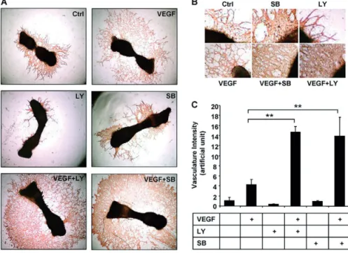
Fig. 1. TGF-β inhibits VEGF-induced formation of the endothelial network in mouse metatarsal assays. Metatarsals
of 17-day-old mouse fetuses were prepared, transferred to cell-culture plates and allowed to adhere, and were then stimulated with VEGF (50 ng/ml), TGF-β3 (5 ng/ml) or both. (A) Cultures were fixed and vessel-like structures were visualized by anti-CD31 staining. Six bones were stimulated per experimental group and one representative picture of each group is shown. Ctrl,control. (B) TGF-β3 did not significantly affect baseline formation of the endothelial network. Incubation with VEGF strongly stimulated the formation of vessel-like structures, which was dramatically decreased by addition of TGF-β3. **P≤0.01.
ALK5 inhibitor and VEGF synergize in inducing EC sprouts in 3D spheroid culture
Fig. 2. Effects of VEGF and SB-431542 on endothelial network formation in mouse metatarsal assays. (A) Metatarsals of
17-day-old mouse fetuses were prepared, transferred to cell-culture plates and allowed to adhere for 4 days. Medium was refreshed and bones were stimulated for 7 days with VEGF (50 ng/ml), SB-431542 [SB; or LY-2157299 (LY)] (10 μM) or both. Cultures were fixed and vessel-like structures were visualized by anti-CD31 staining. Six bones were stimulated per experimental group and one representative picture of each group is shown. Ctrl control. (B) Enlargements of the EC-sheet formation of the images in A. (C) SB-431542 did not significantly affect baseline formation of the endothelial network. LY-2157299 slightly inhibited formation of the endothelial network. Incubation with VEGF strongly stimulated the formation of vessel-like structures, which was dramatically promoted by co-stimulation with SB- 431542 or LY-2157299. **P≤0.01.
Ch
ap
te
r 2
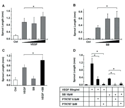
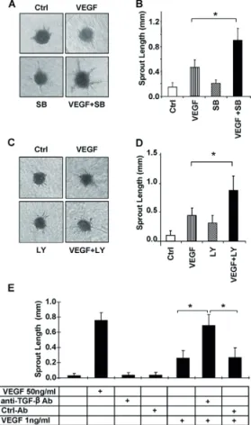
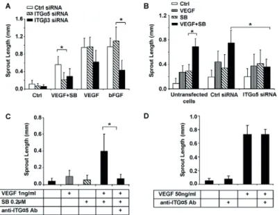
Fig. 3. Synergistic effect of SB-421542 or TGF-β neutralizing antibody with VEGF on EC sprouting. (A) HUVEC spheroids
embedded in collagen were stimulated with increasing amounts of VEGF (10, 25 or 50 ng/ml). (A)HUVEC spheroids embedded in collagen were stimulated with increasing amounts of SB-431542 (SB; 1, 5 or 10 μM). (C) HUVEC spheroids embedded in collagen were stimulated with VEGF (50 ng/ml), SB-431542 (10 μM) or both for 24 hours. (D) EC spheroids were stimulated with VEGF or SB-431542 in the presence or absence of the VEGF-receptor-kinase inhibitor PTK787. (A-D) Quantitative analysis of the mean total sprout length was performed on at least ten spheroids per experimental group. *P≤0.05.
Although stimulation of the spheroids with VEGF (50 ng/ml) and SB-431542 (10 μM) further increased sprouting (Fig. 3C), this effect was additive and not synergistic. Therefore, we hypothesized that the concentrations of VEGF and SB-431542 are near-to-plateau levels, and we analyzed the effect of sub-optimal concentrations of both VEGF and the ALK5 kinase inhibitor. Stimulation of EC spheroids with 1 ng/ml VEGF or 0.2 μM SB-431542 alone resulted in a small induction of sprouting compared with control (Fig. 4A,B). Interestingly, the combination of low levels of VEGF and SB-431542 synergistically enhanced EC sprouting (Fig. 4A,B). Similar results were obtained when the TGF-β type I receptor kinase inhibitor LY-2157299 was used. The combination of sub-optimal concentrations of LY-2157299 and VEGF resulted in enhanced sprouting compared with VEGF or the inhibitor alone (Fig. 4C,D).
isotype-matched control antibody (Fig. 4E). The isotype-isotype-matched control antibody had no effect on basal or VEGF-induced sprouting. Addition of the TGF-β neutralizing antibody did not induce EC sprouting. However, the combination of TGF-β neutralizing antibody with VEGF significantly induced EC sprouting compared with VEGF or TGF-β blocking antibody alone (Fig. 4E). As expected, ectopic expression of a constitutively active form of ALK5 receptor (caALK5) exerted the opposite response of SB-431542 treatment and inhibited VEGF-induced HUVEC sprouting in the 3D spheroid system (supplementary material Fig. S1A). Moreover, caALK5 overexpression inhibited basal cord formation of mouse embryonic endothelial cells (MEECs) (supplementary material Fig. S1B). Taken together, our results suggest that inhibition of the TGF-β signalling pathway significantly enhances VEGF-induced EC sprouting in vitro.
Fig. 4. Synergistic effect of SB-421542 or TGF-β neutralizing antibody with VEGF on EC sprouting. (A,B) HUVEC spheroids embedded
Ch
ap
te
r 2
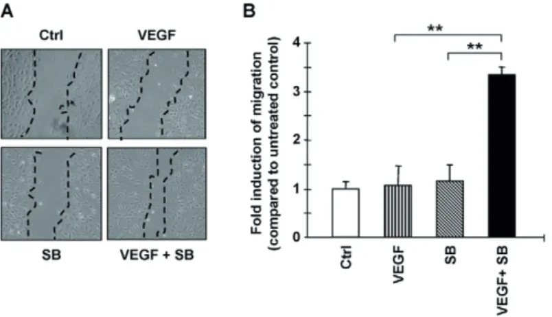
VEGF and ALK5 inhibitor promote EC migration
To further investigate the mechanisms by which VEGF and SB-431542 exert their synergistic effect on angiogenesis, we analyzed their effects on EC migration. To study the effect of VEGF and SB- 431542 on migration, serum-starved monolayers of HUVECs were wounded by scratching and were stimulated for 6 hours. There was no effect on migration when cells were stimulated by VEGF or the ALK5-kinase inhibitor alone. However, the combination of VEGF and SB-431542 significantly induced EC migration (Fig. 5A,B). Consistent with these results, over-expression of caALK5 in MEECs resulted in reduced migration, using the transwell migration assay, and invasion, using a Transwell invasion assay with Matrigel coating (11) (supplementary material Fig. S2A,B).
Fig. 5. Effects of SB-431542 and VEGF on EC migration. VEGF and SB- 431542 (SB) synergistically stimulate HUVEC migration. HUVECs were allowed to grow to confluence and serum-starved for 5 hours. Monolayers were wounded and stimulated with VEGF (1 ng/ml), SB-431542 (0.2 μM) or both for 6 hours. (A) One representative picture of each group is shown. Dashed lines indicate the wound edge. (B) Wound closure was measured after 6 hours using ImageJ software. **P≤0.01.
Transcriptional profiling of the VEGF+SB-431542-induced EC response
pathways that promote EC survival. VEGF and SB-431542 synergistically induced the expression of several other genes implicated in angiogenesis, such as angiotensin receptor I, ACE, CCL5, IL3 and
IL7 (Table 1). Integrins have been shown to play an important role in EC migration and survival,
as well as in capillary sprouting during angiogenesis. In line with this notion, VEGF+SB-431542 stimulation synergistically induced α5 integrin and β3 integrin mRNA expression. To verify these results, we performed quantitative PCR analysis using PCR primer sets different from those used in the array (supplementary material Fig. S3A). Similar results were obtained when mRNA was isolated from EC spheroids embedded in collagen after VEGF and/or SB-431542 stimulation (supplementary material Fig. S3B).
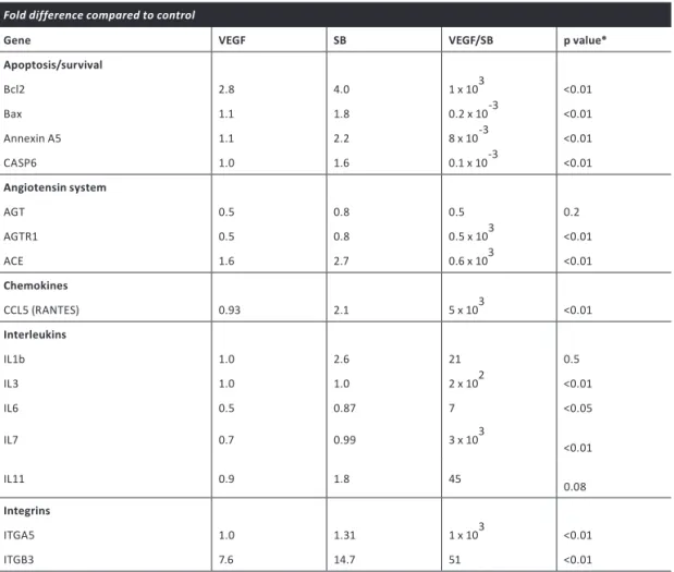
Table 1. Summary of genes identified as significantly altered by VEGF/SB-431542 stimulation of ECs, using an EC function PCR-based array system.
Fold difference compared to control
Gene VEGF SB VEGF/SB p value*
Apoptosis/survival
Bcl2 2.8 4.0 1 x 10³ <0.01
Bax 1.1 1.8 0.2 x 10⁻³ <0.01
Annexin A5 1.1 2.2 8 x 10⁻³ <0.01
CASP6 1.0 1.6 0.1 x 10⁻³ <0.01
Angiotensin system
AGT 0.5 0.8 0.5 0.2
AGTR1 0.5 0.8 0.5 x 10³ <0.01
ACE 1.6 2.7 0.6 x 10³ <0.01
Chemokines
CCL5 (RANTES) 0.93 2.1 5 x 10³ <0.01
Interleukins
IL1b 1.0 2.6 21 0.5
IL3 1.0 1.0 2 x 10² <0.01
IL6 0.5 0.87 7 <0.05
IL7 0.7 0.99 3 x 10³ <0.01
IL11 0.9 1.8 45 0.08
Integrins
ITGA5 1.0 1.31 1 x 10³ <0.01
ITGB3 7.6 14.7 51 <0.01

![Figure was repro- duced from [97], with permission from Dr N Morrell and Lippincott, Williams and Wilkins.](https://thumb-us.123doks.com/thumbv2/123dok_us/8250028.2185997/24.785.149.667.155.363/figure-repro-duced-permission-morrell-lippincott-williams-wilkins.webp)