THE INFLUENCE OF LOCAL MUSCLE VIBRATION DURING FOAM ROLLING ON RANGE OF MOTION, MUSCLE ACTIVATION, PAIN, AND LOWER EXTREMITY
KINEMATICS
Danielle Nicole Enrique
A thesis submitted to the faculty of the University of North Carolina at Chapel Hill in partial fulfillment of the requirements for graduation with honors in the Department of Exercise and
Sport Science.
Chapel Hill 2015
Approved by: Darin Padua
This research was supported by a Tom and Elizabeth Long Excellence Fund for Honors, administered by Honors Carolina.
©2015
iii ABSTRACT
DANIELLE ENRIQUE: The influence of local muscle vibration during foam rolling on range of motion, muscle activation, pain, and lower extremity kinematics
(Under the direction of Darin Padua)
The purpose of this study was to identify the combined effects of foam rolling and
vibration therapy on dorsiflexion range of motion. Determining a more effective treatment for
increasing range of motion will contribute to more effective treatment paradigms for range of
motion restrictions, improvement of lower extremity kinematics, and injury risk reduction.
Healthy, physically active young adults (n=20) with restricted dorsiflexion in the weight-bearing
lunge (<40 degrees) and a minimum of one myofascial trigger point were included. A controlled
crossover experimental design was used. Participants received foam rolling with vibration
during one session and foam rolling without vibration during the other session. Foam rolling
with vibration was more effective in increasing range of motion than foam rolling without
vibration without a resultant increase in measured or perceived pain. Foam rolling with vibration
is an effective clinical tool for improving range of motion restrictions caused by myofascial
trigger points.
TABLE OF CONTENTS
CHAPTER I: INTRODUCTION……….1
VARIABLES……….…..2
RESEARCH QUESTION AND RESEARCH HYPOTHESIS……….4
DEFINITION OF TERMS………..5
CHAPTER II: REVIEW OF THE LITERATURE……….7
CONSEQUENCES OF RESTRICTED ANKLE DORSIFLEXION………..7
CAUSES OF RESTRICTED ANKLE DORSIFLEXION AND RELEVANT ANATOMY………8
INTERVENTIONS FOR RANGE OF MOTION DEFICITS ………10
SUMMARY………...16
CHAPTER III: METHODOLOGY………...18
SUBJECTS………18
INSTRUMENTATION……….19
Range of Motion Instruments……….19
Kinematic Instruments………...19
Other Instruments………..20
PROCEDURES………..20
Screening Session………...20
v
Passive Dorsiflexion Measurements………..24
EMG Setup……….24
3D Motion Analysis………25
Overhead Squat………..25
Single Leg Squat………26
Jump Landing……….26
Foam Roll Intervention………..26
Stretching Intervention………...27
DATA REDUCTION AND ANALYSIS………..28
CHAPTER IV: MANUSCRIPT………30
INTRODUCTION………...30
METHODS………...32
Subjects………..32
Instrumentation………..34
PROCEDURES………..34
Range of Motion Measurements………34
Pain Pressure Threshold and Numerical Rating Scale Measurements………….35
Foam Roll Interventions………36
Stretching Intervention………...37
STATISTICAL ANALYSIS………..37
RESULTS……….38
Range of Motion……….38
DISCUSSION………40
FIGURES………...43
TABLES………49
1 CHAPTER I
Introduction
Restricted ankle dorsiflexion range of motion has been shown to be a predictor of high-risk lower extremity kinematics (Dill, Padua et al., 2014; Fong, Blackburn et al., 2011; Sigward, Ota et al., 2008). These high-risk movement patterns include decreased knee flexion, greater medial knee displacement, and increased ground reaction forces, putting those with decreased dorsiflexion range of motion at risk for knee injury, including patellofemoral pain syndrome and ACL sprain (Bell, Padua et al., 2008; Mauntel, Padua et al., 2012). Identifying a more effective treatment for increasing range of motion will aid clinicians and patients in improving ankle dorsiflexion range of motion. This may contribute to rectifying high-risk movement patterns and decreasing incidence of injury.
Several dependent variables have been used to identify ankle dorsiflexion dysfunction, including range of motion, electromyography, and lower extremity kinematics (Dill, Padua et al., 2014; Macrum, Padua et al., 2012). The combination of electromyography with lower extremity kinematics during functional tasks has been used to identify neuromuscular characteristics differentiating populations with restricted ankle dorsiflexion range of motion and those with normal dorsiflexion range of motion (Padua et al., 2012). The weight-bearing lunge test has been used to identify those with restricted ankle dorsiflexion as a predictor of functional range of motion restriction.
extensibility of the plantar flexors (Terada et al., 2013, Travell, 1999). Myofascial trigger points are loci of hyperirritability in the muscle, and the presence of myofascial trigger points has been linked to muscle pain and decreased range of motion (Travell and Simons, 1993). Myofascial trigger points may result from trauma to the muscle or other causes, and may develop along with fascial adhesions. The adhesions then cause restrictions in the movement of the muscle
underneath the fascia (Barnes, 1997). Several methods of direct compression have been
identified as effective treatments to rectify range of motion deficits caused by myofascial trigger points. The direct compression technique involves finding a locus of hyperirritability within the muscle and applying prolonged pressure to the area (Lewit, 1999). Self-myofascial release is one method of direct compression that has been shown to increase range of motion without decreasing muscle function (MacDonald et al., 2013; Sullivan et al., 2013). One instrument used for self-myofascial release is the foam roller, a cylinder of high-density foam. It has been
suggested that foam rolling applies pressure over these adhesions, helping to reduce the myofascial trigger points and break up adhesions. To further help quantify the effectiveness of myofascial trigger points, pain pressure threshold objectively measures the severity of trigger points pre- and post-treatment (Jaeger and Reeves, 1986; Reeves et al. 1986).
3
and Bosco, 2003; De Gail et al., 1966). There is currently no literature that specifically examines the effect of local vibration therapy on treatment of myofascial trigger points.
There is a gap in the literature regarding the combined effects of direct compression and local vibration therapy. The purpose of this study was to identify the combined effects of self-myofascial release and vibration therapy on dorsiflexion range of motion restriction.
Determining a more effective treatment for increasing range of motion will contribute to developing a more efficient treatment of range of motion restrictions, improvement of lower extremity kinematics, and a reduction of injury.
Independent Variables
• Intervention (Self-myofascial release, Self-myofascial release with vibration) • Time (Pre-intervention, Post-intervention)
Dependent Variables • Range of Motion
o Ankle dorsiflexion with knee straight o Ankle dorsiflexion with knee bent to 90° o Weight-bearing lunge
• Pain Pressure Threshold
• Numerical Rating Scale for PPT
• Numerical Rating Scale for Intervention • Lower Extremity Kinematics
o Tibialis Anterior o Lateral Gastrocnemius o Medial Gastrocnemius
Research Question and Research Hypothesis
1. Research Question 1: Is there a statistically significant difference in change in ankle dorsiflexion range of motion, including weight-bearing lunge, passive knee-bent and passive knee-straight measurements, after self-myofascial release combined with local vibration therapy compared to traditional self-myofascial release?
• Research Hypothesis 1: The self-myofascial release combined with vibration will result in a greater change in ankle dorsiflexion range of motion from pre- to post-intervention measures compared to traditional self-myofascial release.
2. Research Question 2: Is there a statistically significant difference in change in pain pressure threshold between these two interventions?
• Research Hypothesis 2: The self-myofascial release combined with vibration will result in a greater change in pain pressure threshold from pre- to post-intervention compared to traditional self-myofascial release.
3. Research Question 3: Is there a statistically significant difference in change of muscle activation of the gastrocnemius and anterior tibialis after self-myofascial release compared to self-myofascial release combined with vibration therapy?
• Research Hypothesis 3: The self-myofascial release combined with vibration will result in a greater decrease in muscle activation of the gastrocnemius and anterior tibialis compared to traditional self-myofascial release.
5
• Research Hypothesis 4: The change in lower extremity kinematics will be greater for peak knee flexion angles, knee flexion displacement, ankle dorsiflexion displacement, and peak dorsiflexion angles and lesser for peak knee valgus angles, knee valgus displacement, toe-out displacement, and medial knee displacement for self-myofascial release combined with vibration compared to traditional self-myofascial release Statistical Hypothesis
• H0: SMR_V=SMR
• HA: SMR_V≠SMR Definition of Terms
• Weight-bearing lunge dorsiflexion range of motion: Measured with digital inclinometer using weight-bearing lunge technique (Krause, Cloud et al., 2011; Bennell, Techovanich et al., 1998; Denegar, Hertel et al., 2002)
• Gastrocnemius dorsiflexion range of motion: Measured with plastic manual goniometer with the participant supine and knee straight with ankle on a bolster
• Soleus dorsiflexion range of motion: Measured with plastic manual goniometer with the participant supine and knee bent to 90°
• Overhead Squat: Functional task performed with feet shoulder-width apart and toes pointed forward, arms overhead, heels on the ground and squatting to at least 60 degrees of knee flexion
• Jump landing: Functional task performed from a 30cm box placed at a distance equal to 50% of participant’s height away from the landing surface, landing on both feet and immediately jumping again vertically for height
• Dominant limb: Limb used to kick a soccer ball for greatest distance
• Physically active: Participation in at least 30 minutes of physical activity at least three times a week for the last six months
• Myofascial Trigger point: Area within muscle that meets at least 2 of the following criteria: palpable band taught within muscle, nodule/spot of hypersensitivity within a taut band, recognition of pain complaint with pressure on tender nodule, or painful limit to full range of motion (Travell and Simons 1993; Gerwin et al., 1997; Grieve et al., 2011). • Self-myofascial release: Application of pressure to a myofascial trigger point by the
patient using a foam roller (Sullivan et al., 2013; MacDonald et al., 2013)
• Local vibration therapy: Intervention involving vibration on a specific body part in order to effect greater range of motion.
• Pain pressure threshold: Measurement taken using handheld dynamometer over an
7 CHAPTER II
Review of the Literature
Consequences of Restricted Ankle Dorsiflexion
Restricted ankle dorsiflexion can contribute to a multitude of lower extremity injuries. Injury is caused by the alteration of proper mechanics during functional movement.
Approximately 10 degrees of ankle dorsiflexion are required for normal gait, and up to 30
degrees may be required for sport-specific activity (Lindsjö, Sahlstedt, 1985; Segal D, Whitelaw, 1985). When an individual cannot achieve the required amount of ankle dorsiflexion,
compensations occur in movement that can lead to both chronic and acute injuries.
Limited ankle dorsiflexion can lead to increased risk for lateral ankle sprains, plantar fasciitis, medial tibial stress syndrome, and Achilles tendinopathies (Terada et al., 2013; Kaufmun and Cullison, 1999). If the foot remains in a plantarflexed position, there is a higher risk for inversion ankle sprains because the foot remains in a loose-packed position (Hertel, 2002). This allows for greater inversion and internal rotation range of motion, which is the mechanism for lateral ankle sprains (Hertel, 2002). A prior inversion ankle sprain is a predictor for decrease ankle dorsiflexion range of motion, resulting in a detrimental cycle of ankle sprains and loss of motion. Repeated ankle sprains also lead to functional ankle instability. During gait, compensation may occur in the form of internal tibial rotation or increased or prolonged
syndrome, and Achilles tendinitis (Maffulli 2004). These chronic injuries result from repeated stresses caused by an alteration of proper biomechanics.
Compensation in individuals with restricted dorsiflexion range of motion affects the entire kinetic chain. The previously discussed tibial internal rotation is linked to increase internal femoral rotation and increased medial knee displacement (Franco 1987; Levinger, Gilleard et al. 2006). During an overhead squat, compensation occurs in the form of increased knee frontal plant motion (varus or valgus) and decreased knee flexion motion (Dill, Padua et al., 2014; Macrum, Padua et al., 2012). Decreased knee flexion during functional tasks has been shown to increase ground reaction forces, which has been linked to increased incidence of patellofemoral pain syndrome (Macrum, Padua et al., 2012; Nigg 1985). These movement compensations have also been linked to increased risk for anterior cruciate ligament (ACL) sprain (Padua, Marshall et al., 2009; Hewett, Stroupe et al., 1996).
Causes of Restricted Ankle Dorsiflexion and Relevant Anatomy
Bony alignment, ligamentous restrictions, myofascial restrictions, and decreased
extensibility of the plantar flexors can all cause a decrease in ankle dorsiflexion. In order to have successful arthrokinematics, the talus must be able to roll and glide within the mortise of the tibia and fibula. If the talus is displaced anteriorly, it cannot achieve proper arthrokinematics,
resulting in decreased ankle dorsiflexion (Terada et al., 2013). Assuming there is no joint dysfunction, the musculature of the lower leg is often the source of dorsiflexion restrictions.
9
tibialis anterior and foot extensors contract and shorten. When the gastrocnemius and soleus are not extensible, proper ankle dorsiflexion cannot be achieved (Hertel, 2002).
Muscles are composed of muscle tissue wrapped in several layers of fascia. Muscle fibers, or myofibrils, are wrapped in a fascial sheath called endomysium. Fascicles composed of bundles of muscle fibers are wrapped in a layer of fascia called perimysium. Groups of fascicles are then wrapped in the epimysium. The epimysium often blends into fascia on top of the muscle (Saladin, 1998). These sheaths may combine into the fibrous tendon. Both intra- and extra-muscular fascia must be able to slide across the underlying muscle tissue in order to allow for efficient movement (Prentice, 2011). Additionally, it has been demonstrated that fascia may contribute to the process of muscle contraction by actively contracting, rather than just providing passive support (Schleip, 2006). Fascial restrictions may develop in response to microtrauma or acute trauma to the muscle. These fascial restrictions can inhibit muscular function and may develop along with myofascial trigger points. Myofascial trigger points can also develop independently of fascial restrictions (Travell, 1999). A myofascial trigger point is an area of hypersensitivity within skeletal muscle or fascia. A palpable band or nodule may be felt within the muscle. Myofascial trigger points are categorized into active and latent trigger points. An active trigger point produces a clinical complaint when palpated as well as with movement, while a latent trigger point may produce increased muscle tension or shortening and pain upon palpation but does not exhibit pain during movement (Travell, 1999). Myofascial trigger points and fascial restrictions inhibit efficient sliding of the fascia over the muscle. Myofascial
Decreased range of motion caused by myofascial trigger points is not the only issue to be addressed. The pain caused by myofascial trigger points can result in myofascial pain syndrome, in which a patient has regional pain and muscle tenderness caused by the presence of myofascial trigger points. The prevalence of myofascial pain syndrome is approximately 37% in middle-aged males, 65% in middle-middle-aged females, and 85% in those older than sixty-five years old (Desai et al., 2013). The estimated cost of treating myofascial pain syndrome in the United States is approximately $47 billion per year (Desai et al., 2013).
Interventions for Range of Motion Deficits Caused by Myofascial Restrictions
Many interventions have been proposed to increase range of motion. Interventions that have been demonstrated to increase range of motion include static stretching, thermotherapies, warm-up exercises, and vibration (DePino et al., 2000; Wenos, Konin, 2004; Issurin et al., 1994). Some interventions that specifically address myofascial restrictions include manual therapies, therapeutic ultrasound, low-level laser treatment, electrical stimulation, and dry needling
(Kannan, 2012; Sarrafzadeh et al., 2012; Hou et al., 2002; Vulfsons et al., 2012). Vibration and manual therapy in the form of self-myofascial release will be examined as viable methods for decreasing range of motion deficits caused by myofascial restrictions.
Vibration therapy is an intervention that has been proposed to have many different uses, ranging from decreasing delayed onset muscle soreness to improving body composition to
11
increased muscle temperature and viscoelasticity, and induced relaxation (Bosco et al., 1999; Kerschan-Schindl et al., 2001). Relaxation may be effected by stimulating Golgi tendon organs, which causes an autonomic inhibition of the muscle, via the –Ib afferent neuron pathway (Bove et al., 2003). Vibration training may cause altered proprioception by increasing the threshold of proprioceptive sensors (Weerakkody et al., 2007). Decreased musculotendinous stiffness has also been observed as an effect of vibration (Atha, Wheatley, 1976; Herda, Cramer et al., 2010). There has also been an increase in pain threshold observed during and directly after vibration (Lundeberg, 1984; Pantaleo et al.,1986; Issurin et al., 1994; Peer et al., 2009). There is no conclusive evidence that any of these mechanisms is the primary mechanism by which vibration improves flexibility. Vibration therapy may work through a combination of the previously discussed physiological effects. The resultant physiological effects are also somewhat dependent on the frequency and amplitude of the vibration (Cochrane, 2011).
Two different categories of vibration therapy or training have been established. Whole-body vibration involves standing on a platform that delivers vibration of a certain, usually alterable, frequency and amplitude. Local vibration involves a device that administers the vibration to a specific muscle group or body part. Issurin et al. (2005) hypothesized that whole-body vibration causes global metabolic, neuromuscular and hormonal changes that are not necessarily seen in local vibration. There is nothing in the literature to suggest that the
mechanisms and effects of whole-body vibration can or cannot be applied to local vibration as well. As such, research involving local vibration as an acute treatment will predominantly be examined.
training with three training sessions per week. Vibration was administered in oscillations of 3 mm in amplitude and 44 Hz in frequency. The stretching protocol lasted approximately 7
minutes per session. There was a statistically significant increase in range of motion in side-split as well as forward bend for the vibration and stretching group compared to the control group and group that only completed stretching. A study of similar design was completed with twenty-two female gymnasts (age = 11.3 ± 2.6 years) in which a vibration of frequency 30 Hz and
displacement of 2 mm was administered acutely to the vibration-stretching group for 10 seconds with 5 seconds rest for four cycles (Kinser, Stone et al., 2007). The vibration-only group and vibration-stretching groups both had significant increases in flexibility, with greater gains in the vibration-stretching group, and there were no losses in power in either vibration group compared to the stretching-only group.
Sands et al. (2008) examined local vibration and its effect on split flexibility in male gymnasts. A Power-Plate Pro 5 Airdaptive platform was used with a vibration setting of 2 mm displacement and 30-Hz frequency. The 30-Hz frequency was selected based on prior literature suggesting that this frequency was effective in inhibiting the monosynaptic stretch reflex
13
effects may not have been seen because the effects were minimized by the time post-test measurements were taken, the pain mechanism for stretching differed from that which was monitored by the algometer because skin and fascial pain also contributed, or pain pressure reduction was not the primary pathway that allowed increased stretching to occur during vibration training.
Similar increases in range of motion have been observed for local vibration and whole-body vibration in acute and repeated sessions, ranging from shoulder flexibility to plantar flexor extensibility (Feland et al., 2010; Karatrantou et al., 2013; Ferguson et al., 2013). The greatest increase has been observed in combining vibration with stretching regimens (Osawa, Oguma, 2013). The effect of vibration on myofascial trigger points has not been directly observed; however, Lundeberg et al. (1984) noted that the greatest pain reduction occurred when vibration was applied to a painful area or trigger point at a moderate pressure. Further research into the physiological effect of vibration on myofascial trigger points is necessary.
an effective therapy in reducing pain associated with myofascial trigger points as well as increasing range of motion (Grieve et al., 2010; Hou et al., 2002).
Self-myofascial release is a myofascial release technique developed to allow patients to complete myofascial release by themselves, allowing for administration of treatment outside of the clinic and allowing the clinician time to administer other treatments or tend to other patients. Self-myofascial release is completed by the patient using his/her bodyweight to apply pressure to affected soft tissue on a dense foam cylinder, or foam roller. Self-myofascial release can also be completed with a hand-held rolling device, but the pressure applied is dependent on ability to reach a body part as well as upper body strength.
Several techniques have been suggested for self-myofascial release. One technique involves small undulations back and forth, working from distal to proximal over a muscle or proximal to distal (MacDonald et al., 2013). Another technique involves a sweeping, rolling motion all the way from distal to proximal, with no undulations (Halperin et al., 2014; Sullivan et al., 2013). Yet another technique involves rolling the length of the affected muscle or fascia, pausing and applying increased pressure on areas of hypersensitivity (Diehl, 2014). The differences between these techniques are analogous to different methodologies for general myofascial release. The latter technique described combines the theory of trigger point pressure release with general myofascial release.
15
allowing increased benefits. As a result, MacDonald (2013) used a foam roller of increased hardness on the quadriceps of college-aged male participants. Participants were instructed to use the undulating technique while rolling from proximal to distal for one minute with a thirty
second rest for two cycles with as much bodyweight applied as could be tolerated. A statistically significant increase of 10% was seen in the foam-rolling group quadriceps range of motion compared to the control group two minutes after the intervention. An increased range of motion of 8% was still observed ten minutes after the intervention was administered. No change was observed in neuromuscular activation or strength.
Sullivan et al. (2013) examined the use of a handheld roller-massager on hamstrings range of motion. Both male and female participants took part in the study. The roller-massager was applied to the hamstrings with constant force over a 5 second or 10 second intervention. The difference between administering 2 sets versus 1 set of each timeframe was also examined. All intervention types resulted in a statistically significant 4.6% increase in sit-and-reach range of motion compared to the control group. No statistically significant difference was seen
between administering 1 or 2 sets of roller-massager application. Application of the intervention for 10 seconds rather than 5 seconds demonstrated a larger positive change in range of motion. No statistically significant difference was observed between genders.
Self-myofascial release has been proven to cause an increase range of motion without causing neuromuscular deficits. Mohr et al. (2014) demonstrated a greater increase in hip flexion range of motion in those that underwent both a foam-rolling and stretching regimen for six daily sessions compared to those that received just stretching or just foam-rolling.
There has not been investigation into pain pressure threshold changes that result immediately from self-myofascial release. However, Pearcey et al. (2015) examined pain pressure threshold in relation to foam rolling and delayed-onset muscle soreness. Participants that foam rolled the lower extremities for 20 minutes immediately after exercise, 24 hours after exercise, and 48 hours after exercise demonstrated an increase in pain pressure threshold than those that did not foam roll. MacDonald et al. (2013) demonstrated a decrease in self-reported muscle soreness 24, 48, and 72 hours after intense exercise followed by foam rolling. There is no evidence regarding the effects of foam rolling on pain pressure threshold immediately after the intervention.
Summary
17
CHAPTER III
Methodology
Subjects
Twenty healthy, physically active, college-aged students were recruited from the UNC-Chapel Hill community. Recruitment was performed through flyers posted on campus and university email services. All participants were between the ages of 18 and 26 years and were physically active a minimum of 30 minutes, 3 times per week for the past 6 months. Participants all had a weight-bearing lunge measuring less than 40 degrees and the presence of at least 1 active myofascial trigger point in the gastrocnemius or soleus of the dominant leg. The dominant leg was used as the testing leg for all measurements and motion analysis. Participants were excluded if they had any history of lower extremity surgical procedure or any history of lower extremity injury within the past 6 months that limited physical activity for more than 2 days. Any participants with a known neurological disorder were also excluded.
19 Instrumentation
Range of Motion Instruments
Digital Inclinometer
A digital inclinometer was used to record measurements during the weight-bearing lunge (Saunders Group, Inc., Chaska, MN). The intra-rater reliability of the weight-bearing lunge procedure of the investigator taking the measures in this study was calculated with intra-class coefficients (ICC) and standard error of the measurement (SEM) for the weight-bearing lunge measurement (ICC (3, k) = 0.976; SEM = 0.864).
Standard Goniometer
A standard 12-inch plastic goniometer was used to measure gastrocnemius and soleus range of motion. Intra-rater reliability of the passive range of motion testing procedure of the investigator taking the measures in this study were calculated with intra-class coefficients (ICC) and standard error of the measurement (SEM) for each range of motion measurement (ICC (3, k) range 0.966-0.989; SEM range 0.804-1.587).
Kinematic Instruments
Electromagnetic Motion Capture System
A Motion Star electromagnetic motion capture system (Ascension Technologies, Inc, Burlington, VT) controlled by the Motion Monitor v8.0 (Innovation Sports Training, Inc, Chicago, IL) was used to capture lower extremity kinematics during an overhead squat task, single leg squat task, and jump-landing task. The Motion Star system measured and recorded the position and orientation of the sensors about the x, y, and z axes. The system includes an
range transmitter produces changes in the electromagnetic field, which are recorded by electromagnetic sensors sampling at 140 Hz.
Other Instruments
Electromyography
A surface electromyography (EMG) system (Bagnoli-8; Delsys, Inc, Boston, MA) with an interelectrode distance of 10 mm, amplification factor of 1,00 (20-45 Hz), common-mode rejection ratio of 60 Hz (>80 dB), and input impedance > 1015//0.2 Ω//pF was used to record lower extremity muscle activity.
Isometric Dynamometer
A handheld digital dynamometer (Manual Muscle Tester Model 01163, Lafayette Instrument Co., Lafayette, IN) was used to measure pain pressure threshold. Intra-rater
reliability of the pain pressure threshold testing procedure of the investigator taking the measures in this study were calculated with intra-class coefficients (ICC) and standard error of the
measurement (SEM) for the pain pressure threshold measurement (ICC (3, k) = .916; SEM = 0.477).
Procedures
Screening Session
21
confirmed inclusion and exclusion criteria. Participants also completed a general medical health questionnaire.
Participants then completed a 5-minute bike warm-up at a moderate intensity equal to 3 out of 10 on a rating of perceived exertion scale (RPE). Following warm-up, participants underwent a weight-bearing lunge dorsiflexion range of motion test and were examined for myofascial trigger points to confirm inclusion/exclusion criteria. The remainder of the data collection session was completed barefoot, excepting the jump-landing task.
For the weight-bearing lunge test, the researcher marked a point 15 cm below the middle of the tibial tuberosity, serving as the reference for the middle of the digital inclinometer to be placed on the tibia. The participant removed his/her shoes and then stood on the dominant leg and held onto the wall for balance and rested the non-dominant leg in a comfortable position on the floor. The participant bent the dominant knee and lunged forward as far as possible while keeping the dominant foot in line with the long axis of the leg and the heel on the ground. The examiner held the heel on the ground to ensure that it did not lift off the ground. The foot was then moved posteriorly until the maximum range of dorsiflexion was reached, which was identified by the heel lifting off the ground (Figure 2). The inclinometer measurement was
recorded when the participant was in the position of maximum dorsiflexion (Krause, Cloud et al., 2011; Bennell, Techovanich et al., 1998; Denegar, Hertel et al., 2002). Participants who had an average dorsiflexion range of motion measurement greater than 40 degrees were excluded from the study at this point. Participants with an average of less than 40 degrees of dorsiflexion motion continued with the screening procedure.
myofascial trigger point: a palpable band taught within the skeletal muscle, a hypersensitive tender spot/nodule within a taut band, recognition of current pain complaint by pressure on the tender nodule, and painful limit to full stretch range of motion (Travell and Simons, 1993; Gerwin et al., 1997). The researcher palpated the gastrocnemius and soleus to identify all myofascial trigger points on both legs. At least 2 of the above criteria had to be satisfied to identify a myofascial trigger point (Grieve et al., 2011). The location of the myofascial trigger points and criteria satisfied were documented. If no myofascial trigger point was found, the participant was excluded from the study at this time.
Testing Session
Participants meeting the inclusion and exclusion criteria returned for a testing session within 4-7 days of the screening session. The participant’s anthropometric measurements of height (cm) and body mass (kg) were taken at the beginning of the session.
Participants began with a five-minute bike warm-up at a moderate intensity equal to 3 out of 10 on a rating of perceived exertion scale (RPE). Testing included pre-treatment
measurements, treatment, and treatment measurements. The order of the pre- and post-treatment measurements were not counterbalanced and were taken in the order they are written in order to limit the effects of fatigue.
The first pre-treatment measurement was the weight-bearing lunge test, in the same manner as in the screening session (Figure 2) (Krause, Cloud et al., 2011; Denegar, Hertel et al., 2002). Three measurements were taken and averaged. If a participant no longer met the
23
measurements, and overhead squat, single leg squat, and jump landing kinematics. Participants then received a treatment based on random assignment to one of two treatments: traditional foam rolling or vibrational foam rolling. After the treatment, participants were re-tested for range of motion measurements. They then completed a stretching protocol on a slant board with each leg with knee bent and knee straight for 3 sets of 30 seconds each and were re-tested for all pre-treatment measurements.
Pressure Pain Threshold
Passive Dorsiflexion Measurements
Participants were placed in a supine position on a treatment table with a foam roller under the distal shank and knee in full extension. The investigator marked the center of the lateral malleolus and the fibular head. Motion was measured with a standard goniometer while the researcher moved the foot into ankle dorsiflexion until restriction was felt. The axis of the goniometer was centered over the lateral malleolus, the stationary arm was aligned with the fibular head, and the mobile arm aligned with the 5th metatarsal (Figure 4) (Bell, Padua et al. 2008; Cosby 2011). Three measurements were taken and averaged. Next, passive motion was measured with the knee flexed to 90 degrees (Bell, Padua et al. 2008; Cosby 2011; Fong, Blackburn et al. 2011). Participants lay supine on a treatment table and a block was used to
maintain 90 degrees of knee flexion. Motion measurement was taken with a standard goniometer, with the same landmarks as the knee extended measurement (Figure 5). Three measurements were taken and averaged.
EMG Setup
Before application of surface electrodes, electrode sites were identified, shaved if
necessary, lightly abraded, and cleaned with isopropyl alcohol. On the dominant leg, the surface electrodes were applied to the muscle bellies of the lateral gastrocnemius, medial gastrocnemius, and anterior tibialis, and the reference electrode was placed on the tibial tuberosity. The
25
proper electrode placement. The participant was instructed to point their toes and resist the researcher moving them into dorsiflexion in order to test the medial gastrocnemius and lateral gastrocnemius sensor placement. The participant was then placed into ankle dorsiflexion and eversion and was instructed to resist the researcher pulling them into plantarflexion and inversion in order to test anterior tibialis sensor placement. Static trials were used for baselines instead of MVICs in order to limit the effects of fatigue.
3D Motion Analysis of Squatting and Jump Landing Tasks
Electromagnetic motion-tracking sensors were placed on the participant’s dominant leg at the sacrum, lateral thigh, anterior shank, and dorsal surface of the foot using double sided tape, pre-wrap, and athletic tape. Global and segment axis systems were established with the X-axis designated as positive forward/anteriorly, the Y-axis positive leftward, and the Z-axis positive upward/superiorly. Data indicating the orientation and position of each sensor relative to an extended range transmitter were conveyed back to a personal computer. These data were used to measure hip, knee, and ankle displacement in the sagittal, frontal, and transverse planes during overhead squat, single-leg squat, and jump-landing tasks. Participants were videotaped during motion analysis from the front and side in order to analyze movement patterns.
Overhead Squat
Participants were asked to perform a double leg squat maneuver, beginning with their feet shoulder-width apart, toes pointing straight ahead, and arms extended over their head.
the participant did not complete at least 3 successful squats, another set of 5 squats were completed.
Single Leg Squat
Participants performed a series of single leg squats. Participants were instructed to stand on their dominant leg with their hands on their waist and their non-dominant leg flexed to 45 degrees at the hip and 90 degrees at the knee. After 1-3 practice trials, participants completed 5 consecutive squats. A squat was considered successful when the peak knee flexion angle was at least 60 degrees, toes and heels remained on the ground, and the participant did not lose balance or touch down with the other foot. If the participant did not complete at least 3 successful squats, another set of 5 squats were completed.
Jump Landing
Participants put shoes on for this task to ensure movement was not affected by discomfort. The jump-landing task consisted of participants jumping from a 30cm box towards a force plate placed a distance of 50% of their standing height away from the box. Participants jumped straight from the box to the target on a force plate, rather than jumping upwards. After landing the participants were instructed to immediately perform a second jump for maximum vertical height (Padua, Marshall et al., 2009). Participants performed 5 trials with a 30 second rest period between each trial.
Foam Roll Intervention
A random number generator was used to determine which treatment was
administered. The VYPER™ foam roller was used for the intervention. The VYPER™ is a cylinder of high-density foam, measuring 18” long and 6” round. A vibrating motor is
27
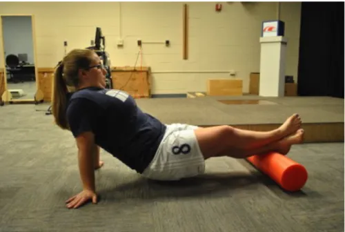
the self-myofascial release treatment without the vibrational component used the VYPER™ with the vibration feature turned off. For the self-myofascial release with vibration treatment, the participants used the foam roller with the vibration feature turned on to setting 2. For both treatments, the participant was instructed to place the foam roller under the calf of one leg, applying pressure using their full body weight. Participants were instructed to place the non-rolling leg on top of the other leg and to keep hips off of the ground (Figure 6). If this position was too painful to maintain, participants were instructed to flex their knee and put the foot of the non-rolling leg on the ground. This position was then used for both data collection sessions. Participants were instructed to slowly roll from ankle to knee for 5 seconds, and then return quickly to ankle and repeat the process. They foam rolled in this manner for 30 seconds. Then they focused on the pre-identified trigger points, holding pressure over each trigger point for 45 seconds. The participant then switched to the other leg, using the same methods.
After the foam-rolling intervention was completed, NRS was used to determine how much pain was caused by the treatment. The range of motion measurements of passive ankle dorsiflexion for knee extended and knee flexed as well as the measurements for weight-bearing lunge were taken for the second time.
Stretching Intervention
Post-treatment measurements followed the same procedures and order as the
pre-treatment measurements. The post-pre-treatment measurements were not counterbalanced and were taken in the order written for the pre-measurements section in order to limit the effects of fatigue. Measurements included pressure pain threshold, pain level on NRS after pain pressure threshold, ankle dorsiflexion range of motion of the weight-bearing lunge; passive, knee extended; and passive, knee flexed to 90 degrees range of motion. Additional measurements consisted of motion analysis with EMG, as previously described.
The participants returned 7 days after the first data collection session to complete the second data collection session. Seven days were more than enough to eliminate the possibility of a carryover effect from the first treatment (Sullivan et al., 2013). During the second testing session, the same procedures were followed, with the only change being the intervention. If the self-myofascial release with vibration component was used the first testing session, the vibration component was not used during the second testing session.
Data Reduction and Analysis
29
descent phase of each trial, and peak values were identified. Displacements were calculated by subtracting the start value from the peak value of the descent phase for all variables. The three middle trials were used for analysis of each task, and displacements and peak values were averaged across the three trials.
For range of motion values, change scores were calculated between pre-intervention and post-foam rolling as well as pre-intervention and post-stretching. These change scores from both foam-rolling interventions were then used for 2x2 repeated measures ANOVA. A Tukey post-hoc comparisons test was used for pairwise comparisons if there was a significant intervention by time interaction.
Raw EMG was exported into a custom MATLAB program (The MathWorks, Inc, Natick, MA) and passively demeaned, bandpass (10 to 350 Hz) and notch (59.5 to 60.5 Hz) filtered, and smoothed using a 25- millisecond root mean square sliding window. Mean EMG amplitudes during the descent phase of each task were normalized to the mean amplitude during a 1 second interval in the middle of the static trial recorded at the beginning of motion analysis. Both pre- and post-intervention EMG values were normalized to the pre-intervention static trial.
For kinematic, EMG, and pain pressure threshold data, a change score was calculated between pre-intervention and post-stretching. Dependent t-tests were used for the change score of each variable to compare the two foam-rolling interventions. Dependent t-tests will also be used to complete baseline comparisons for kinematic, EMG, ROM, and pain pressure threshold data to ensure no carryover effect between data collection sessions 1 and 2. Statistical
CHAPTER IV
Manuscript
Introduction
Restricted ankle dorsiflexion range of motion has been shown to be a predictor of high-risk lower extremity biomechanics (Dill, Padua et al., 2014; Fong, Blackburn et al., 2011; Sigward, Ota et al., 2008). These high-risk biomechanical patterns include less knee flexion, greater medial knee displacement, and greater ground reaction forces, putting those with restricted dorsiflexion range of motion at risk for knee injury, including patellofemoral pain syndrome and anterior cruciate ligament (ACL) sprain (Bell, Padua et al., 2008; Mauntel, Padua et al., 2012). Evidence based interventions to improve ankle dorsiflexion range of motion may be an important component of corrective exercise programs aimed to modify high-risk lower extremity biomechanics and reduce injury risk. Identifying the most effective treatment options for increasing range of motion will aid in improving ankle dorsiflexion range of motion.
31
identified as effective treatments to rectify range of motion deficits caused by myofascial trigger points (Lewit, 1999; Travell and Simons, 1993). The direct compression technique involves finding a locus of hyperirritability within the muscle and applying prolonged pressure to the area (Lewit, 1999). Self-myofascial release is one method of direct compression that has been shown to increase range of motion without decreasing muscle function (MacDonald et al., 2013; Sullivan et al., 2013). A foam roller is a commonly-used instrument for self-myofascial release. Foam rolling applies pressure over the myofascial adhesions, which helps to reduce the myofascial trigger points and break up the adhesions. To further help quantify the effectiveness of myofascial trigger points, pain pressure threshold objectively measures the severity of trigger points pre- and post-treatment (Jaeger and Reeves, 1986; Reeves et al. 1986).
Vibration training has also been proposed to increase range of motion without muscle deficiency. Several studies have shown vibration to increase both flexibility and muscle function (Cochrane and Stannard, 2005; Rhea, Bunker et al., 2009; Jacobs and Burns, 2009). The repeated eccentric and concentric contractions caused by the vibration therapy effect changes in the muscle tissue, including increased blood flow, increased muscle temperature and viscoelasticity (Bosco et al., 1999; Kerschan-Schindl et al., 2001), increase in pain threshold (Issurin et al., 1994), and improved neural efficiency (Cardinale and Bosco, 2003; De Gail et al., 1966). There is currently no literature that specifically examines the effect of local vibration therapy on treatment of myofascial trigger points.
developing a more efficient treatment of range of motion restrictions, improvement of lower extremity kinematics, and a reduction of injury risk.
Methods
A crossover study design was used. Participants were randomly allocated using a random number generator into 2 different groups (n1=10, n2=10). Each group received both interventions and underwent the same testing procedures. One group received self-myofascial release with vibration during the first testing session, while the other group received self-myofascial release with vibration during the second testing session.
Subjects
33
that limited physical activity for more than 2 days. Any participants with a known neurological disorder were also excluded. Participants completed a general medical health questionnaire and confirmed all inclusion and exclusion criteria were met. Participants read and signed an
Institutional Review Board approved informed consent form. Participants wore their own athletic shorts, t-shirt, and athletic shoes throughout the screening and data collection sessions.
Prior to data collection, all participants completed a screening session. During the screening session, participants completed a weight-bearing lunge dorsiflexion range of motion test and were examined for myofascial trigger points. Myofascial trigger points were identified by placing the participants in a prone position with the knees extended on a treatment table. Four criteria were used to identify a myofascial trigger point: 1) a palpable band taught within the skeletal muscle, 2) a hypersensitive tender spot/nodule within a taut band, 3) recognition of current pain complaint by pressure on the tender nodule, and 4) painful limit to full stretch range of motion (Travell and Simons, 1993; Gerwin et al., 1997). At least 2 of the above criteria had to be satisfied to identify a myofascial trigger point (Grieve et al., 2011). The primary investigator palpated the gastrocnemius and soleus to identify all myofascial trigger points on both legs. The location of the myofascial trigger points and criteria satisfied were documented. If no
myofascial trigger point was found in the dominant limb, the participant was excluded from the study at this time. Participant inclusion was approximately 50%, with 39 possible participants screened to identify 20 participants.
completed a stretching intervention, and then completed range of motion measurements and pain pressure threshold a third time. Participants reported 7 days later for the second data collection session and completed the same procedures with the other foam rolling intervention (Figure 1). Instrumentation
A digital inclinometer (Saunders Group, Inc., Chaska, MN) was used to record
measurements during the weight-bearing lunge. The intra-rater reliability of the weight-bearing lunge measurement by the primary investigator was calculated with intra-class coefficients (ICC3,k = 0.976) and standard error of the measurement (SEM = 0.864).
A standard 12-inch plastic goniometer was used to measure gastrocnemius and soleus range of motion. Intra-rater reliability of the passive range of motion measurement by the primary investigator was calculated with intra-class coefficients (ICC3,k = 0.966-0.989) and standard error of the measurement (SEM = 0.804-1.587) for each range of motion measurement.
A handheld digital dynamometer (Manual Muscle Tester Model 01163, Lafayette Instrument Co., Lafayette, IN) was used to measure pain pressure threshold. Intra-rater
reliability of the pain pressure threshold measurement by the primary investigator was calculated with intra-class coefficients (ICC3,k = 0.916) and standard error of the measurement (SEM = 0.477) for the pain pressure threshold measurement.
Procedures
Range of Motion Measurements
35
forward as far as possible while keeping the dominant foot in line with the long axis of the leg and the heel on the ground. The primary investigator held the heel in place on the ground. The foot was then moved posteriorly until the maximum range of dorsiflexion was reached, which was identified by the heel lifting off the ground (Figure 2). The inclinometer measurement was recorded when the participant was in the position of maximum dorsiflexion (Krause, Cloud et al., 2011; Bennell, Techovanich et al., 1998; Denegar, Hertel et al., 2002). Three measurements were taken and averaged.
Ankle dorsiflexion measurements were recorded using a 12-inch goniometer and standard range of motion techniques for the gastrocnemius and soleus (Figures 4 and 5). Measurements were taken at the first point of restriction in dorsiflexion with the knee bent to 90 degrees as well as straight (Bell, Padua et al. 2008; Cosby 2011, Fong, Blackburn et al. 2011). Three
measurements were taken and averaged. All range of motion measurements were counterbalanced.
Pain Pressure Threshold and Numerical Rating Scale Measurements
was then asked to identify how much pain was caused by the pain pressure threshold
measurement on a Numerical Rating Scale (NRS). Only whole numbers were recorded on a scale from 0 to 10. Zero was delineated as no pain, 5 was delineated as moderate pain, and 10 was delineated as unbearable pain. The numerical pain values were verbally stated while the participant was shown a number line displaying the same values and definitions. The NRS was administered in the same manner after the foam rolling treatment (Jensen et al., 1999; Gemmell, Hilland, 2011).
Foam Roll Interventions
A random number generator was used to determine which treatment was administered. The VYPER™ (Hyperice, Irvine, CA) foam roller was used for the
intervention. The VYPER™ is a cylinder of high-density foam, measuring 18” long and 6” round. A vibrating motor is embedded within the high-density foam to facilitate local muscle vibration. During the self-myofascial release treatment without the vibrational component used the VYPER™ with the vibration feature turned off. For the self-myofascial release with
37
seconds. After 30 seconds the participants were instructed to focus on the pre-identified trigger points, holding pressure over the three most painful trigger points for 45 seconds.
After the foam-rolling intervention was completed, the NRS was used to determine how much pain was caused by the treatment. The range of motion measurements of passive ankle dorsiflexion for knee extended and knee flexed as well as the measurements for weight-bearing lunge were taken for the second time.
Stretching Intervention
All participants performed the same stretching intervention. The stretching intervention took place immediately after the second round of range of motion measurements. Participants performed weight bearing gastrocnemius and soleus stretching using a slant board with the knee straight (Figure 7) and knee bent (Figure 8) respectively. Three sets of a thirty-second static stretch were performed for both positions on both legs.
Statistical Analysis
For all range of motion values, change scores were calculated between pre-intervention and post-foam rolling as well as pre-intervention and post-stretching. A 2x2 repeated measures ANOVA with time defined as pre- to post-stretching vs. pre- to post-foam rolling and
intervention defined as traditional or vibrational foam rolling analyzed differences. A Tukey post-hoc comparisons test was used for pairwise comparisons if there was a significant
intervention by time interaction. Examination of 95% confidence intervals was also performed to determine if the change scores (pre- to post-foam rolling and pre- to post-stretching)
For pain pressure threshold and NRS scores for pain pressure threshold, a change score was calculated between pre-intervention and post-stretching. Dependent t-tests were used for the change score of each variable to compare the two foam-rolling interventions. A dependent t-test was used to compare the NRS score for each intervention. Dependent t-tests were also used to complete baseline comparisons for ROM, NRS, and pain pressure threshold data to ensure no carryover effect between data collection sessions 1 and 2. Statistical significance was set at α<0.05. Examination of 95% confidence intervals was also performed to determine if the change scores (pre-intervention to post-stretching) were significant. Statistical significance was defined as the change score’s 95% confidence interval not crossing zero. Data were analyzed using SPSS 19.0 statistical software (SPSS, Inc., Chicago, IL).
Results
There were no differences between baseline measurements for range of motion, pain pressure threshold, or NRS based on 95% confidence intervals for pre-measurements recorded in Tables 1 and 2. There was no carryover effect between days one and two of data collection. Range of Motion
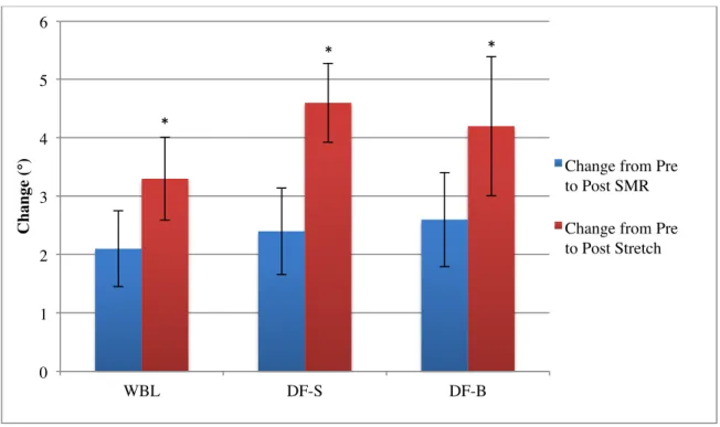
Range of motion descriptive statistics are recorded in Table 1. There was no significant intervention by time interaction for the weight-bearing lunge (F1,18 = 1.10, p = 0.307), ankle dorsiflexion with knee straight (F1,18 = 0.77, p = 0.392), or ankle dorsiflexion with knee bent (F1,18 = 0.77, p = 0.392). Thus, the amount of change in ankle dorsiflexion range of motion measures was similar between interventions following foam rolling and static stretching.
There was a significant main effect for intervention, defined as foam rolling with
39
p = 0.028), ankle dorsiflexion knee straight (F1,18 = 6.54, p = 0.020) and ankle dorsiflexion knee bent (F1,18 = 12.29, p = 0.003) measures. The change in ankle dorsiflexion range of motion was greater following foam rolling with vibration compared to traditional foam rolling. Examination of 95% confidence intervals demonstrated that change scores for all ankle dorsiflexion range of motion measures did not cross zero. These findings indicate that both traditional and vibration foam rolling improved ankle dorsiflexion range of motion measures, but the change was greater following vibration foam rolling (significant intervention main effect) (Figure 9).
A significant main effect for time, defined as the change in range of motion resulting from foam rolling versus the change in range of motion resulting foam rolling plus stretching, was noted for the weight-bearing lunge (F1,18 = 27.93, p < 0.001), ankle dorsiflexion knee straight (F1,18 = 46.18, p < 0.001) and ankle dorsiflexion knee bent (F1,18 = 42.37, p < 0.001) measures. Examination of 95% confidence intervals revealed that all change scores of ankle dorsiflexion range of motion did not cross zero. These combined findings indicate that ankle dorsiflexion range of motion measures were significantly improved after foam rolling (95% CI did not cross zero) and there were significant improvements with the addition of static stretching following foam rolling (significant time main effect) (Figure 10).
Pain Pressure Threshold and Numerical Rating Scale
Participants demonstrated an increase in pain-pressure threshold after both traditional and vibrational foam rolling (mean difference = 1.6 lbs, 95% CI = [1.0, 2.1]). However, there was no difference in change in pain-pressure threshold between the interventions (p = 0.66).
vibrational foam rolling (mean difference = -0.4, 95% CI = [-1.0, 0.2]) on the numerical rating scale (p = 0.874). Descriptive statistics for PPT and NRS are recorded in Table 2.
There was no difference between reported NRS for the traditional foam rolling treatment and the vibrational foam rolling treatment (p = 0.39). Participants reported a mean NRS of 5.8 (SD = 2.0, 95% CI = [4.8, 6.7]) for traditional foam rolling and a mean NRS of 6.1 (SD = 1.7, 95% CI = [5.2, 6.9]).
Discussion
The purpose of this study was to determine the effect of combined self-myofascial release and vibration on dorsiflexion range of motion and myofascial pain. We hypothesized that self-myofascial release combined with vibration would result in greater range of motion increases and greater increase in pain pressure threshold than traditional self-myofascial release. The results support the hypothesis that foam rolling with vibration leads to a greater increase in all ankle dorsiflexion range of motion measures than traditional foam rolling. However, the clinical significance of a 2º increase compared to a 3º to 4º increase in dorsiflexion range of motion has not been determined.
41
needed to determine if increased range of motion will allow for improved movement patterns. Additionally, a greater increase in range of motion was demonstrated between the pre- to post-foam rolling change in range of motion compared to the pre- to post-stretching change range of motion. This result supports previous findings that static stretching results in an increased range of motion when combined with vibration (Sands et al., 2008) and when
combined with self-myofascial release (Škarabot et al., 2015). A lack of significant intervention by time interaction for range of motion measurements indicates there is no difference in increase in range of motion resulting from combining vibrational foam rolling and stretching compared to combining traditional foam rolling and stretching. Vibration was not shown to increase the effects of stretching, suggesting that vibration may not cause increased viscoelastic changes or that such changes are only present for a short time after treatment (Mizuno et al., 2013). The mechanism for increased range of motion due to vibration combined with self-myofascial release could be neural in origin. It has been observed that muscle spindles at myofascial trigger points are more sensitive, so relaxation could be effected by inhibiting the muscle spindle by
stimulating golgi tendon organs (Ge, Serrao et al., 2009; Bove et al., 2003).
rather than a standard amount of pressure. The objective increase seen in pain pressure threshold implies that there was a significant reduction in pain resulting from both foam rolling and foam rolling with vibration. Previous literature suggests that pain itself causes restrictions in range of motion (Gerber et al., 2013). A reduction in pain could be contributing to the observed increase range of motion. Pain also contributes to compensatory movement patterns (Seeley et al., 2013), so a reduction in pain caused by myofascial trigger points could allow for increases in range of motion as well as improved movement patterns.
The clinical significance of the increase in range of motion caused by the foam roller with vibration is compounded by the lack of difference between reported pain for the two foam-rolling interventions. This means patients can receive a greater increase in range of motion without a resultant increase in pain. This helps with patient compliance as well as efficiency in treatment.
Further investigation is necessary to determine the effects of prolonged use of self-myofascial release with vibration, rather than just an acute application. Examination of lower-extremity kinematics, kinetics, and muscle activation would also be beneficial in determining the clinical importance of these findings. Results from this study are only generalizable to college-aged, physically active individuals with a dorsiflexion range of motion restriction. It is unknown whether self-myofascial release with vibration would have an effect on non-pathologic
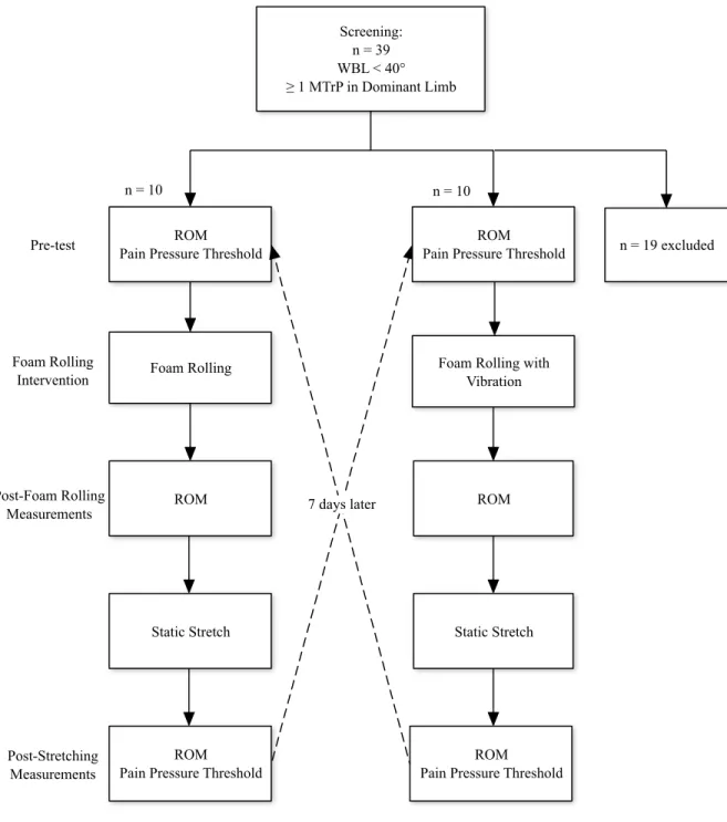
43 FIGURES
Figure 1: Experimental Procedures. Testing sessions were completed 7 days apart,
depicted by the dashed lines.
n = 10
Screening: n = 39 WBL < 40° ≥ 1 MTrP in Dominant Limb
n = 19 excluded ROM
Pain Pressure Threshold
Foam Rolling Foam Rolling with Vibration
ROM ROM
Pre-test
Foam Rolling Intervention
Post-Foam Rolling Measurements
Static Stretch Static Stretch
Post-Stretching Measurements
n = 10
7 days later
ROM Pain Pressure Threshold
ROM Pain Pressure Threshold
Figure 2: Measurement of Weight-bearing Lunge
45
Figure 4: Measurement of Ankle Dorsiflexion with Knee Straight
Figure 6: Foam Rolling Procedure
47 Figure 8: Stretching Protocol with Knee Bent
Figure 9: Graph of Range of Motion Changes Comparing Foam Rolling to Foam Rolling with Vibration
Error bars represent 95% CI
* Significant difference between traditional and vibrating foam rolling (p < 0.05)
0 1 2 3 4 5 6 7
WBL DF-S DF-B
C
h
an
ge
(°
)
Traditional Vibration
*
*
Figure 10: Graph of Range of Motion Changes Comparing Foam Rolling to Foam Rolling Plus Stretch
Error bars represent 95% CI
* Significant difference between pre to post SMR and pre to post SMR plus stretch (p < 0.05)
0 1 2 3 4 5 6
WBL DF-S DF-B
C
h
an
ge
(°
) Change from Pre
to Post SMR Change from Pre to Post Stretch
*
TABLES
Table 1: Range of Motion Descriptive Statistics
Variable Intervention
Pretest Mean° (SD°) [95% CI] Post SMR Mean° (SD°) [95% CI] Post Stretch Mean° (SD°) [95% CI]
Δ Pre to Post-SMR Mean° (SD°)
[95% CI]
Δ Pre to Post-Stretch Mean° (SD°) [95% CI] p Weight-Bearing Lunge Traditional SMR 33.9 (4.6) [31.8, 36.0] 35.7 (5.1) [33.3, 38.1] 36.7 (5.0) [34.3, 39.0] 1.8 (1.4) [1.1, 2.4] 2.7 (1.5)
[2.0, 3.4] 0.307 Vibrating SMR 34.0 (4.0) [32.1, 35.9] 36.6 (5.1) [34.1, 39.1] 37.8 (4.7) [35.6, 40.1] 2.5 (1.9) [1.6, 3.4] 3.9 (2.1) [2.9, 4.9] Ankle Dorsiflexion Knee Straight Traditional SMR -2.7 (6.2) [-5.6, 0.2] -0.9 (6.1) [-3.7, 2.0] 0.6 (5.7) [-2.1, 3.2] 1.8 (2.7) [0.5, 3.1] 3.2 (2.6) [1.9, 4.4] 0.392 Vibrating SMR -2.5 (5.5) [-5.1, 0.1] 1.1 (5.6) [-1.7, 3.9] 2.5 (6.0) [-0.3, 5.3] 3.4 (1.4) [2.7, 4.1] 5.2 (1.7) [4.3, 6.0] Ankle Dorsiflexion Knee Bent Traditional
SMR [0.3, 6.8] 3.6 (7.0) [2.4, 8.6] 5.5 (6.6) [4.4, 10.5] 7.5 (6.5) [1.0, 2.2] 1.6 (1.3) [2.2, 4.9] 3.6 (2.8)
0.392 Vibrating
SMR [1.3, 7.2] 4.3 (6.3) [4.5, 10.4] 7.4 (6.1) [7.3, 12.7] 10.0 (5.7) [2.0, 4.5] 3.2 (2.6) [4.3, 7.2] 5.7 (3.0)
Table 2: Pain Pressure Threshold and NRS Descriptive Statistics
Variable Intervention
Pretest Mean (SD)
[95% CI]
Posttest Mean (SD)
[95% CI]
Difference Mean (SD) [95% CI]
p
Pain Pressure Threshold*
Traditional SMR [4.5, 9.5] 7.0 (5.3) [5.8, 11.7] 8.7 (6.3) [1.0, 2.4] 1.7 (1.5)
0.660 Vibrating SMR [4.1, 8.4] 6.2 (4.6) [5.1, 10.1] 7.6 (5.3) [0.4, 2.4] 1.4 (2.1)
NRS for Pain Pressure Threshold
Traditional SMR [3.5, 5.2] 4.4 (1.9) [3.1, 4.7] 3.9 (1.7) [-1.0, 0.1] -0.5 (1.3)
0.874 Vibrating SMR [3.4, 5.0] 4.2 (1.8) [3.0, 4.6] 3.8 (1.8) [-1.0, 0.2] -0.4 (1.2)
51
REFERENCES
Atha J, Wheatley DW. Joint mobility changes due to low frequency vibration and stretching exercise. Br. J. Sports Med. 1976;10:26–34.
Avela J, Kyrolainen H, Komi, PV. Altered reflex sensitivity after repeated and prolonged passive muscle stretching. Journal of Applied Physiology. 1999;8:1283–1291.
Barnes M. The basic sciences of myofascial release: Morphologic change in connective tissue. Journal of Bodywork & Movement Therapies. 1997;1:231-238.
Bell DR, Oates DC, Clark MA, Padua DA. Two- and 3-dimensional knee valgus are reduced after an exercise intervention in young adults with demonstrable valgus during squatting. Journal of athletic training 2013;48(4):442-449.
Bell DR, Padua DA, Clark MA. Muscle strength and flexibility characteristics of people displayed excessive medial knee displacement. Archives of physical medicine and rehabilitation. 2008;89(7):1323-1328.
Bosco C, Colli R, Introini E, Cardinale M, Tsarpela O, Madella A, et al. Adaptive responses of human skeletal muscle to vibration exposure. Clinical Physiology. 1999;19:183–187.
Bove M, Nardone A, Schieppati M. Effects of leg muscle tendon vibration on group Ia and group II reflex responses to stance perturbation in humans. J Physiol 2003;550:617-30.
Cardinale M, Bosco C. The use of vibration as an exercise intervention. Exercise and Sport Science Reviews. 2003;31:3–7.
Church JB, Wiggins MS, Moode FM, Crist R. Effect of Warm-Up and Flexibility Treatments on Vertical Jump Performance: Journal of Strength and Conditioning Research. 2001
Aug;15(3):332–6.
Cochrane DJ. Acute whole body vibration training increases vertical jump and flexibility performance in elite female field hockey players. British Journal of Sports Medicine. 2005 Nov 1;39(11):860–5.
Cochrane DJ. The potential neural mechanisms of acute indirect vibration. J Sports Sci Med. 2011 Mar 1;10(1):19-30.
Cosby NL, Hertel J. Relationships Between Measures of Posterior Talar Glide and Ankle Dorsiflexion Range of Motion. Athletic Training & Sports Health Care 2010.
Curran PF, Fiore RD, Crisco JJ. A comparison of the pressure exerted on soft tissue by 2 myofascial rollers. J Sport Rehabil 17:432-442, 2008.
motion, posterior talar glide, and joint laxity. J Orthop Sports Phys Ther 2002;32(4):166 -173.
DePino G, Webright WG, Arnold BL. Duration of maintained hamstring flexibility after cessation of an acute static stretching protocol. J Athl Train. 2000;35:56–59.
Desai MJ, Bean MC, Heckman TW, Jayaseelan D, Moats N, Nava A. Treatment of myofascial pain. Pain Manag. 2013 Jan;3(1):67-79.
De Gail P, Lance W, Neilson PD. Differential effects on tonic and phasic reflex mechanisms produced by vibration of muscles in man. Journal of Neurology, Neurosurgery and Psychiatry. 1966;29:1–11.
Diehl A. Increase hamstring flexibility: Combination of self-myofacial release and static stretching programs. Ann Arbor: Lamar University - Beaumont; 2014.
Dill KE, Begalle R, Frank B, Zinder S, Padua DA. Altered Knee and Ankle Kinematics During Squatting in Those With Limited Weight-Bearing Lunge Ankle-Dorsiflexion Range of Motion. J Athl Train. 2014.
Feland JB, Hawks M, Hopkins JT, Hunter I, Johnson AW, Eggett DL. Whole body vibration as an adjunct to static stretching. Int J Sports Med. 2010 Aug;31(8):584-9.
Ferguson SL, Kim E, Seo DI, Bemben MG. Comparing the effects of 3 weeks of upper-body vibration training, vibration and stretching, and stretching alone on shoulder flexibility in college-aged men. J Strength Cond Res. 2013 Dec;27(12):3329-34.
Fong CM, Blackburn JT, Norcross MF, McGrath M, Padua DA. Ankle-dorsiflexion range of motion and landing biomechanics. Journal of Athletic Training. 2011;46(1):5-10.
Franco AH. Pes cavus and pes planus: analyses and treatment. Phys 1987;6(5):688--694.
Ge HY, Serrao M, Andersen OK, Graven-Nielsen T, Arendt-Nielsen L. Increased H-reflex response induced by intramuscular electrical stimulation of latent myofascial trigger points. Acupunct Med. 2009 Dec;27(4):150-4.
Gemmell H, Hilland A. Immediate effect of electric point stimulation (TENS) in treating latent upper trapezius trigger points: a double blind randomised placebo-controlled trial. J Bodyw Mov Ther. 2011;15(3):348-54.
Gerber LH, Sikdar S, Armstrong K, Diao G, Heimur J, Kopecky J, et al. A systematic


![Table 2: Pain Pressure Threshold and NRS Descriptive Statistics Variable Intervention Pretest Mean (SD) [95% CI] Posttest Mean (SD) [95% CI] Difference Mean (SD) [95% CI] p Pain Pressure Threshold * Traditional SMR 7.0 (5.3) [4.5, 9.5] 8.7](https://thumb-us.123doks.com/thumbv2/123dok_us/8332534.2210908/56.918.100.799.148.405/pressure-threshold-descriptive-statistics-intervention-difference-threshold-traditional.webp)
