Open Access
Research
Microarray identifies ADAM family members as key responders to
TGF-
β
1 in alveolar epithelial cells
Dominic T Keating
1,2, Denise M Sadlier
1, Andrea Patricelli
1,
Sinead M Smith
3, Dermot Walls
3, Jim J Egan
2and Peter P Doran*
1Address: 1General Clinical Research Unit, Mater Misericordiae University Hospital, School of Medicine and Medical Sciences, University College Dublin, Dublin 7, Ireland, 2Advanced Lung Disease Programme and Lung Transplant Unit, Mater Misericordiae University Hospital and 3School of Biotechnology, Dublin City University, Dublin, Ireland
Email: Dominic T Keating - [email protected]; Denise M Sadlier - [email protected]; Andrea Patricelli - [email protected]; Sinead M Smith - [email protected]; Dermot Walls - [email protected]; Jim J Egan - [email protected];
Peter P Doran* - [email protected] * Corresponding author
Abstract
The molecular mechanisms of Idiopathic Pulmonary Fibrosis (IPF) remain elusive. Transforming Growth Factor beta 1(TGF-β1) is a key effector cytokine in the development of lung fibrosis. We used microarray and computational biology strategies to identify genes whose expression is significantly altered in alveolar epithelial cells (A549) in response to TGF-β1, IL-4 and IL-13 and Epstein Barr virus.
A549 cells were exposed to 10 ng/ml TGF-β1, IL-4 and IL-13 at serial time points. Total RNA was used for hybridisation to Affymetrix Human Genome U133A microarrays. Each in vitro time-point was studied in duplicate and an average RMA value computed. Expression data for each time point was compared to control and a signal log ratio of 0.6 or greater taken to identify significant differential regulation. Using normalised RMA values and unsupervised Average Linkage Hierarchical Cluster Analysis, a list of 312 extracellular matrix (ECM) proteins or modulators of matrix turnover was curated via Onto-Compare and Gene-Ontology (GO) databases for baited cluster analysis of ECM associated genes.
Interrogation of the dataset using ontological classification focused cluster analysis revealed coordinate differential expression of a large cohort of extracellular matrix associated genes. Of this grouping members of the ADAM (A disintegrin and Metalloproteinase domain containing) family of genes were differentially expressed. ADAM gene expression was also identified in EBV infected A549 cells as well as IL-13 and IL-4 stimulated cells. We probed pathologenomic activities (activation and functional activity) of ADAM19 and ADAMTS9 using siRNA and collagen assays. Knockdown of these genes resulted in diminished production of collagen in A549 cells exposed to TGF-β1, suggesting a potential role for these molecules in ECM accumulation in IPF.
Published: 01 September 2006
Respiratory Research 2006, 7:114 doi:10.1186/1465-9921-7-114
Received: 29 May 2006 Accepted: 01 September 2006
This article is available from: http://respiratory-research.com/content/7/1/114
© 2006 Keating et al; licensee BioMed Central Ltd.
Background
Idiopathic pulmonary fibrosis (IPF) is a progressive and lethal pulmonary fibrotic lung disease. It is the most com-mon form of the idiopathic interstitial pneucom-monias and is unresponsive to treatment resulting in a median survival from diagnosis of 2.9 years [1]. Although the pathogenesis of IPF remains elusive, a number of conditions and risk factors are associated with the disease, including cigarette smoking, several viral proteins, and genetic predisposi-tion to IPF [2].
During both lung development and fibrogenesis, mesen-chymal signaling alters alveolar epithelial cell phenotype and regulates pneumocyte differentiation [3,4]. Effective cell function in both the epithelium and the mesenchyme is dependent on signals originating in both compart-ments, acting in a complimentary axis. In disease the unchecked signaling emanating from these compartments establishes persistent fibroblast migration and extracellu-lar matrix deposition with resultant pulmonary fibro-sis[5].
Injured alveolar epithelail cells release a number of profi-brotic cytokines including transforming growth factors-beta-1, platelet derived growth factor tumour necrosis fac-tor-alpha and interleukin-1 [6]. As a result of these medi-ators being released there a chemoattraction gradient for fibroblasts toward these areas of lung, with subsequent phenotypic differentiation.
TGF-β1 is a prominent mediator in normal wound repair without the development of fibrosis[8]. Excess produc-tion of latent TGF-β1 and active TGF-β1 has been associ-ated with the development of temporary inflammation, however, only TGF-β1 overexpression results in fibroblast migration and proliferation with increased deposition of extracellular matrix. This suggests that inflammation in IPF is not crucial for pathogenesis but may instead be an associated phenomenon [9].
Targeted overexpression of TGF-β1 is associated with aug-mented fibrosis, while antagonism of the growth factor results in abrogation o the fibrotic process. TGF-β1 knock-out mice die prematurely due to developmental retarda-tion and progressive inflammaretarda-tion[10]; however, treatment with TGF-β1 specific antagonists in mice did not result in a significant disturbance of the immune sys-tem[11]. TGF-β1 has been shown to augment epithelial cell apoptosis and inhibition of this process has been shown to reduce fibrosis in animal models[12,13]. In other studies instillation of apoptotic cells into inflamed lungs has accelerated healing in a TGF-β1 dependent manner[14].
TGF-β1 is consistently associated with progressive fibrosis with increased expression being associated with a variety of fibrotic lung disease [15-17]. Adenovirus-mediated gene transfer of TGF-β1 resulted in severe fibrosis in ani-mal models[15]; while αVβ6 integrin (a TGF-β1 activator) knockout mice developed lung inflammation but not fibrosis in response to bleomycin[18]. TGF-β1 displays a pivotal role in the development of a fibrotic process in animal models, however the reasons surrounding its over-expression and the predilection towards a fibrotic pheno-type in this setting remains unexplained.
Interleukin (IL)-13, a Th2 cytokine, has been shown to be increased in IPF [19], while the lungs of mice injured with bleomycin display increased 13 and its receptor 13Ralpha2 [20]. The vehicle for fibrosis in response to IL-13 is activated TGF-β1 [21]. In asthmatic individuals the overexpression of IL-13 is associated with subepithelial fibrosis which has obvious implications for the develop-ment of idiopathic fibrosis [22]. IL-4 is also increased in the lungs of IPF patients and in bleomycin murine models [23]. The role for IL-4 in IPF may be two fold, limiting T-cell migration and stimulating fibrosis. IL-4 transforms fibroblasts into myofibroblasts inferring a role in the fibrotic process [24]. Through the release of TGF-β2 IL-4 initiates the release of matrix proteins by myofibroblasts while inhibition of its receptor in bleomycin-injured mice attenuates the fibrotic response [20,22].
Epstein-Barr virus (EBV) is a ubiquitous human herpesvi-rus associated with various diseases including infectious mononucleosis, Burkitt's lymphoma and Hodgkin's dis-ease. A link between EBV and IPF has been suggested since Vergnon and colleagues demonstrated an elevation in the IgA levels against viral capsid antigen in IPF patients [25]. EBV usually infects the upper respiratory tract but has also been shown to infect and replicate in the lower respiratory tract. Immunohistochemical studies suggest that this rep-lication occurs in the type II alveolar epithelial cells [26]. Further evidence for EBV involvement in IPF comes from the observation of a poorer prognosis in these patients when associated with EBV latent membrane protein 1 (LMP1) in epithelial cells [27]. LMP1 is an EBV associated protein expressed on the surface of EBV Infected cells in the latent and replicating phase [28]. Aberrant DNA found in lung tissue and serum of IPF patients suggested a mechanism by which a persistent virus can change from a latent to a productive phase via recombinatorial events [29].
Methods
Cell culture and EBV Infection in vitro
Human alveolar epithelial cells (A549) were obtained from European Collection of Cell Cultures (Salisbury, United Kingdom) and grown in vitro in Hams F12 (Life Technologies, Paisley, Scotland) supplemented with 10% fetal calf serum, 146 mg/L L-glutamine, 1% penicillin and 1% streptomycin. For stimulation experiments, cells were serum starved overnight before exposure to 10 ng/ml
TGF-β1, IL-4 or IL-13 (Sigma) for the indicated time points, control samples were maintained in serum free condi-tions.
To effect virus infection, A549 cells were co-cultured with Akata cells (an Epstein-Barr virus-negative cell line infected with recombinant EBV carrying the neomycin resistance gene) [30]. The Akata cell line was maintained in RPMI 1640 (Life Technologies, Paisley, Scotland) sup-plemented with 10% FBS, 2 mM L-glutamine, 100 U/ml Penicillin, 100 µg/ml streptomycin and 700 µg/ml G418. The viral lytic cycle was induced in the Akata cells by add-ing goat anti-human serum Immunoglobulin G (Sigma) at 100 µg/ml. After four hours the Akata cells were added to the A549 wells at a concentration of 5 × 10⁵/ml. After two days incubation half the medium was replaced with medium containing 5% FCS. Following a further 4 days incubation the media containing the Akata cells was removed, pelleted down and resuspended in M5 media (Calcium free DMEM supplemented with 5% Horse Serum, 2 mM glutamine, cortisol, 2 ng/ml EGF, 10 mg/ml Insulin, 100 ng/ml cholera toxin, 100 U/ml Penicillin and 100 µg/ml streptomycin). 2 mls of this cell suspension was then placed onto the wells and left to incubate for 2 days. The wells were washed with PBS and the media replaced with fresh medium containing 10% FCS. Follow-ing incubation for 24 hours 700 ng/ml G418 (Sigma) was added to select for EBV-infected A549 cells.
Western blot analysis
Latent infection of the A549 cells with EBV was confirmed by the detection of latent membrane protein 1 (LMP1) by Western blot. After removal of EBV/A549 cells from flasks protein lysates were prepared by boiling for 10 minutes in
2% SDS, 100 mM NaCl, 0.01 M Tris-HCl, 5% β
-Mercap-toethanol, 1 mM EDTA, 100 µg of phenylmethylsulfonyl fluoride/ml, and 2 µg of leupeptin/ml. The product was then sonicated on ice and clarified at room temperature by centrifugation at 12,000 rpm for 10 min. Protein frac-tionated by discontinuous SDS-5–10% polyacrylamide gel electrophoresis and blotted onto a nitrocellulose filter. Anti-LMP1 CS1-4 antibody (University of Wales, Cardiff) cocktail diluted to 1:100 in Blotto (5% skim milk and 0.1% Tween 20 in Tris-buffered saline) was used to probe the filters at 4°C overnight. Alkaline phosphatase-conju-gated sheep anti-mouse immunoglobulin G (IgG)
(Promega) was used to detect immunocomplexes, which were visualized using 5-bromo-4-chloro-3-indolylphos-phate (BCIP)-nitroblue tetrazolium liquid substrate (Sigma).
RNA extraction and gene array analysis
Following stimulation of A549 cells with 10 ng/ml TGF-B1 for 15 mins, 30 mins, 2 hour and 4 hours, RNA isola-tion, cDNA synthesis, in vitro transcription and microarray analysis were performed as previously reported [31] and in accordance with Affymetrix protocols(Affymetrix, Santa Clara, California). Arrays were scanned with a confocal scanner (Affymetrix). Each RNA sample derived from an individual well and 15 min, 30 min, 2 hour and 4 hour in vitro exposures were microarrayed in duplicate on HU133 Affymetrix chips. Image files were obtained through Affymetrix GeneChip software (MAS5) and subsequently robust multichip analysis (RMA) was performed. To ensure the average was statistically significant a t-test and p-value were generated. Only those genes with a p-value ≤ 0.01 were included in subsequent bioinformatic analysis. Expression data was further probed to identify those genes whose expression is altered. Expression data for each time point was compared to control and a signal log ratio of 0.6 or greater (equivalent to a fold change in expression of 1.5 or greater) was taken to identify significant differential regulation. Using normalised RMA values, unsupervised average linkage hierarchical cluster analysis was per-formed using an Eisen software program [32]. Cluster analysis is a group of mathematical techniques for the identification of patterns in large datasets. Briefly, a dis-tance metric is used to calculate the similarity between the expression profiles of a group of genes. The more similar the expression profiles of genes are, the closer they are placed together on a dendrogram or tree. A list of 312 extracellular matrix proteins or modulators of matrix turn-over was curated via the publicly available Onto-Compare and Gene-Ontology databases [33].
Real-time PCR
Reverse transcription was carried out using the Promega reverse transcription system. 1 µl of Oligo(dT)15 (0.5 µg/
2 min in order to inactivate the enzyme. The subsequent cDNA was stored at 4°C until required.
Real time RT-PCR was performed on a TaqMan ABI 7700 Sequence Detection System® (AppliedBiosystems, Weiter-stadt, Germany) using heat activated AmpliTaq Gold, DNA polymerase (Amplitaq Gold, Applied Biosystems, Weiterstadt, Germany) as previously described [34]. The ribosomal 18S was used as an endogenous control for normalisation of the target genes. Its primer and probe were supplied as a PDAR (predeveloped assay reagent) from Applied Biosystems with the probe labelled with VIC at the 5' end. Primer and probes for the genes of interest were designed in PrimerExpress® version 2.0(AppliedBio-systems). The probes for the target genes were labelled with fluorescent dye, FAM on the 5' end and the quencher TAMRA onthe 3' end. PCR reactions were set up in sepa-rate tubes with TaqMan Universal PCR Master Mix from Applied Biosystems. Optimal concentration of primers and probes were 200 nM for probe, 300 nM for its prim-ers, and 100 nM reaction mix for PDARs. cDNA was amplified on the 7700HT detection system (Applied bio-science) at default thermal conditions: 2 min @50°C, 10 min @95°C for enzyme activation and the 40 cycles of 15 sec @95°C for denaturation and 1 min @60°C for annealing and extension. Controls consisting of distilled H2O were negative in all runs. All measurements were per-formed in triplicate for each time point.
Following cycling, to ensure specificity, melt curve analy-sis was carried out to verify the amplification of PCR prod-ucts starting at 65°C and ramping to 90°C at .1°C/sec. One peak in the melt curve indicated no secondary, non-specific products were formed. All results were compared to those for unstimulated A549 cells and analysed using the delta delta Ct method. All experiments were per-formed in triplicate for each time point.
Gene silencing by RNA interference
Knock down of gene expression was achieved using RNA interference. Two siRNA duplexes were designed and syn-thesised for silencing ADAM19 and ADAMTS9. (Qiagen Inc. CA, USA). A chemically synthesized non-silencing siRNA duplex that had no known homology with mam-malian genes was used to control for non-specific silenc-ing events. 2 × 10⁵ A549 cells were added to each well of a 6-well plate in 3 ml growth media and incubated under the standard conditions of 37°C and 5 % CO2 in a humid incubator for 24 hr. A sufficient amount of growth medium was added to 5 µg siRNA and 30 µl RNAifect (Qiagen) to bring the final volume to 100 µl. Following incubation, media was removed from the cells and this mix was added drop-wise. 3 ml growth medium was added and the cells were incubated for 48 hr under stand-ard conditions. Following this, all growth media was
removed and cells were washed with sterile PBS. 1 ml TRI-zol™ (Sigma) was added to each well and left for 10 min at room temperature with occasional shaking. 200 µl chloroform was added and the mixture was shaken, left at room temperature for 15 min and centrifuged at 13,000 g at 4°C for 15 min. The upper aqueous layer was trans-ferred to a fresh 1.5 ml tube. 0.5 ml ice-cold isopropanol was added to the aqueous phase, shaken and left to stand on ice for 10 min before it was centrifuged at 13000 g at 4°C for 10 min. The supernatant was removed and 1 ml of sterile 75 % ethanol was added to wash the pellet by gentle vortexing and centrifugation at 7500 g for 5 min. The ethanol was removed and the pellet was allowed to air-dry for 5 min. Pellets were resuspended in 50 µl 0.1 % DEPC treated H2O by heating at 60°C for 15 min. All RNA was stored at -80°C.
Collagen Assay
Sircol collagen assay (Biocolour) was performed as per manufacturer's guidelines. The dye reagent contains sirius red in picric acid. Sirius Red is an anionic dye with sul-phonic acid side chain groups. These groups react with the side chain groups of the basic amino acids present in col-lagen under specific conditions permitting determination of mammalian collagens types I to V. Briefly 100 µl aliq-uots of cell culture supernatant were incubated with the dye reagent by gentle mixing for 30 mins at room temper-ature. The dye-bound collagen was pelleted by centrifuga-tion at 10000 g for 10 mins. Unbound dye was removed by aspiration of the supernatant following centrifugation. The collagen dye complex was washed with 500 µl etha-nol to ensure complete removal of unbound dye. The col-lagen bound dye pellet was recovered by solubilization in an alkaline solution. Absorbance of bound collagen at 540 nm was determined using a spectrophotometer. Bound collagen concentration was determined by com-parison with absorbance standard curve of known con-centration samples.
Results
Global changes in gene expression in response to TGF-β1
pat-Global changes in alveolar epithelial cell gene expression following exposure to 10 ng/ml TGF-β1
Figure 1
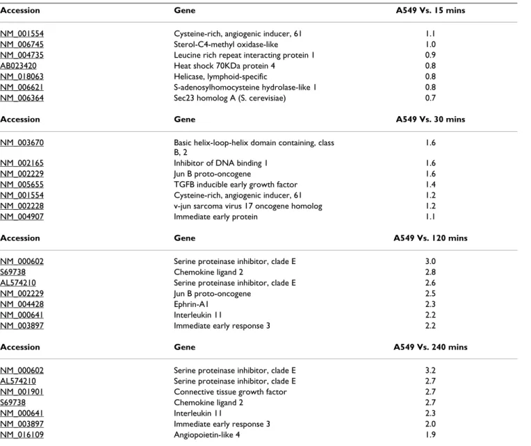
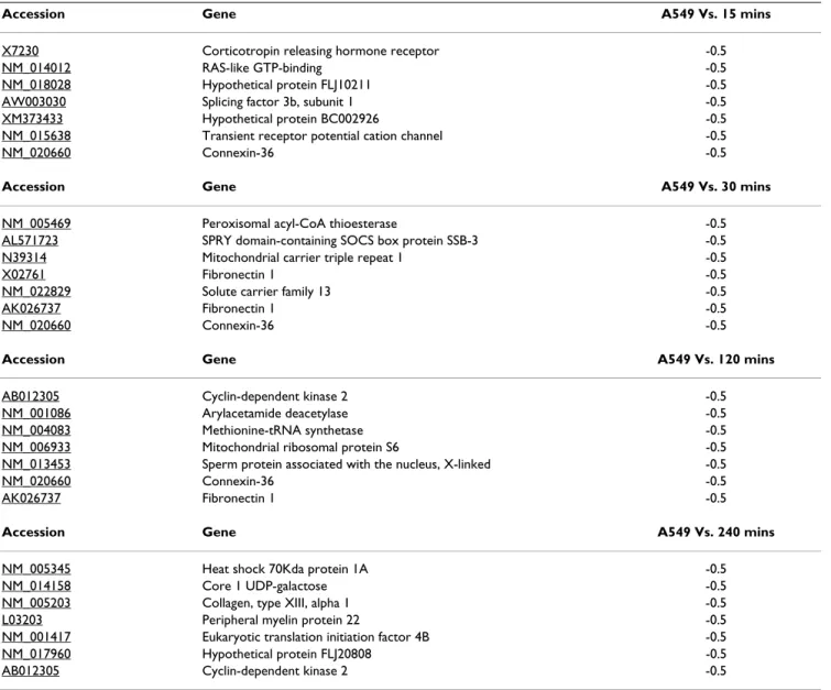
tern of gene expression alterations was observed for both up and down regulated transcripts. Of the 22,216 gene sequences represented on the Affymetrix HGU133A oligo-nucleotide microarray 2.9% (649) genes), 1.7% (383 genes), 2.89% (643 genes) and 6.01% (1339 genes) were significantly altered following 15 minutes, 30 minutes, 2 hour and 4 hour exposure to TGF-β1 respectively (Figure 1, Panel B). Tables 1 and 2 highlight the genes whose mRNA levels were most strikingly altered at 15, 30, 120 and 240 minutes post TGF-β1 exposure.
Baited Cluster analysis Identifies Extracellular Matrix Associated Genes as major responders to TGF-β1 exposure
Figure 2, panel A shows the result of unsupervised hierar-chical cluster analysis of all alveolar epithelial cell genes
whose expression is significantly altered in response to TGF-β1. As can be seen groups of genes are found to clus-ter together depending on the kinetics of their alclus-tered expression. Having delineated the global transcriptomic response of alveolar epithelial cells to TGF-β1, we catego-rized the significantly perturbed genes according to their biological function. This approach permits rapid annota-tion of large datasets for the identificaannota-tion of funcannota-tional patterns of dysregulation. All significantly perturbed genes were used as input in classification searches. Figure 2 Panel B shows the overall pattern of regulation of key functional families throughout the time course exposure. All gene families studied were found to increase over time, reflecting the increased transcriptomic activity in the latter time points.
Table 1: Genes undergoing most striking up-regulation at 15, 30, 120 and 240 minutes post TGF-β1 exposure.
Accession Gene A549 Vs. 15 mins
NM_001554 Cysteine-rich, angiogenic inducer, 61 1.1
NM_006745 Sterol-C4-methyl oxidase-like 1.0
NM_004735 Leucine rich repeat interacting protein 1 0.9
AB023420 Heat shock 70KDa protein 4 0.8
NM_018063 Helicase, lymphoid-specific 0.8
NM_006621 S-adenosylhomocysteine hydrolase-like 1 0.8
NM_006364 Sec23 homolog A (S. cerevisiae) 0.7
Accession Gene A549 Vs. 30 mins
NM_003670 Basic helix-loop-helix domain containing, class B, 2
1.6
NM_002165 Inhibitor of DNA binding 1 1.6
NM_002229 Jun B proto-oncogene 1.6
NM_005655 TGFB inducible early growth factor 1.4
NM_001554 Cysteine-rich, angiogenic inducer, 61 1.2
NM_002228 v-jun sarcoma virus 17 oncogene homolog 1.2
NM_004907 Immediate early protein 1.1
Accession Gene A549 Vs. 120 mins
NM_000602 Serine proteinase inhibitor, clade E 3.0
S69738 Chemokine ligand 2 2.8
AL574210 Serine proteinase inhibitor, clade E 2.6
NM_002229 Jun B proto-oncogene 2.5
NM_004428 Ephrin-A1 2.3
NM_000641 Interleukin 11 2.2
NM_003897 Immediate early response 3 2.2
Accession Gene A549 Vs. 240 mins
NM_000602 Serine proteinase inhibitor, clade E 3.2
AL574210 Serine proteinase inhibitor, clade E 2.7
NM_001901 Connective tissue growth factor 2.7
S69738 Chemokine ligand 2 2.7
NM_000641 Interleukin 11 2.3
NM_003897 Immediate early response 3 2.0
Of the 312 ECM genes displayed on the microarray 95 were significantly altered in this setting. Figure 3 illus-trates the extracellular matrix associated genes whose expression was altered in response to TGF-β1 including matrix proteins, such as members of the collagen family and growth factors known to be involved in matrix regu-lation, including connective tissue growth factor and transforming growth factor. Figure 3, Panel A and B show the expression patterns of up and downregulated tran-scripts respectively.
TGF-β1 stimulation drives ADAM family gene expression in alveolar epithelial cells
Of note with respect to the mechanisms of fibrotic lung injury was the finding of coordinate differential regula-tion of ADAM gene family members. We focused on four ADAM family members identified in the ECM cluster of
our oligonucleotide microarrays. ADAM19 and
ADAMTS9 were increased in response to TGF-β1
expo-sure, whilst mRNA levels of ADAM28 and ADAMTS8 were reduced.
Microarray findings were validated using quantitative real time PCR. ADAM19 expression was significantly enhanced by 6 fold at 4 hours (figure 4, panel A). ADAMTS9 analysis showed an increase in response to TGF-β1 exposure after 15 minutes and reaching a signifi-cantly elevated level of 2 fold at 4 hours (figure 4, panel B).
ADAMTS8 was identified as being downregulated in response to TGF-β1. Real time quantitative PCR con-firmed the TGF-β1 responsiveness of this gene in alveolar epithelial cells at all but the 4 hour exposure time points, Table 2: Genes undergoing most striking down-regulation at 15, 30, 120 and 240 minutes post TGF-β1 exposure.
Accession Gene A549 Vs. 15 mins
X7230 Corticotropin releasing hormone receptor -0.5
NM_014012 RAS-like GTP-binding -0.5
NM_018028 Hypothetical protein FLJ10211 -0.5
AW003030 Splicing factor 3b, subunit 1 -0.5
XM373433 Hypothetical protein BC002926 -0.5
NM_015638 Transient receptor potential cation channel -0.5
NM_020660 Connexin-36 -0.5
Accession Gene A549 Vs. 30 mins
NM_005469 Peroxisomal acyl-CoA thioesterase -0.5
AL571723 SPRY domain-containing SOCS box protein SSB-3 -0.5
N39314 Mitochondrial carrier triple repeat 1 -0.5
X02761 Fibronectin 1 -0.5
NM_022829 Solute carrier family 13 -0.5
AK026737 Fibronectin 1 -0.5
NM_020660 Connexin-36 -0.5
Accession Gene A549 Vs. 120 mins
AB012305 Cyclin-dependent kinase 2 -0.5
NM_001086 Arylacetamide deacetylase -0.5
NM_004083 Methionine-tRNA synthetase -0.5
NM_006933 Mitochondrial ribosomal protein S6 -0.5
NM_013453 Sperm protein associated with the nucleus, X-linked -0.5
NM_020660 Connexin-36 -0.5
AK026737 Fibronectin 1 -0.5
Accession Gene A549 Vs. 240 mins
NM_005345 Heat shock 70Kda protein 1A -0.5
NM_014158 Core 1 UDP-galactose -0.5
NM_005203 Collagen, type XIII, alpha 1 -0.5
L03203 Peripheral myelin protein 22 -0.5
NM_001417 Eukaryotic translation initiation factor 4B -0.5
NM_017960 Hypothetical protein FLJ20808 -0.5
Functional classification of global gene expression changes in alveolar epithelial cells elicited by TGF-β1
Figure 2
(Figure 4, Panel C). Downregulation of ADAM 28 mRNA was confirmed by quantitative real-time PCR at 4 hours (Figure 4, Panel D).
These data confirm the microarray-identified alterations in ADAM family members in alveolar epithelial cells in response to TGF-β1.
mRNA levels of ADAM Family members are altered in response to endogenous and exogenous stimuli
Having determined that exposure of alveolar epithelial cells to TGF-β1 resulted in coordinate regulation of ADAM family members we explored the effect of other fibrotic stimuli on ADAM expression particularly IL-13 and IL-4.
Alveolar epithelial cells were exposed to 10 ng/ml IL-13 for 15, 30, 60, 120 and 240 mins and ADAM gene expres-sion assessed by quantitative Real time PCR. Figure 5 Panel A and B demonstrates the induction of the TGF-β1
upregulated genes, ADAM19 and ADAMTS9 in response to IL-13 stimulation. ADAM 19 was found to be signifi-cantly induced at the 60 min time point post IL-13 expo-sure, then the levels returning to almost baseline by 240 min. ADAMTS9 was significantly enhanced at all time points, with maximal induction seen in the 240 min set-ting. In contrast IL-13 was found to have little effect on the
TGF-β1 downregulated genes, ADAM28 and ADAMTS 8
(Figure 5, Panel C and D).
Interleukin 4 exposure had no significant effect on the expression of ADAM 19 or ADAMTS9. There was a general trend towards downregulation of these transcripts, the opposite effects to that seen with TGF-β1. Suppression of
the TGF-β1 downregulated genes ADAM 28 and
ADAMTS8 by IL-4 was interrogated by real time PCR. However only the latter time points of IL-4 exposure pro-duced a statistically significant change in ADAM 28 expression (Figure 6, panel C).
Extracellular matrix family gene expression in response to TGF-β1
Figure 3
EBV infection of A549 cells was confirmed by western blot expression of latent membrane protein 1 (LMP1) in A549 infected cells (Figure 7). Having confirmed the viral infec-tion of A549 cells with EBV we determined the effect of this infection on ADAM gene expression. ADAM19 and ADAMTS9 were found to be significantly induced in virus infected alveolar epithelial cells. Stimulation of these infected cells with TGF-β1 resulted in further enhanced expression of these genes, suggesting a synergistic activity of EBV and TGF-β1 in the fibrotic lung (Figure 8, panel A and B). Of note was the finding that EBV infection had no statistically significant effect on ADAM 28 gene expression either alone or in conjunction with TGF-β1. Decreased expression of ADAMTS8 was found in virus infected
TGF-β1 exposed alveolar epithelial cells (Figure 8, Panel C and D).
ADAM 19 and ADAMTS9 Gene Silencing inhibits lung fibrosis in vitro
To determine the biological importance of enhanced ADAM 19 and ADAMTS9 expression in response to
TGF-β1 exposure we evaluated the effect of gene knock down on the cellular phenotype. To achieve this goal, specific small interfering RNA oligonucleotide [35] probes were designed and transfected into A549 alveolar epithelial cells using the lipofectamine strategy as described. Follow-ing transfection knockdown of the genes was confirmed by quantitative PCR (Figure 9, Panel A). Transfected cells were exposed to 10 ng/ml TGF-β1 for four hours as previ-ously described and collagen (Types I-V) deposition, as a hallmark of fibrosis, was determined using the Sircol assay kit. Figure 9 panel B demonstrates reduced collagen deposition in both ADAM 19 and ADAMTS9 siRNA
trans-ADAM mRNA expression levels in TGF-β1 stimulated alveolar epithelial cells
Figure 4
fected cells. These data suggest that the upregulation of ADAM 19 and ADAMTS9 by TGF-β1 in the setting of lung fibrosis plays a role in the deposition of collagen in the cellular milieu, thereby contributing to extracellular matrix deposition and lung scarring.
Discussion
To delineate the molecular events in the pathogenesis of pulmonary fibrosis we utilised an oligonucleotide micro-array strategy to identify alveolar epithelial genes and gene clusters whose expression is altered in response to TGF-β1. These studies identified a large number of genes whose expression was altered in a temporal fashion. Functional classification of the transcriptomic response identified coordinate expression of important functional groups of genes in response to TGF-β1. Particular focus was made on genes that are associated with extracellular matrix and it's remodelling. We curated the Affymetrix Human Genome HU133a microarray to obtain a list of such genes
represented on the chip. Approximately 30% of this gene cohort was found to be differentially regulated further underpinning the relative contribution of matrix mole-cules and mediators to the response to TGF-β1. Represent-ative among these extracellular matrix genes were growth factors, collagens and members of the ADAM gene family. The interaction of epithelial cells with other local and infiltrating cell types drives the further articulation of the fibrotic response in the lung. The ability of ADAM family members to regulate the interaction of these cell types and indeed to regulate their interaction with the extracellular matrix raises the hypothesis that these mediators are important in driving the local fibrotic milieu in the lung.
The ADAM gene family encompasses the ADAM (A disin-tegrin and metalloproteinase domain containing protein) and ADAMTS (A disintegrin-like and metalloproteinase with thrombospondin type I motif) proteins. The ADAMTSs are soluble proteins that can bind to the ECM
ADAM mRNA expression in response to interleukin-13
Figure 5
through the TS motifs. To date 34 members of the ADAMs family have been described, of which approximately 50% have been demonstrated to have protease activity. The substrates of ADAM protease activity are either integral membrane proteins or extracellular matrix proteins. In addition to their proteolytic activity, a number of ADAMs have been shown to bind integrins, via their disintegrin domain [36]. This dual activity of ADAMs suggests that these molecules are key mediators of biological processes such as cell-cell adhesion, ectodomain shedding, myob-last fusion and development. In the context of lung dis-ease, this family is noteworthy. The possible role for ADAM family genes in lung fibrosis has been intimated by the observation that ADAM 10 autoantibody is associated with dermatomyositis related lung fibrosis and was shown to be present in one patient with IPF [37]. ADAM33 polymorphisms have also been shown to play a
role in the development of asthma and also in disease progression via effects on airway remodelling [38].
The ADAM gene family is characterised by members that possess both proteolytic and cell-cell and cell-matrix inter-action promoting activities. Thus the matrix proteolytic activities of ADAMs may represent an important facet of the development of pulmonary fibrosis, as their differen-tial expression may alter the balance of matrix turnover. ADAM genes identified in this study have previously been associated with ventricular septal defects and valvular defects (ADAM19) [39], fibrotic eye disease (ADAMTS9) [40], disrupting angiogenesis (ADAMTS8) [41], and cell adhesion (ADAM 28) [42]. Functional assessment of the role of these genes in the setting of the lung was described using a gene knock down based strategy. These investiga-tions showed an effect on collagen deposition in cells
ADAM mRNA expression in response to interleukin-4
Figure 6
where the ADAM gene was knocked down, thus illustrat-ing the importance of dysregulation of this gene family in the setting of lung fibrosis.
Having demonstrated the responsiveness and functional activity of ADAM family members in response to TGF-β1 we explored their induction in response to other endog-enous and exogendog-enous pro-fibrotic insults. We determined the effect of IL-4 and IL-13 on alveolar epithelial cell ADAM production. Whilst the specific dysregulation of ADAM family members by these cytokines was different to that elicited by TGF-β1, the general trend of ADAM response remained the same. These data suggest that the ADAM family response may be more indicative of the overall fibrotic activity rather than being a stimulus spe-cific effect. These findings lend further weight to the
argu-ADAM mRNA expression levels in Epstein Barr Virus infected alveolar epithelial cells
Figure 8
ADAM mRNA expression levels in Epstein Barr Virus infected alveolar epithelial cells. Using real time PCR the effect of EBV infection and TGF-β1 costimulation on ADAM expression was determined. ADAM expression in A549 cells (C), A549 cells stimulated with TGF-β1 (TGF), EBV infected A549 cells with (VTGF) and without (V) TGF-β1 are shown. Expres-sion levels of ADAM 19 (Panel A), ADAM 28 (Panel B), ADAMTS9 (Panel C) and ADAMTS8 (Panel D) are shown. All expres-sion values were normalised to 18S rRNA to control for equivalence of loading. All measurements were completed in triplicate. Data are quoted relative to control, which has a value of 1.
Epstein Barr Virus infection of alveolar epithelial cells
Figure 7
Epstein Barr Virus infection of alveolar epithelial cells. EBV infection of A549 alveolar epithelial cells was con-firmed by western blotting for the viral protein LMP1. LMP1 was detected in Akata cells (Control) as a positive control. Lanes V1 and V2 demonstrate LMP-1 protein expression in infected A549 alveolar epithelial cells. Each lane represents protein taken from separate experiments containing 4 × 10⁵
ment that ADAM genes are central in mediating fibrosis as opposed to being an artefact of the experimental model.
Of particular note was the finding that viral infection itself resulted in ADAM family dysregulation in these cells, an effect that was further enhanced in experiments where repeated injury consisting of both EBV infection and
TGF-β1 exposure induced ADAM gene expression. Whilst the ADAM gene expression was not as great in EBV infected cells as it was in A549 cells stimulated with TGF-β1, a sig-nificant rise in ADAM19 and ADAMTS9 expression was observed. The combined stimulation of A549 cells with both EBV and TGF-β1 resulted in an additional increase in ADAM19 and ADAMTS9 expression.
These data strengthen the hypothesis that the initiation and progression of lung fibrosis is due to injury of the epi-thelial cell with abnormal healing. Repetitive injury due to EBV infection is one plausible mediator for the
devel-opment of fibrosis. It is of note that the ADAM response was consistently observed following different experimen-tal exposures, underpinning the fact that these observed changes in ADAM expression and functionality may be of clinical importance and are not merely representative of experimental and model derived artefacts.
The data highlight the complex mechanism underpinning the initiation and progression of IPF and underline mech-anisms for both the multifactorial nature of disease initi-ation and the mediators of deposition of extracellular matrix.
References
1. American Thoracic Society/European Respiratory Society
International Multidisciplinary Consensus Classification of the Idiopathic Interstitial Pneumonias. This joint statement of the American Thoracic Society (ATS), and the European Respiratory Society (ERS) was adopted by the ATS board of directors, June 2001 and by the ERS Executive Committee, June 2001. Am J Respir Crit Care Med 2002, 165(2):277-304.
ADAM 19 and ADAMTS9 gene knockdown abrogate TGF-β1 induced collaged production
Figure 9
ADAM 19 and ADAMTS9 gene knockdown abrogate TGF-β1 induced collaged production. Successful knockdown
of ADAM19 and ADAMTS9 mRNA levels are shown in panel A using real time PCR. In panel B a standard curve for quantifica-tion is shown along with a bar chart illustrating reduced collagen deposiquantifica-tion in response to TGF-β1 in ADAM19 and
2. Crystal RG, Bitterman PB, Mossman B, Schwarz MI, Sheppard D, Almasy L, Chapman HA, Friedman SL, King TE Jr, Leinwand LA, et al.:
Future research directions in idiopathic pulmonary fibrosis: summary of a National Heart, Lung, and Blood Institute working group. Am J Respir Crit Care Med 2002, 166(2):236-246. 3. Warburton D, Zhao J, Berberich MA, Bernfield M: Molecular
embryology of the lung: then, now, and in the future. Am J Physiol 1999, 276(5 Pt 1):L697-704.
4. Woodcock-Mitchell JL, Burkhardt AL, Mitchell JJ, Rannels SR, Rannels DE, Chiu JF, Low RB: Keratin species in type II pneumocytes in culture and during lung injury. Am Rev Respir Dis 1986,
134(3):566-571.
5. Selman M, King TE, Pardo A: Idiopathic pulmonary fibrosis: pre-vailing and evolving hypotheses about its pathogenesis and implications for therapy. Ann Intern Med 2001, 134(2):136-151. 6. Noble PW, Homer RJ: Idiopathic pulmonary fibrosis: new
insights into pathogenesis. Clin Chest Med 2004, 25(4):749-758. vii
7. Ramos C, Montano M, Garcia-Alvarez J, Ruiz V, Uhal BD, Selman M, Pardo A: Fibroblasts from idiopathic pulmonary fibrosis and normal lungs differ in growth rate, apoptosis, and tissue inhibitor of metalloproteinases expression. Am J Respir Cell Mol Biol 2001, 24(5):591-598.
8. Letterio JJ, Roberts AB: Transforming growth factor-beta1-defi-cient mice: identification of isoform-specific activities in vivo.
J Leukoc Biol 1996, 59(6):769-774.
9. Xu YD, Hua J, Mui A, O'Connor R, Grotendorst G, Khalil N: Release of biologically active TGF-beta1 by alveolar epithelial cells results in pulmonary fibrosis. Am J Physiol Lung Cell Mol Physiol 2003, 285(3):L527-539.
10. Kulkarni AB, Huh CG, Becker D, Geiser A, Lyght M, Flanders KC, Roberts AB, Sporn MB, Ward JM, Karlsson S: Transforming growth factor beta 1 null mutation in mice causes excessive inflammatory response and early death. Proc Natl Acad Sci USA 1993, 90(2):770-774.
11. Yang YA, Dukhanina O, Tang B, Mamura M, Letterio JJ, MacGregor J, Patel SC, Khozin S, Liu ZY, Green J, et al.: Lifetime exposure to a soluble TGF-beta antagonist protects mice against metasta-sis without adverse side effects. J Clin Invest 2002,
109(12):1607-1615.
12. Kuwano K, Kunitake R, Maeyama T, Hagimoto N, Kawasaki M, Mat-suba T, Yoshimi M, Inoshima I, Yoshida K, Hara N: Attenuation of bleomycin-induced pneumopathy in mice by a caspase inhib-itor. Am J Physiol Lung Cell Mol Physiol 2001, 280(2):L316-325. 13. Kuwano K, Hagimoto N, Kawasaki M, Yatomi T, Nakamura N, Nagata
S, Suda T, Kunitake R, Maeyama T, Miyazaki H, et al.: Essential roles of the Fas-Fas ligand pathway in the development of pulmo-nary fibrosis. J Clin Invest 1999, 104(1):13-19.
14. Huynh ML, Fadok VA, Henson PM: Phosphatidylserine-depend-ent ingestion of apoptotic cells promotes TGF-beta1 secre-tion and the resolusecre-tion of inflammasecre-tion. J Clin Invest 2002,
109(1):41-50.
15. Sime PJ, Xing Z, Graham FL, Csaky KG, Gauldie J: Adenovector-mediated gene transfer of active transforming growth fac-tor-beta1 induces prolonged severe fibrosis in rat lung. J Clin Invest 1997, 100(4):768-776.
16. Giri SN, Hyde DM, Hollinger MA: Effect of antibody to trans-forming growth factor beta on bleomycin induced accumu-lation of lung collagen in mice. Thorax 1993, 48(10):959-966. 17. Corrin B, Butcher D, McAnulty BJ, Dubois RM, Black CM, Laurent GJ,
Harrison NK: Immunohistochemical localization of trans-forming growth factor-beta 1 in the lungs of patients with systemic sclerosis, cryptogenic fibrosing alveolitis and other lung disorders. Histopathology 1994, 24(2):145-150.
18. Munger JS, Huang X, Kawakatsu H, Griffiths MJ, Dalton SL, Wu J, Pit-tet JF, Kaminski N, Garat C, Matthay MA, et al.: The integrin alpha v beta 6 binds and activates latent TGF beta 1: a mechanism for regulating pulmonary inflammation and fibrosis. Cell 1999, 96(3):319-328.
19. Hancock A, Armstrong L, Gama R, Millar A: Production of inter-leukin 13 by alveolar macrophages from normal and fibrotic lung. Am J Respir Cell Mol Biol 1998, 18(1):60-65.
20. Jakubzick C, Choi ES, Joshi BH, Keane MP, Kunkel SL, Puri RK, Hog-aboam CM: Therapeutic attenuation of pulmonary fibrosis via targeting of IL-4- and IL-13-responsive cells. J Immunol 2003,
171(5):2684-2693.
21. Lee CG, Homer RJ, Zhu Z, Lanone S, Wang X, Koteliansky V, Shipley JM, Gotwals P, Noble P, Chen Q, et al.: Interleukin-13 induces tis-sue fibrosis by selectively stimulating and activating trans-forming growth factor beta(1). J Exp Med 2001, 194(6):809-821. 22. Richter A, Puddicombe SM, Lordan JL, Bucchieri F, Wilson SJ, Dju-kanovic R, Dent G, Holgate ST, Davies DE: The contribution of interleukin (IL)-4 and IL-13 to the epithelial-mesenchymal trophic unit in asthma. Am J Respir Cell Mol Biol 2001,
25(3):385-391.
23. Ando M, Miyazaki E, Fukami T, Kumamoto T, Tsuda T: Interleukin-4-producing cells in idiopathic pulmonary fibrosis: an immu-nohistochemical study. Respirology 1999, 4(4):383-391. 24. Saito A, Okazaki H, Sugawara I, Yamamoto K, Takizawa H: Potential
action of IL-4 and IL-13 as fibrogenic factors on lung fibrob-lasts in vitro. Int Arch Allergy Immunol 2003, 132(2):168-176. 25. Vergnon JM, Vincent M, de The G, Mornex JF, Weynants P, Brune J:
Cryptogenic fibrosing alveolitis and Epstein-Barr virus: an association? Lancet 1984, 2(8406):768-771.
26. Lung ML, Lam WK, So SY, Lam WP, Chan KH, Ng MH: Evidence that respiratory tract is major reservoir for Epstein-Barr virus. Lancet 1985, 1(8434):889-892.
27. Tsukamoto K, Hayakawa H, Sato A, Chida K, Nakamura H, Miura K:
Involvement of Epstein-Barr virus latent membrane protein 1 in disease progression in patients with idiopathic pulmo-nary fibrosis. Thorax 2000, 55(11):958-961.
28. Liebowitz D, Wang D, Kieff E: Orientation and patching of the latent infection membrane protein encoded by Epstein-Barr virus. J Virol 1986, 58(1):233-237.
29. Kelly BG, Lok SS, Hasleton PS, Egan JJ, Stewart JP: A rearranged form of Epstein-Barr virus DNA is associated with idiopathic pulmonary fibrosis. Am J Respir Crit Care Med 2002,
166(4):510-513.
30. Takada K, Horinouchi K, Ono Y, Aya T, Osato T, Takahashi M, Hay-asaka S: An Epstein-Barr virus-producer line Akata: establish-ment of the cell line and analysis of viral DNA. Virus Genes
1991, 5(2):147-156.
31. Sadlier DM, Connolly SB, Kieran NE, Roxburgh S, Brazil DP, Kairaitis L, Wang Y, Harris DC, Doran P, Brady HR: Sequential extracellu-lar matrix-focused and baited-global cluster analysis of serial transcriptomic profiles identifies candidate modulators of renal tubulointerstitial fibrosis in murine adriamycin-induced nephropathy. J Biol Chem 2004, 279(28):29670-29680. 32. Irizarry RA, Bolstad BM, Collin F, Cope LM, Hobbs B, Speed TP:
Summaries of Affymetrix GeneChip probe level data. Nucleic Acids Res 2003, 31(4):e15.
33. Draghici S, Khatri P, Bhavsar P, Shah A, Krawetz SA, Tainsky MA:
Tools, the toolkit of the modern biologist: Onto-Express, Onto-Compare, Onto-Design and Onto-Translate.
Nucleic Acids Res 2003, 31(13):3775-3781.
34. Fink L, Seeger W, Ermert L, Hanze J, Stahl U, Grimminger F, Kummer W, Bohle RM: Real-time quantitative RT-PCR after laser-assisted cell picking. Nat Med 1998, 4(11):1329-1333.
35. Parkes JG, Liu Y, Sirna JB, Templeton DM: Changes in gene expression with iron loading and chelation in cardiac myo-cytes and non-myocytic fibroblasts. J Mol Cell Cardiol 2000,
32(2):233-246.
36. White JM: ADAMs: modulators of cell-cell and cell-matrix interactions. Curr Opin Cell Biol 2003, 15(5):598-606.
37. Fujita J, Takeuchi T, Dobashi N, Ohtsuki Y, Tokuda M, Takahara J:
Detection of anti-ADAM 10 antibody in serum of a patient with pulmonary fibrosis associated with dermatomyositis.
Ann Rheum Dis 1999, 58(12):770-772.
38. Jongepier H, Boezen HM, Dijkstra A, Howard TD, Vonk JM, Koppel-man GH, Zheng SL, Meyers DA, Bleecker ER, Postma DS: Polymor-phisms of the ADAM33 gene are associated with accelerated lung function decline in asthma. Clin Exp Allergy 2004,
34(5):757-760.
39. Zhou HM, Weskamp G, Chesneau V, Sahin U, Vortkamp A, Horiuchi K, Chiusaroli R, Hahn R, Wilkes D, Fisher P, et al.: Essential role for ADAM19 in cardiovascular morphogenesis. Mol Cell Biol 2004,
24(1):96-104.
Publish with BioMed Central and every scientist can read your work free of charge "BioMed Central will be the most significant development for disseminating the results of biomedical researc h in our lifetime."
Sir Paul Nurse, Cancer Research UK
Your research papers will be:
available free of charge to the entire biomedical community
peer reviewed and published immediately upon acceptance
cited in PubMed and archived on PubMed Central
yours — you keep the copyright
Submit your manuscript here:
http://www.biomedcentral.com/info/publishing_adv.asp
BioMedcentral 41. Vazquez F, Hastings G, Ortega MA, Lane TF, Oikemus S, Lombardo
M, Iruela-Arispe ML: METH-1, a human ortholog of ADAMTS-1, and METH-2 are members of a new family of proteins with angio-inhibitory activity. J Biol Chem 1999, 274(33):23349-23357. 42. Bridges LC, Tani PH, Hanson KR, Roberts CM, Judkins MB, Bowditch RD: The lymphocyte metalloprotease MDC-L (ADAM 28) is a ligand for the integrin alpha4beta1. J Biol Chem 2002,