Open Access
Research
Intra- and inter-observer analysis in the morphological assessment
of early-stage embryos
Goedele Paternot*, Johanna Devroe, Sophie Debrock, Thomas M D'Hooghe
and Carl Spiessens
Address: Leuven University Fertility Centre, UZ Leuven, Campus Gasthuisberg, Herestraat 49, 3000 Leuven, Belgium
Email: Goedele Paternot* - [email protected]; Johanna Devroe - [email protected]; Sophie Debrock - [email protected]; Thomas M D'Hooghe - [email protected]; Carl Spiessens - [email protected]
* Corresponding author
Abstract
Background: The aim of this study was to determine the intra- and inter-observer variability in the evaluation of embryo quality. Multilevel images of embryos on day 1, day 2 and day 3, were analysed using different morphological parameters.
Methods: Multilevel images of embryos on day 1, day 2 and day 3, were analysed using a standard scoring system. The kappa coefficient was calculated to measure intra- and inter-observer variability before and after training sessions.
Results: Good to excellent intra-observer agreement was present for most parameters exceptions being scoring the position of pronuclei and the presence of a cytoplasmic halo on day 1, multinucleation on day 2 and the size of fragments on day 3. Inter-observer agreement was only good to excellent for the number of blastomeres on day 2 and day 3 and the orientation of the cleavage axes on day 2. Training sessions had a positive impact on inter-observer agreement.
Conclusion: In conclusion, assessment of morphological characteristics of early stage embryos using multilevel images was marked by a high intra-observer and a moderate inter-observer agreement. Training sessions were useful to increase inter-observer agreement.
Background
Reproductive outcome in IVF depends on an interplay of different factors including embryo quality [1,2]. Many dif-ferent classification and scoring systems, based on cleav-age and morphological parameters, have been proposed to evaluate embryo quality [2-4]. However, none of these grading systems has been completely validated [5,6]. Reli-ability is one of the most important conditions for the val-idation of a scoring system. Currently, the scoring of cleavage stage embryos involves the direct evaluation of
the embryo by an embryologist assessing the morpholog-ical parameters to determine embryo quality [1].
Some variation in scoring of morphological parameters can be expected among different embryologists (inter-observer variability) and within the same embryologist (intra-observer variability) [7]. However, only limited and controversial data [8,9] are available with respect to intra-and inter-observer variability in the evaluation of mor-phological parameters assessing embryo quality. No data
Published: 29 September 2009
Reproductive Biology and Endocrinology 2009, 7:105 doi:10.1186/1477-7827-7-105
Received: 21 July 2009 Accepted: 29 September 2009
This article is available from: http://www.rbej.com/content/7/1/105 © 2009 Paternot et al; licensee BioMed Central Ltd.
are available with respect to this variability by means of multilevel images. The use of these multilevel images allows embryologists to assess embryo quality in the same way as an exploration at the inverted microscope, but without a limitation in evaluation time.
Therefore, the aim of this study was to determine the intra- and inter-observer agreement in the assessment of embryo quality on day 1, 2 and 3 by means of multilevel images.
Methods
Image collection
A computer system (FertiMorph, IHMedical A/S, Copen-hagen, Denmark) was used to record image sequences of 50 embryos on day 1, day 2 and day 3 of their develop-ment. This system allowed investigators to record image sequences that consisted of 26 images of the same oocyte or embryo by automatically focussing through the com-plete embryo (5 μm intervals). In our study, embryos (N = 50) from 6 IVF/ICSI patients with an oocyte retrieval on the same day were included in order to have a random sample of embryos representing the embryos in daily rou-tine. The mean age of the 6 patients was 29.5 (SD+/-3.27) years. All embryos were evaluated for each parameter.
Embryo scoring system
The multilevel images were presented to the embryolo-gists on a patient-by-patient basis and shown in a chron-ological order of embryo development. Embryo quality was scored according to the criteria defined in the Stand-ard Operating Procedures of the Leuven University Fertil-ity Centre (LUFC) scoring system. The following parameters were evaluated: day 1: the size (equal or une-qual (>25% difference in size)) and position (central or peripheral) of the pronuclei, the presence of a cytoplasmic halo (a light zone over at least 75% of the circumference of the zygote); day 2 and day 3: the number and size (equal, slightly unequal (25-50% difference in size) or unequal (>50% difference in size)) of the blastomeres, the degree of fragmentation (grade 0: no fragmentation, grade 1: <10%, grade 2: 10-25%, grade 3: 25-50%, grade
4:>50% fragmentation), the position (local or dispersed) and the size (small or large) of the fragments. Further-more, the position of the cleavage axes (perpendicular or not) on day 2 (if a 4 cell embryo was available) and the presence of mono- or multinucleation on day 2 and day 3 were evaluated. Since there was no visible nucleus in the majority of the embryos on day 3, this parameter was excluded.
Intra-observer variability
Five embryologists (Table 1) were asked to evaluate the embryos at two different time points with a time interval of minimum one month and maximum two months. Each embryologist was blinded with respect to the assess-ment of the embryo quality in their first evaluation and to the results of the assessments of embryo quality by the other embryologists.
Inter-observer variability
Inter-observer variability in embryo scoring was assessed among 7 embryologists (Table 1). Each embryologist was blinded to the assessments of the other embryologists.
Training session: scoring of "experts" versus "trainees"
In a next step, three trainees who had no experience in the scoring of embryos were asked to score the same embryos with the LUFC scoring system. Only the definition of the parameters used in the LUFC scoring system was given to them as foreknowledge. Two weeks later, these three train-ees and three embryologists (with respectively 3.25, 5 and 6.33 years of experience) (Table 1) were given three train-ing sessions in one week. Durtrain-ing these three traintrain-ing ses-sions, embryo images were shown to visually demonstrate the definitions of all parameters. In this way, a consensus was reached regarding the way the parameters had to be scored. Based on these definitions, a manual was created with a clear description and some examples of each parameter. During the two weeks following the training sessions, the same six persons (three trainees and three trained embryologists) scored the embryos again, this time they used the manual in order to assess the impact of training and pre-existing experience on the inter-observer
Table 1: Participation (yes/no) of individual embryologists (number of years of experience) to the three components of our study (intra-observer variability, inter-observer variability, training sessions)
Participant
(N years of experience)
Intra-observer variability
Inter-observer variability
Training sessions
Embryologist 1 (6.33) Yes Yes Yes
Embryologist 2 (5) Yes Yes Yes
Embryologist 3 (3.25) Yes Yes Yes
Embryologist 4 (5.5) Yes Yes No
Embryologist 5 (1.5) Yes Yes No
Embryologist 6 (16) No Yes No
variability in the assessment of embryo quality. The aim of these training sessions was to increase the number of parameters with a kappa coefficient value higher than 0.60.
Statistics
The kappa coefficient was calculated to measure intra-observer (comparison of embryo scoring given at two dif-ferent time points by the same embryologist) and inter-observer (comparison of embryo scoring by different embryologists) variability. This kappa coefficient was interpreted as an indicator of either excellent (≥ 0.80), good (0.60-0.79), moderate (0.40-0.59), poor (0.20-0.39) and very poor (<0.20) intra- and inter-observer agreement [10]. The number of observations necessary to do kappa statistics is calculated by the equation: 2n2; with n the number of categories for each characteristic. In this study, the degree of fragmentation has the highest number of categories (n = 5) indicating the need for at least 2*(5)2 = 50 embryos [11]. The major disadvantage of using this kappa coefficient is that no statistics are availa-ble to compare different kappa values.
Results
Intra-observer variability
Excellent intra-observer agreement was observed for the number of blastomeres on day 2 and day 3 and for the ori-entation of the cleavage axes on day 2 (Table 2). Good intra- observer agreement was observed for the following parameters: position of the pronuclei, the degree, the pat-tern and size of fragmentation on day 2, the size of the blastomeres on day 2 and day 3, the degree and pattern of fragmentation on day 3 (Table 2). Moderate intra-observer agreement was observed for the evaluation of the
size of the pronuclei and the presence of a cytoplasmic halo on day 1, multinucleation on day 2, and the size of the fragments on day 3 (Table 2).
Inter-observer variability
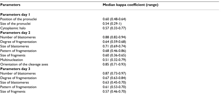
The inter-observer agreement was excellent (Table 3) for the scoring of the number of blastomeres on day 2 and day 3 and good (Table 3) for the evaluation of the orien-tation of the cleavage axes. The inter-observer agreement was moderate for the scoring of all the other embryo qual-ity parameters except for the scoring of the size of pronu-clei and the presence of a cytoplasmic halo where the inter-observer agreement was poor (Table 3).
Training session: scoring of "trainees" versus "experts"
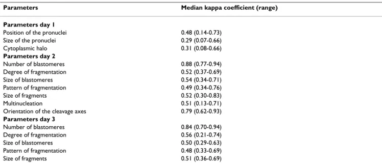
Inter-observer variability before training sessions (Table 4)
Inter-observer agreement was excellent when experts scored the number of blastomeres on day 2 and on day 3, but was only good when trainees scored the number of blastomeres on day 2. When the experts were compared with the trainees, there was a good agreement between groups with respect to the assessment of the number of blastomeres and the orientation of the cleavage axes on day 2. For the other parameters the inter-observer agree-ment was moderate, poor or very poor.
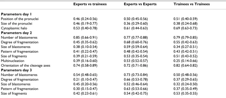
Inter-observer variability after training sessions (Table 5)
Training sessions had a positive impact on inter-observer agreement, as becomes evident when comparing the data from table 5 (after training) with those from table 4 (before training). After training good instead of moderate inter-observer agreement was reached by experts for three extra parameters (cytoplasmic halo, degree and pattern of fragmentation on day 3) and by the trainees for two extra
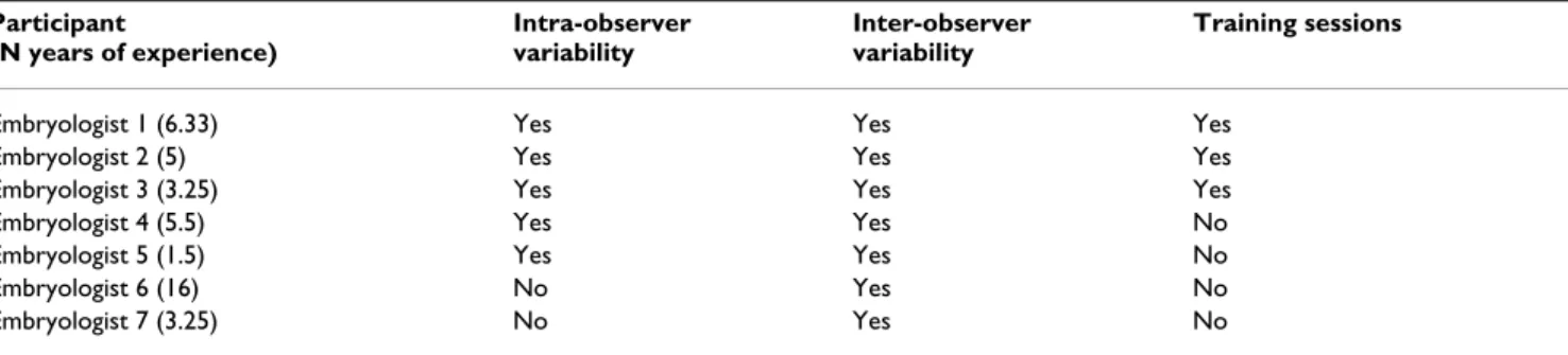
Table 2: Median (range) of kappa coefficient as measurement of intra-observer agreement in embryo evaluation
Parameters Median kappa coefficient (range)
Parameters day 1
Position of the pronuclei 0.60 (0.48-0.64)
Size of the pronuclei 0.54 (0.29-1)
Cytoplasmic halo 0.57 (0.33-0.77)
Parameters day 2
Number of blastomeres 0.88 (0.82-0.94)
Degree of fragmentation 0.64 (0.59-0.68)
Size of blastomeres 0.71 (0.69-0.74)
Pattern of fragmentation 0.68 (0.46-0.86)
Size of fragments 0.60 (0.36-0.65)
Multinucleation 0.51 (0.32-0.79)
Orientation of the cleavage axes 0.85 (0.71-0.93)
Parameters day 3
Number of blastomeres 0.87 (0.73-0.97)
Degree of fragmentation 0.67 (0.63-0.84)
Size of blastomeres 0.63 (0.45-0.70)
Pattern of fragmentation 0.61 (0.53-0.70)
parameters (cytoplasmic halo and orientation of the cleavage axes).
Discussion
To the best of our knowledge, this is the first study where intra- and inter-observer variability in embryo scoring has been assessed using multilevel images. The use of these multilevel images allows embryologists to assess embryo quality in the same way as an exploration at the inverted microscope, but without a limitation in evaluation time. Multilevel images allow the embryologist to evaluate among different focus planes within one embryo.
Previ-ous studies used 2D pictures which limits the evaluation to one focus plane in the embryo [8,9].
Intra-observer agreement was good to excellent for most parameters demonstrating that one individual embryolo-gist is consistent in scoring the same embryo at different time points which is in line with previous publications [8,9]. Indeed, good intra-observer agreement has been reported with respect to the pattern of embryo fragmenta-tion on day 2 (median kappa: 0.75), and with respect to number and size of blastomeres and degree of fragmenta-tion on both day 2 (median kappa values of 0.79, 0.69 Table 3: Inter-observer agreement indicated by the median (range) value of the kappa coefficient
Parameters Median kappa coefficient (range)
Parameters day 1
Position of the pronuclei 0.48 (0.14-0.73)
Size of the pronuclei 0.29 (0.07-0.66)
Cytoplasmic halo 0.31 (0.08-0.66)
Parameters day 2
Number of blastomeres 0.88 (0.77-0.94)
Degree of fragmentation 0.52 (0.37-0.69)
Size of blastomeres 0.54 (0.34-0.71)
Pattern of fragmentation 0.49 (0.34-0.76)
Size of fragments 0.52 (0.30-0.83)
Multinucleation 0.51 (0.13-0.71)
Orientation of the cleavage axes 0.79 (0.62-0.93)
Parameters day 3
Number of blastomeres 0.84 (0.70-0.94)
Degree of fragmentation 0.56 (0.21-0.74)
Size of blastomeres 0.50 (0.29-0.63)
Pattern of fragmentation 0.48 (0.33-0.69)
Size of fragments 0.51 (0.36-0.69)
Table 4: Inter-observer variability among and between experienced embryologists and embryology trainees before embryology training sessions indicated by the median (range) value of the kappa coefficient
Experts vs trainees Experts vs Experts Trainees vs Trainees
Parameters day 1
Position of the pronuclei 0.27 (0.16-0.52) 0.28 (0-0.49) 0.29 (0.21-0.61)
Size of the pronuclei 0.31 (0.12-0.63) 0.27 (0.23-0.39) 0.14 (0.08-0.43)
Cytoplasmic halo 0.16 (0.03-0.67) 0.44 (0.36-0.52) 0.09 (0.02-0.10)
Parameters day 2
Number of blastomeres 0.76 (0.70-0.88) 0.88 (0.82-0.88) 0.67 (0.63-0.79)
Degree of fragmentation 0.43 (0-0.70) 0.60 (0.53-0.61) 0.34 (0.28-0.52)
Size of blastomeres 0.25 (0.02-0.66) 0.52 (0.45-0.53) 0.43 (0.22-0.44)
Pattern of fragmentation 0.36 (0.19-0.42) 0.47 (0.45-0.48) 0.32 (0.20-0.49)
Size of fragments 0.22 (0.01-0.34) 0.52 (0.38-0.83) 0.04 (0-0.14)
Multinucleation 0.16 (0.07-0.32) 0.32 (0.27-0.64) 0.15 (0.07-0.16)
Orientation of the cleavage axes 0.60 (0.32-0.82) 0.79 (0.71-0.79) 0.38 (0.33-0.62)
Parameters day 3
Number of blastomeres 0.52 (0.31-0.65) 0.86 (0.73-0.86) 0.44 (0.43-0.51)
Degree of fragmentation 0.18 (0-0.56) 0.35 (0.22-0.56) 0.05 (0.03-0.44)
Size of blastomeres 0.31 (0.06-0.70) 0.40 (0.39-0.50) 0.27 (0.23-0.48)
Pattern of fragmentation 0.28 (0.19-0.49) 0.47 (0.39-0.51) 0.23 (0.22-0.28)
and 0.64 respectively) and day 3 (median kappa values of 0.66, 0.63 and 0.68 respectively) [9]. Other researchers [8] also demonstrated a good intra-observer agreement with respect to embryo scoring. However, their study was lim-ited by the fact that only supernumerary embryos were used (not representative for daily embryology practice) and that different scoring systems with combined embryo quality parameters were used, whereas in our study embryos from routine practice were evaluated and all embryo quality parameters were scored individually.
Only moderate inter-observer agreement (median kappa 0.40-0.60) was achieved in our study for most embryo quality parameters on day 1, 2 and 3 that were not related to the number of blastomeres or the orientation of the cleavage axes. These results cannot be compared to poor inter-observer agreement (median kappa 0.24) in the assessment of embryo quality reported by some other investigators [8] for the reasons mentioned in the para-graph above. Moreover, the scoring of the most experi-enced person was considered to be the golden standard in that study [8]. However this consideration is not scientifi-cally justified, as our study shows that experience is not necessarily linked to good inter-observer agreement. Our data demonstrating excellent inter-observer agreement for the number of blastomeres (day 2) and moderate inter-observer agreement in scoring the degree and pattern of fragmentation (day 2 and day 3) are in line with those from other investigators using a similar study design with individual assessment of embryo quality parameters [9]. However, evaluation of blastomere size (day 2), multinu-cleation (day 2) and blastomere number (day 3) had a lower inter-observer agreement in our study based on
multilevel images than in the other study [9] based on 2D images. Embryo evaluation in 2D is limited by the fact that superimposed fragments may be invisible and are more difficult to assess. The inter-observer variability for some embryo characteristics, like the size of fragments or blastomeres, is possibly related to volume estimation which remains rather subjective. Furthermore the use of categorical variables, instead of continuous variables (per-centages), may also contribute to the inter-observer varia-bility.
Clearly, more consistency is needed in the evaluation of embryo morphology in order to investigate the impact of these parameters on embryo implantation. Both training and multilevel imaging may be beneficial to achieve that goal.
Inter-observer agreement was better among experienced embryologists than among trainees in our study and improved after training, not only among trainees but also among experienced embryologists. However, after train-ing the kappa coefficient increased not for every parame-ter and inparame-ter-observer variability was still high among the trainees, suggesting that training sessions were not suffi-cient for the trainees in order to achieve reliable scoring of all embryo parameters. The need for training sessions in order to improve inter-observer agreement in the assess-ment of bovine or human embryo quality has been emphasized by other investigators [9,12]. Another idea to increase the inter-observer agreement [8] is to limit embryo scoring to be done by only one person, but this is not an option in the daily routine of larger centres. Table 5: Inter-observer agreement among and between experienced embryologists and embryology trainees after embryology training sessions indicated by the median (range) value of the kappa coefficient
Experts vs trainees Experts vs Experts Trainees vs Trainees
Parameters day 1
Position of the pronuclei 0.46 (0.24-0.56) 0.50 (0.45-0.56) 0.51 (0.40-0.59)
Size of the pronuclei 0.46 (0.19-0.77) 0.36 (0.29-0.60) 0.38 (0.24-0.68)
Cytoplasmic halo 0.53 (0.40-0.78) 0.61 (0.44-0.63) 0.69 (0.63-0.73)
Parameters day 2
Number of blastomeres 0.85 (0.66-0.91) 0.77 (0.77-0.88) 0.79 (0.79-0.85)
Degree of fragmentation 0.45 (0.35-0.62) 0.68 (0.60-0.76) 0.55 (0.42-0.63)
Size of blastomeres 0.38 (0.10-0.54) 0.59 (0.59-0.64) 0.34 (0.27-0.51)
Pattern of fragmentation 0.41 (0.22-0.47) 0.48 (0.42-0.54) 0.43 (0.42-0.51)
Size of fragments 0.39 (0.21-0.59) 0.53 (0.35-0.54) 0.51 (0.43-0.52)
Multinucleation 0.39 (0.16-0.60) 0.53 (0.52-0.57) 0.25 (0.14-0.66)
Orientation of the cleavage axes 0.74 (0.58-0.89) 0.72 (0.71-0.86) 0.82 (0.64-0.82)
Parameters day 3
Number of blastomeres 0.54 (0.48-0.65) 0.73 (0.73-0.84) 0.50 (0.48-0.56)
Degree of fragmentation 0.21 (0.10-0.47) 0.66 (0.53-0.78) 0.37 (0.29-0.65)
Size of blastomeres 0.45 (0.20-0.56) 0.52 (0.46-0.64) 0.32 (0.24-0.50)
Pattern of fragmentation 0.30 (0.15-0.47) 0.63 (0.53-0.66) 0.37 (0.35-0.49)
Publish with BioMed Central and every scientist can read your work free of charge
"BioMed Central will be the most significant development for disseminating the results of biomedical researc h in our lifetime."
Sir Paul Nurse, Cancer Research UK
Your research papers will be:
available free of charge to the entire biomedical community
peer reviewed and published immediately upon acceptance
cited in PubMed and archived on PubMed Central
yours — you keep the copyright
Submit your manuscript here:
http://www.biomedcentral.com/info/publishing_adv.asp
BioMedcentral The fact that our study was carried out in a single centre
may be perceived as a limitation. Indeed, a multi-centre study is the ideal design to evaluate intra- and inter-observer variability in embryo assessment. However, mul-ticentre design was not possible when our study was initi-ated because the technology used is still in development and only a few centres over the world work with the spe-cific computer system and multilevel images used in our study. Furthermore, before a multicentre study is possible, the technical limitations arising when multilevel images are used have to be solved, to ensure that each centre uses this technology in the same way.
Our study design evaluated the intra- and inter-observer variability of individual morphological characteristics in embryo selection for intrauterine transfer. The final deci-sion regarding the clinical use of the embryo was not con-sidered since this is based on a combination of different characteristics.
In a next multicentre study, the variability in the clinical decision made for each embryo will be analysed in order to know if the variability in embryo scoring would influ-ence this decision.
Conclusion
In conclusion, the results of this study show that assess-ment of morphological characteristics of early stage embryos using multilevel images was marked by high intra-observer agreement and moderate inter-observer variability. Furthermore it demonstrates that training ses-sions were useful to increase inter-observer agreement.
Competing interests
The authors declare that they have no competing interests.
Authors' contributions
GP carried out the measurements of the study, performed the statistical analysis and drafted the manuscript. JD, SD and TD participated in the design of the study. CS partici-pated in the design of the study and coordination and helped to draft the manuscript. All authors read and approved the final manuscript.
References
1. Baczkowski T, Kurzawa R, Glabowski W: Methods of embryo scoring in in vitro fertilization. Reprod Biol 2004, 4:5-22. 2. Fisch JD, Rodriguez H, Ross R, Overby G, Sher G: The graduated
embryo score (GES) predicts blastocyst formation and preg-nancy rate from cleavage-stage embryos. Hum Reprod 2001,
16:1970-1975.
3. Desai N, Goldstein J, Rowland D, Goldfarb JM: Morphological eval-uation of human embryos and derivation of an embryo qual-ity scoring system specific for day 3 embryos: a preliminary study. Hum Reprod 2000, 15:2190-2196.
4. Holte J, Berglund L, Milton K, Garello C, Gennarelli G, Revelli A, Bergh T: Construction of an evidence-based integrated mor-phology cleavage embryo score for implantation potential of
embryos scored and transferred on day 2 after oocyte retrieval. Hum Reprod 2007, 22:548-557.
5. Altman DG, Royston P: What do we mean by validating a prog-nostic model? Stat Med 2000, 19:453-473.
6. Mol BW, Lijmer JG, Evers JL, Bossuyt PM: Characteristics of good diagnostic studies. Semin Reprod Med 2003, 21:17-25.
7. Roux C, Joanne C, Agnani G, Fromm M, Clavequin MC, Bresson JL:
Morphometric parameters of living human in-vitro fertiliza-tion embryos: importance of asynchronous division process.
Hum Reprod 1995, 10:1201-1207.
8. Baxter Bendus AE, Mayer JF, Shipley SK, Catherino WH: Interob-server and intra-obInterob-server variation in day 3 embryo grading.
Fertil Steril 2006, 86:1608-1615.
9. Arce JC, Ziebe S, Lundin K, Janssens R, Helmgaard L, Sorensen P:
Interobserver agreement and intraobserver reproducibility of embryo quality assessments. Hum Reprod 2006,
21:2141-2148.
10. Landis JR: The Measurement of Observer Agreement for Cat-egorical Data. Biometrics 1977, 3:159-174.
11. Chicchetti DV: Assessing Inter-Rater Reliability for Rating Scales: Resolving some basic issues. Br J Psychiatry 1976,
129:452-456.