MJCCA9 – 719 ISSN 1857-5552 e-ISSN 1857-5625
Received: March 25, 2016 DOI: doi.org/10.20450/mjcce.2016.895
Accepted: June 15, 2016 Original scientific paper
CHARACTERIZATION OF Co(II)-PAMAM DENDRIMER COMPLEXES
WITH POLYPROPYLENE OXIDE CORE BY USING UV-VIS SPECTROSCOPY
Ali Serol Ertürk1,*, Mustafa Ulvi Gürbüz2, Metin Tülü2, Abdürrezzak Emin Bozdoğan2
1Department of Basic Pharmaceutical Sciences, Faculty of Pharmacy, 02040, Adıyaman University,
Adıyaman, Turkey
2Department of Chemistry, Yıldız Technical University, 34210, Istanbul, Turkey
[email protected]; [email protected]
The aim of this study was to characterize poly(amidoamine) (PAMAM) dendrimer complexes of
Co(II) ions by using UV-Vis spectroscopy. For this aim, a generation-4 polypropylene oxide (Jeffamine®
T-403) cored and amine-terminated PAMAM dendrimer (P4) was used as a model complexation agent.
Optimum complexation condition for P4 was determined by potentiometric and spectroscopic titration
studies. Extent of protonation (EOP) of the amino groups of P4 was determined from the potentiometric
titration data and validated with spectroscopic titration data. The results indicated that pH 8 was the
opti-mum dendrimer aqueous solution media, where the available number of tertiary amines present in P4 was
the highest for possible metal complexation. At the optimized conditions, UV-Vis characterization of the
Co(II)-P4 complexes was performed and 585–635 nm d-d transition bands were observed as the
character-istic complexation bands of the tertiary amine groups of P4 with Co(II) ions. Obtained Co(II)-P4
complex-es might be considered for the development of magnetic rcomplex-esonance image (MRI) improvers or MRI con-trast agents.
Keywords: PAMAM dendrimer; Jeffamine; complexation; UV-Vis spectroscopy; MRI contrast agents
КАРАКТЕРИЗАЦИЈА НА PAMAM-ДЕНДРИМЕРНИ КОМПЛЕКСИ НА Co(II) СО ПРОПИЛЕН-ОКСИДЕН КОСТУР СО КОРИСТЕЊЕ НА UV-VIS СПЕКТРОСКОПИЈА
Целта на ова истражување е да се карактеризираат поли(амидоамински) (PAMAM) дендримерни комплекси на јоните на Co(II) со помош на UV-Vis спектроскопија. За таа цел беа
користени полипропилено оксиден костур од генерацијата-4 (Jeffamine® T-403) и амински
терминиран PAMAM дендример (P4) како модел за средство за комплексирање. Оптималните
услови за P4 беа определени со помош на потенциометриски и спектроскопски титрациски
испитувања. Степенот на протонирање (EOP) на аминогрупите на P4 беше определен со
потенциометриски титрации, кои потоа беа валидирани со спектроскопски титрации. Резултатите укажуваат дека оптимален медиум се водни раствори со рН 8 кога достапниот број на терцијарни
амини присутни во P4 беше највисок за можно комплексирање на металот. Во оптимизираните
услови беше извршена UV-Vis карактеризација на комплексите на Co(II)-P4. Беа регистрирани
585–635 nm d-d транзициски ленти како карактеристични комплексирачки ленти на терцијарните
амински групи на P4 со јоните на Co(II). Добиените комплекси на Co(II)-P4 можат да се земат
предвид за развивање на подобрувачи на магнетно резонатни слики (MRI) и како контрастни средства за MRI.
Клучни зборови: PAMAM дендримери; комплексирање; UV-Vis спектроскопија;
1. INTRODUCTION
Cobalt is a heavy metal that is found rarely in small quantities (25 μg/g) in the earth's crust [1]. It is biodegradable and accumulated in living cells [2]. There are both benefits and harms of cobalt for living organisms. While there are proofs of bene-fits of cobalt for the growth of plants [3], high concentrations of Co(II) cations in aqueous solu-tions are toxic for livings on land and in water, and can cause DNA strand breakage [4, 5]. On the other hand, cobalt as a B12 complex vitamin com-ponent is essential to human beings, in trace amounts to other mammals, and Co-60 radioactive isotope is used in nuclear medicine in the treatment of cancer as a tracer [6]. In addition, extended co-balt complexes with an amine ligand, carboxylate ligand, and a multidentate thiol-containing organic ligand can be used as magnetic resonance imaging (MRI) improvers [7]. Thus, the characterization of Co(II) complexes is important.
Poly(amidoamine) (PAMAM) dendrimers are three dimensional, highly branched, monodis-persive synthetic polymers. Their unique structures give them the ability of behaving like a chelating agent by binding metal ions from interior tertiary amine groups and surface primary amines [8]. Ter-tiary and primary amine groups of PAMAM den-drimers are Lewis bases, and they can bind most metals simultaneously. However, primary amines of dendrimers cannot bind metal ions depending on the pH characteristic of the media. When the extent of protonation (EOP) of amine groups is out of the pH range of metal binding or all amine groups are protonated, they are not available for metal ion coordination [9].
Polymeric core centered or dendronized dendrimers come to denser packets as their genera-tion size increases. They have more active func-tional groups and can exhibit distinctive physical and chemical properties, and find a wide range of applications [10]. Jeffamine® T-403 is a commer-cially available molecule and has a trifunctional primary amine with a molecular average weight of approximately 440.
In the present study, starting from a Jeffam-ine® T-403 polymer based molecule, a generation-4 Jeffamine® T-403 cored PAMAM dendrimer (P4) was synthesized by the successive addition of the repeating units of monomers, methyl acrylate and ethylenediamine to successive generations of den-drimers. Metal binding abilities of P4 in aqueous solutions were investigated by UV-Vis
spectrosco-py. While investigating the EOP of P4 in the pH range of 2–12 with potentiometric titrations, it was observed that protonation of the tertiary amine groups of P4 was correlated with the maximum absorption band range of 288–292 nm. Finally, depending on this observation, binding ability of P4 was studied and absorption bands of Co(II)-dendrimer complexes where the complexation occurs with different dendrimer to Co(II) metal ion ratios were shown.
2. EXPERIMENTAL
2.1. Materials
Jeffamine® T-403 and ethylenediamine were purchased from Sigma-Aldrich. Methyl acrylate, n -butanol, sodium hydroxide, 37% hydrochloric acid, cobalt(II) chloride hexahydrate, and potassium hydrogen phthalate were purchased from Merck. 18.2 MΩ Millipore Milli-Q deionized water was used in the experiments. All chemicals used in experiments were of analytical grade and used without further purification unless otherwise stat-ed. Dendrimer solutions were stored at 4ºC. Liq-uid-phase polymer-based retention (LPR) ultrafil-tration membranes, Amicon 8000 Stirred Cell and dialysis membranes having the molecular cut of size (MWCO) 500, 1000, and 3000 Da were sup-plied from Millipore.
2.2. Instrumentation and software
The CEM Focused Microwave™ Synthesis System, Model Discover (CEM Corporation, North Carolina, USA) with a continuous microwave power delivery system with the operator selectable power output from 0–300 watts (± 30 watts) pro-grammable in 1-watt increments, infrared tempera-ture control system programmable from 25–250ºC, and 5–125 ml vessel capacity was used as a mi-crowave reactor during the synthesis of PAMAM dendrimers.
iodine/iodide reference system. Glass electrode was calibrated with Merck pH 4.0, 7.0, and 11.0 buffer solutions. The IR spectra (4000–400 cm−1, resolution 4 cm−1) were recorded with a Perkin Elmer Spectrum One (Serial No: C68739) in ATR. NMR spectra were recorded on a Bruker Avance 400 MHz Spectrometer.
Spectroscopic titrations were carried out au-tomatically by using a TitroLine® 7000 (SI Ana-lytics GmbH, Hattenbergstraße, Germany) au-totitrator equipped with thermostated titration ves-sel under nitrogen media and PG TG 70 UV-Vis spectrophotometer equipped with UVWin5 Soft-ware v5.0.5, together.
2.3. Synthesis of Jeffamine@ T-403 cored PAMAM
dendrimers (JCPDs)
JCPDs were synthesized by following the procedures reported in our recent studies [11, 12]. The synthesis involves two consecutive reactions, which are Michael addition and the amidation re-action, respectively. Michael addition of excess methyl acrylate (2.5 M eq. per terminal amine) to Jeffamine@ T-403 core in methanol gives the ester-terminated half-generation PAMAM dendrimers (PAMAM-OCH3), Pn.5. The successive amidation reaction of PAMAM-OCH3 with excess ethylene-diamine (EDA) (10 M eq. of EDA per ester branched half-generation) in methanol under ap-propriate microwave irradiation produces amine-terminated full generation PAMAM dendrimers (PAMAM-NH2) referred to Pn. Repetition of Mi-chael addition and amidation reactions gives next higher generation. By repeating the Michael addi-tion and amidaaddi-tion reacaddi-tions, we synthesized both PAMAM-OCH3 (P0.5-P3.5) and PAMAM-NH2 (P1 -P4) dendrimers. Purification of the synthesized dendrimers were succeeded by using 1-3 kDa dial-ysis membranes in methanol-water (1:1) depending on the relative molecular weight of Pn.5 and Pn by LPR technique. While resulting pure PAMAM-OCH3 dendrimers were water insoluble, PAMAM-NH2 dendrimers were water-soluble. Thus, the highest generation P4, which has the highest number of available amino groups for binding with Co(II) ions, was used in complexation studies (Table 1).
2.4. Potentiometric titrations
Acid base titrations were conducted accord-ing to the literature [13, 14]. Here, 56.10 mg P4 was weighted and diluted to 60.0 ml with 0.10 M NaCl solution. The resulting solution was
trans-ferred into a 100 ml thermostated vessel and tem-perature was kept at 25 ± 0.1oC. Titrations were carried out by using 0.258 N HCl (forward titra-tion) and 0.248 N NaOH (back titratitra-tion) (standard-ized against to KHP). First of all, amine groups of P4 were fully protonated by forward titrations up to pH ~2.0. Afterwards, investigation of the deproto-nation pH ranges of the tertiary and primary amine groups of P4 was performed by back titrations up to pH ~12.0.
2.5. Spectroscopic titrations
Spectroscopic titrations were carried out ac-cording to our previous study [11]. All of the UV-Vis measurements were taken between the wave-lengths of 250–350 nm with 10 mm quartz UV cells. Precisions of titrations were increased ± 0.01 pH unit by using a TitroLine® 7000 (SI Analytics GmbH, Hattenbergstraße, Germany) autotitrator supported with Titrisoft® 2.73 software. The pH meter was calibrated with at least three buffer solu-tions at pH 4.0, 7.0 and 10.0 before each experi-ment. The titrant was 0.1015 N HCl. All the spectra were obtained using a 1.00 cm quartz cuvette and all were referenced to water. After each pH adjustment, dendrimer solution was transferred into a cuvette and the absorption spectrum was recorded.
2.6. Preparation of Co(II)-PAMAM dendrimer complexes
3. RESULTS AND DISCUSSION
3.1. Preparation and characterization of JCPDs
The JCPDs were synthesized via divergent approach (Scheme 1) and characterized with 1H
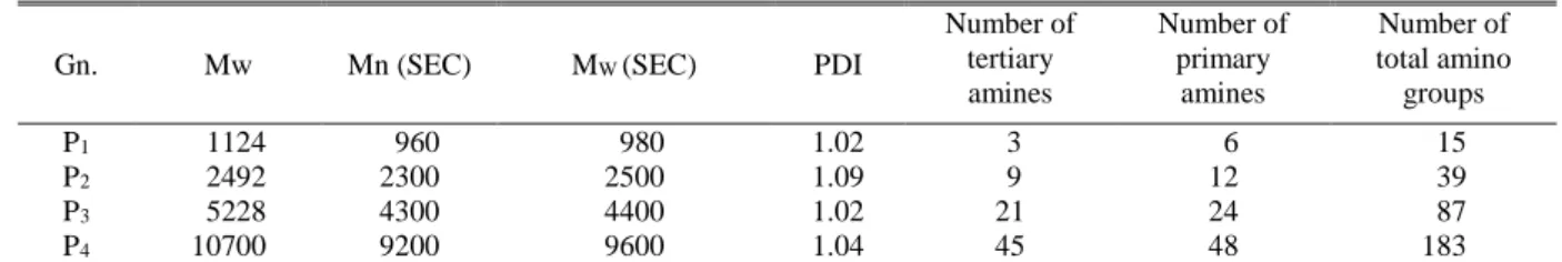
NMR, 13C NMR, FTIR-ATR, and GPC, and the results were in good agreement with the literature [11, 12]. The prepared dendrimers were stored in methanolic solution and stored at ±4ºC. Some characteristic and characterization data of the dif-ferent generation JCPDs are listed in Table 1.
T a b l e 1
Selected physicochemical properties of P1–P4 [11]a
Gn. Mw Mn (SEC) MW (SEC) PDI
Number of tertiary amines
Number of primary
amines
Number of total amino
groups
P1 1124 960 980 1.02 3 6 15
P2 2492 2300 2500 1.09 9 12 39
P3 5228 4300 4400 1.02 21 24 87
P4 10700 9200 9600 1.04 45 48 183
aGn.: generation; Mw: theoretical molecular weight (g/mol); Mn (SEC): nominal molecular weight measured by size exclusion
chromatography; Mw (SEC): molecular weight measured by size exclusion chromatography; PDI: polydispersity index.
3.2. Potentiometric measurements
Tertiary and primary amine groups of PA-MAMs are Lewis bases and they can bind most metals simultaneously (Table 1) [8]. However, the primary amines of PAMAMs cannot bind metal ions depending on the pH characteristic of the me-dia when the EOP of amine groups is out of the pH range of metal binding or all amine groups are protonated. Therefore, they are not available for metal ion coordination. Figure 1 shows the titration curves obtained from direct titration with 0.258 N HCl volumetric solution and back titration with 0.248 N NaOH. Titration curves exhibited three end points. The first is for excess acid added to dendrimer solution during direct titration, while the other two are for tertiary and primary amines, re-spectively. The first derivative of the back potenti-ometric titration curve of P4 was overlapped to show end points. Diallo et al. [9] accepted the maximum points in the second derivative curve as the inflection points referring to pKa values of the
tertiary and primary amines of PAMAM den-drimers. By using the same approach, we calculat-ed the pKa values of tertiary and primary amine groups of P4 to be 6.71 and 9.84, respectively (Ta-ble 2). Inspection of Ta(Ta-ble 2 shows that the pKa values we observed were in good agreement with the literature.
Fig. 1. Illustration of forward and back potentiometric titration curves of P4 with the second derivative curve of back titration
curve. a is the end point for excess HCl, b and c are the end points for tertiary and primary amines
T a b l e 2
pKa values for the tertiary and primary amine groups of P4
Dendrimer pKa of tertiary N groups
pKa of terminal amine groups
pKa of tertiary N groups (literature)
pKa of terminal amine groups (literature)
P4 6.71 9.84 6.85a–6.70b–6.30c 10.29a–9.00b–9.23c
aData taken from Diallo et al. [9]. bData taken from Cakara et al. [15]. cData taken from Niu et al. [16]. pKa values were calculated
by taking the inflection points of the second derivative of titration curves as it is stated in the study of Diallo et al. [9]. Acid base titration of the aqueous solutions of the P4 can be seen in Fig.1.
3.3. Effect of extent of protonation (EOP) on metal binding ability of PAMAM dendrimers
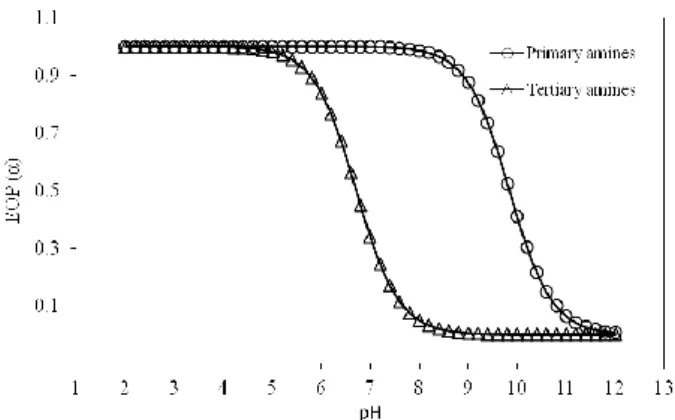
Figure 2 shows the predicted extent of pro-tonation (α) values of tertiary and primary amine groups of P4 dendrimer in aqueous solutions within the pH range of 2–12. These values were calculat-ed by using Hendersen-Hasselbech equation (1) with the calculated pKa values on Table 2 [17].
(1)
where α is extent of protonation, pKa is the acidity constants for tertiary and primary amine groups, and pH is the measurement of the hydronium ion concentration of the aqueous media.
a
Fig. 2. Extent of protonation (α) of primary and tertiary amine groups of P4
Some of the selected predicted EOP values of the amino groups of the P4 dendrimer are given in Table 3 in order to look closely at the available metal binding sites and behavior of P4 at low, neu-tral and high pH values. Results indicated that all amine groups of P4 became almost protonated at low pH (≤5) while at neutral pH 7, only the tertiary amine groups remained unprotonated (α = 0.339). On the other hand, at higher pH 9, the tertiary amine groups just remained deprotonated (α = 0.05). These results are correlated with those reported by Diallo et al. [9] (Table 2).
T a b l e 3
Extent of protonation (EOP) values of tertiary and primary amine groups of P4 as a function of pHa
A. Fraction of protonated tertiary amine groups Dendrimer
P4
pH 3 0.999
pH 5 0.980
pH 7 0.339
pH 9 0.005
B. Fraction of protonated primary amine groups Dendrimer
P4
pH 3 0.999
pH 5 0.999
pH 7 0.998
pH 9 0.873
aData obtained from the EOP graph of P
4 (Fig. 2)
Structures of dendrimers can be controllable and the mass of dendrimers increases as the gener-ation (Gn.) increases (Table 1). Tertiary and prima-ry amine groups of P4 are the possible proton bind-ing sites. Figure 3 shows the proton bindbind-ing per-cent values of P4 in the pH range of 2–12. These values were driven from EOP values presented in Figure 2. During the derivation of these values, it was benefited from core shell protonation method-ology [15, 18, 19]. This methodmethod-ology asserts that dendrimers start to protonate at higher pH and continue to protonate as the pH decreases. During this protonation, amino groups present in each core protonate together (Fig. 3).
Fig. 3. Proton binding (%) curve for P4
Taking into consideration Figure 2 and Fig-ures 3 together, it could be easily seen that the pro-tonation of the tertiary amine groups lasts approxi-mately in the pH range of 5–8, while primary amine groups are found to be in the deprotonated confor-mation over pH ~8. On the other hand, all of the amino groups are protonated under pH ~5. Almost 55% of the primary amine groups are protonated with a change in pH 5 (99.07%) to pH 8 (53.23%). That is, at a pH of about 8, most primary, but few tertiary, amines are protonated and such information is difficult to obtain from titration data alone.
The pH of the aqueous dendrimer solutions are desired to be at the pH at which all primary amine groups are in the protonated conformation and all of the tertiary amine groups are deprotonat-ed, in other words, free for binding in terms of coordination chemistry [9]. Investigation of Figure 3 reveals that pH 8 is the most desired pH, where the metal binding ability of P4 is expected to be highest and P4 behaves like hosts to many metal ions with the deprotonated inner tertiary amine groups. For this reason, to attain maximum com-plexation, we evaluated the pH of the aqueous dendrimer solutions at pH 8 to gain maximum complexation between P4 and Co(II) ions.
3.4. Spectroscopic titrations and UV-Vis characterization of PAMAM dendrimers
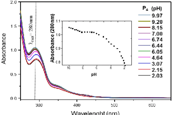
investi-gation of the inset of Figure 4 also indicates that P4 dendrimer can exhibit interesting ligand properties as chelating agents, especially in the pH range of 6.0–8.0. Interestingly, spectroscopic titration re-sults validated the potentiometric titration studies where pH 8 is the most favorable pH for tertiary amine groups of P4 to have the highest opportunity to bind Co(II) ions.
Fig. 4. Representative absorption spectra of P4
as a function of pH. The inset graph shows how the band at 280 nm changes as a function of pH..
3.5. UV-Vis characterization of Co(II)-P4
complex
Characterization of the Co(II) binding ability of PAMAMs in aqueous solutions was performed by UV-Vis spectroscopy. Normally, hexa-co-ordinated Co(II) aqua complexes in aqueous solu-tions are observed at λmax = 510–512 nm [21]. UV-Vis measurements were performed between the wavelength ranges of 200–750 nm with 1.00 nm interval against the dendrimer solutions. Displace-ment of the 512 nm band with 585–635 nm d-d
transition bands, confirmed the formation of Co(II)-P4 complexes (Fig. 5).
500 600 700
0.0 0.1 0.2 0.3 0.4
Ab
sor
ban
ce
Wavelength (nm)
Co(II): P4.NH2 = 60
Co(II): P4.NH2 = 30
Co(II): P4.NH2 = 20
Co(II): P4.NH2 = 15 pH=8.00
Fig. 5. UV-Vis spectra of Co(II)-P4 complexation bands
at 585–635 nm
Complexation studies were conducted in the pH range of 6–8 and best observations were per-formed at pH 8, as predicted by potentiometric and spectroscopic studies conducted on the P4 den-drimer. In addition, the color of dendrimer solu-tion, when the pH was adjusted to 8, immediately turned dark green after the addition of Co(II) ions. This color change and absorption bands could be attributed to the formation of tetra amine complex-es of Co(II) with dendrimer internal tertiary amine groups (Fig. 5).
4.CONCLUSION
In the present study, Co(II) complexes of a model generation-4 Jeffamine® cored PAMAM dendrimer (P4) were synthesized. The optimum complexation conditions for the P4 was tried to determine with potentiometric and spectroscopic titration studies. pH data driven from potentiom-etric titration were used to form EOP profiles in order to investigate the protonation behavior of P4. Investigation of EOP and proton binding profiles showed that pH 8 was the optimum dendrimer aqueous solution media, where the available num-ber of tertiary amines present in P4 was highest for possible metal complexation. The new Co(II)-P4 complexes obtained could be used in possible fu-ture applications as MRI improvers or MRI con-trast agents.
Acknowledgements.This research has been supported
by Yıldız Technical University Scientific Research Projects Coordination Department. Project Numbers (2011-01-02-KAP04, 2011-01-02-KAP05, 2011-01-02- KAP06 and 2012-01-02-DOP05).
REFERENCES
[1] J. H. Kim, H. J. Gibb, P. D. Howe, M. Sheffer, U. N. E. Programme, I. L. Organisation, W. H. Organization, I.-O.P.f.t.S.M.o. Chemicals, Cobalt and Inorganic Cobalt Compounds, World Health Organization (2006).
[2] S. E. Bailey, T. J. Olin, R. M. Bricka, D. D. Adrian, A review of potentially low-cost sorbents for heavy metals,
Water Res.,33, 2469–2479 (1999). DOI: 10.1016/s0043-1354(98)00475-8.
[3] A. Kabata-Pendias, Trace Elements in Soils and Plants, Third Edition, Taylor & Francis, (2010).
DOI: 10.1201/9781420039900.ch5.
[5] K. Yamamoto, S. Inoue, A. Yamazaki, T. Yoshinaga, S. Kawanishi, Site-specific DNA damage induced by cobalt(II) ion and hydrogen peroxide: role of singlet oxygen, Chem. Res. Toxicol.,2, 234–239 (1989). DOI: 10.1021/tx00010a004.
[6] A. Sigel, H. Sigel, R. K. O. Sigel, Organometallics in Environment and Toxicology, RSC Publishing, (2010). DOI: 10.1039/9781849730822..
[7] J. F. Hainfeld, Extended organic cobalt and nickel magnetic complexes, in, Google Patents (2003).
[8] F. Zeng, S. C. Zimmerman, Dendrimers in supra-molecular chemistry: From supra-molecular recognition to self-assembly, Chem. Rev. (Washington, D. C.), 97, 1681–1712 (1997). DOI: 10.1021/CR9603892.
[9] M. S. Diallo, S. Christie, P. Swaminathan, L. Balogh, X. Shi, W. Um, C. Papelis, W. A. Goddard, III, J. H. Johnson, Jr., Dendritic Chelating Agents. 1. Cu(II) Binding to Ethylene Diamine Core Poly(amidoamine) Dendrimers in Aqueous Solutions, Langmuir,20, 2640– 2651 (2004).
DOI: 10.1021/la036108k.
[10] A. D. Schluter, J. P. Rabe, Dendronized polymers: synthesis, characterization, assembly at interfaces, and manipulation, Angew. Chem., Int. Ed.,39, 864–883 (2000). DOI: 10.1002/(SICI)1521-3773(20000303)39:5<864:: AID-ANIE864>3.0.CO;2-E.
[11] A. S. Ertürk, M. Tülü, A. E. Bozdoğan, T. Parali, Microwave assisted synthesis of Jeffamine cored PAMAM dendrimers, Eur. Polym. J., 52, 218–226 (2014). DOI: 10.1016/j.eurpolymj.2013.12.018.
[12] A. S. Erturk, M. U. Gurbuz, M. Tulu, A. E. Bozdogan, Water-soluble TRIS-terminated PAMAM dendrimers: microwave-assisted synthesis, characterization and Cu(ii) intradendrimer complexes, RSC Adv.,5, 60581– 60595 (2015). DOI: 10.1039/C5RA11157A.
[13] I. J. Majoros, B. Keszler, S. Woehler, T. Bull, J. R. Baker, Acetylation of Poly(amidoamine) Dendrimers,
Macromolecules,36, 5526–5529 (2003). DOI: 10.1021/ma021540e.
[14] I. J. Majoros, T. P. Thomas, C. B. Mehta, J. R. Baker, Poly(amidoamine) Dendrimer-Based Multifunctional Engineered Nanodevice for Cancer Therapy, J. Med. Chem.,48, 5892–5899 (2005). DOI: 10.1021/jm0401863. [15] D. Cakara, J. Kleimann, M. Borkovec, Microscopic
Protonation Equilibria of Poly(amidoamine) Dendrimers from Macroscopic Titrations, Macromolecules, 36, 4201–4207 (2003). DOI: 10.1021/ma0300241.
[16] Y. Niu, L. Sun, R. M. Crooks, Determination of the Intrinsic Proton Binding Constants for Poly (amidoamine) Dendrimers via Potentiometric pH Titration, Macromolecules,36, 5725–5731 (2003). DOI: 10.1021/ma034276d.
[17] V. Kabanov, A. Zezin, V. Rogacheva, Z. G. Gulyaeva, M. Zansochova, J. Joosten, J. Brackman, Polyelectrolyte behavior of astramol poly(propyleneimine) dendrimers,
Macromolecules,31, 5142–5144 (1998). DOI: 10.1021/ma971643a.
[18] L. Sun, R. M. Crooks, Interactions between Dendrimers and Charged Probe Molecules. 1. Theoretical Methods for Simulating Proton and Metal Ion Binding to Symmetric Polydentate Ligands, J. Phys. Chem. B,106, 5864–5872 (2002). DOI: 10.1021/jp020189w.
[19] M. H. Kleinman, J. H. Flory, D. A. Tomalia, N. J. Turro, Effect of Protonation and PAMAM Dendrimer Size on the Complexation and Dynamic Mobility of 2-Naphthol,
J. Phys. Chem. B,104, 11472–11479 (2000). DOI: 10.1021/jp001882r.
[20] S. Pande, R. M. Crooks, Analysis of poly(amidoamine) dendrimer structure by UV–Vis spectroscopy,
Langmuir,27, 9609–9613 (2011). DOI: 10.1021/la201882t.