44
ANATOMICAL ASPECTS OF
BEGONIAHYDROCOTYLIFOLIA
OTTO EX
HOOK. (BEGONIACEAE) LEAF
RODICA BERCU Faculty of Natural and Agricultural Sciences,”Ovidius” University, Constantza University Alley, No. 1, B, 900470, Constantza E-mail: [email protected]
Key words: anatomy, leaf, petiole, mesophyll, Begonia hydrocotylifolia
ABSTRACT
The paper presents anatomical aspects concerning the leaf structure of Begonia hydrocotylifolia Otto ex Hook. belonging to Begoniaceae family. Anatomically, the petiole has a unistratous epidermis and a differentiated mesophyll. The vascular system is fascicular type with a large number of collateral bundles placed into a basic tissue. The blade consits of an upper epidermis with a many-layerd hypodermis, a lower epidermis and the mesophyll. The mesophyll is differentiated into palisade and spongy tissue with the same vascular bundle structure such as those of the petiole but with foliar arrangement of the conductive tissues. Stomata are present to the lower epidermis. Remarkable are the cytological elements represented by prismatic oxalate crystals, druses, crystalline sand and tannin cells. The mechanical tissue is represented by collencyma cellls, presented in the petiole and blade structure.
INTRODUCTION
The family Begoniaceae consists of two genera: Begonia Linnaeus, with approximately 1.500 species and pantropical distribution (spread in Central and Southern America, Asia, Africa, the Pacific Isles) and the monospecific Hillebrandia Oliver, from the Hawaiian Islands (Jacques and Mamede, 2005). Begoniaceaefamily comprises perennial herbaceous plants, suffrutescent or frutescent plants, alternateleafed plants, sometimes asymmetrical, stipellated, whole or with a lobed side, lobed or divided, variously colored (Cruceru, 2011). The family is characterized by threewinged capsular fruits, bifid styluli and peculiar seed micromorphology (Forrest et al., 2005).
Most Begoniaceae are monoecious perennials with very few dioecious exceptions. Begonias are widely spread in the rainforest in the humid mountain areas, inside the woods, on the edge of water courses, on rock walls, where water drops. They most likely originated in the mid-Eocene to late Oligocene and reached their current distribution by multiple intercontinental dispersal events (Goodall-Copestake et al., 2009). Over 10000 begonias hybrids and cultivars have been introduced by commercial growers. Many begonias are popular ornamentals (Awal et al, 2008).
Analele Universităţii din Craiova, seria Agricultură Montanology, Cadastre Series)Vol. XLVII 201
Stratu et al, 2011), chromosome cytology (Zeilinga, 1962; Peng et al, 2014), phylogenetic relationships (Tebbitt et al., 2006), somatic e
et al, 2014), the effect of potassium silicate on the growth and leaf epidermal characteristics (Lim et al., 2012).
Many species are observed to have a hypoderm and abnormal stomatal patterning (“stomatal cluster”) (Dehnel, 1961; Tang et al., 2002). Medullary and cortical vascular bundles in the petiole and stem represent an anatomical pattern more like monocotyledons than dicots. The stem has superficial cork
the numbers and size of trichomes were examined (Mclellan, 2005). Physiologically, Begonia is distinct for the presence of oxalic acid in cytoliths, another characterist
in the angiosperms (Pireyre, 1961). Calcium oxalate crystals are most widespread storage material in plant (druses and prismatic types) (Ianovici, 2010: Niculescu, 2009).
species are examples of plants with paedomorphic secondary xylem c walled, wide libriform fibers (Dulin, 2008).
Begoniahydrocotylifolia Otto ex Hook. is
spontaneously in nature. In our country is known as a decorative species especially by it leaves. It is a plant both inside and outside, being used in decorating gardens. If is cultivated as ornamental plant in
and a constant humidity as Cruceru (2011) reported for
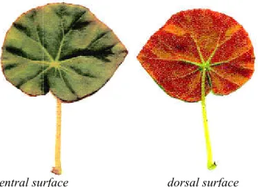
blade has a slightly asymmetrical shape, with cordate base and full red hair to the margins. The blade is discolored, the upper face is dark green, very glossy and glabrous, with slightly wrinkled and discolored ribs (Fig, 2, A, B). The lower face is reddish brick, with red hairs. The consistency of the blade is fleshy. The cylindrical petiole is long, fleshy and yellowish (Fig. 1, A). It possess all over the surface fine red hairs. The hairs of this species are different by the numerous brownish hairs at the junction of the petiole with the blade described for other species of
arranged in 4-5 in dichasium inflorescence (Fig. 1, B).
Fig. 1. Begonia hydrocotylifolia Otto ex Hook.: leaves (A) and inflorescence (B) (A
The aim of this paper is to analyze the some amatomical aspects of the petiole and blade of Begonia hydrocotylifolia. In literature
our country almost lack. This is the way we believe that the present paper brings added knowledge about this group of plants (genus
Begoniahydrocotylifolia. A A
Agricultură – Montanologie – Cadastru (Annals of the University of Craiova I 2017
45
Stratu et al, 2011), chromosome cytology (Zeilinga, 1962; Peng et al, 2014), phylogenetic relationships (Tebbitt et al., 2006), somatic embryogenesis and plant regeneration (Rosilah et al, 2014), the effect of potassium silicate on the growth and leaf epidermal characteristics (Lim et al., 2012).
Many species are observed to have a hypoderm and abnormal stomatal patterning (“stomatal cluster”) (Dehnel, 1961; Tang et al., 2002). Medullary and cortical vascular bundles in the petiole and stem represent an anatomical pattern more like monocotyledons han dicots. The stem has superficial cork-cambium. Correlations between leaf shape and the numbers and size of trichomes were examined (Mclellan, 2005). Physiologically,
is distinct for the presence of oxalic acid in cytoliths, another characterist
in the angiosperms (Pireyre, 1961). Calcium oxalate crystals are most widespread storage material in plant (druses and prismatic types) (Ianovici, 2010: Niculescu, 2009).
species are examples of plants with paedomorphic secondary xylem c walled, wide libriform fibers (Dulin, 2008).
Otto ex Hook. is originating from Mexico where it can be found spontaneously in nature. In our country is known as a decorative species especially by it leaves. It is a plant both inside and outside, being used in decorating gardens. If is cultivated as ornamental plant in pots, the plant prefers an indirect action of the sun's rays and a constant humidity as Cruceru (2011) reported for Begonia species. This species blade has a slightly asymmetrical shape, with cordate base and full red hair to the margins. colored, the upper face is dark green, very glossy and glabrous, with slightly wrinkled and discolored ribs (Fig, 2, A, B). The lower face is reddish brick, with red hairs. The consistency of the blade is fleshy. The cylindrical petiole is long, fleshy and yellowish (Fig. 1, A). It possess all over the surface fine red hairs. The hairs of this species are different by the numerous brownish hairs at the junction of the petiole with the blade described for other species of Begonia by Foster (1958). The flower
5 in dichasium inflorescence (Fig. 1, B).
hydrocotylifolia Otto ex Hook.: leaves (A) and inflorescence (B) (A Web 1; B-Web 2).
The aim of this paper is to analyze the some amatomical aspects of the petiole and blade In literature are few data about this plant leaf anatomy and in This is the way we believe that the present paper brings added knowledge about this group of plants (genus Begonia) in general and in particular for
B B
Annals of the University of Craiova - Agriculture,
Stratu et al, 2011), chromosome cytology (Zeilinga, 1962; Peng et al, 2014), phylogenetic mbryogenesis and plant regeneration (Rosilah et al, 2014), the effect of potassium silicate on the growth and leaf epidermal
Many species are observed to have a hypoderm and abnormal stomatal patterning (“stomatal cluster”) (Dehnel, 1961; Tang et al., 2002). Medullary and cortical vascular bundles in the petiole and stem represent an anatomical pattern more like monocotyledons cambium. Correlations between leaf shape and the numbers and size of trichomes were examined (Mclellan, 2005). Physiologically, is distinct for the presence of oxalic acid in cytoliths, another characteristic limited in the angiosperms (Pireyre, 1961). Calcium oxalate crystals are most widespread storage material in plant (druses and prismatic types) (Ianovici, 2010: Niculescu, 2009). Begonia species are examples of plants with paedomorphic secondary xylem containing thin
originating from Mexico where it can be found spontaneously in nature. In our country is known as a decorative species especially by it leaves. It is a plant both inside and outside, being used in decorating gardens. If is pots, the plant prefers an indirect action of the sun's rays species. This species blade has a slightly asymmetrical shape, with cordate base and full red hair to the margins. colored, the upper face is dark green, very glossy and glabrous, with slightly wrinkled and discolored ribs (Fig, 2, A, B). The lower face is reddish brick, with red hairs. The consistency of the blade is fleshy. The cylindrical petiole is long, fleshy and yellowish (Fig. 1, A). It possess all over the surface fine red hairs. The hairs of this species are different by the numerous brownish hairs at the junction of the petiole with the blade by Foster (1958). The flowers are mostly pink,
hydrocotylifolia Otto ex Hook.: leaves (A) and inflorescence (B) (A-
Fig. 2. Frunză de Begonia
The plant mature leaves of
International S.R.L in august, 2016. Small pieces of leaves (petiole and blade) were fixed in FAA (formalin: glacial acetic acid: alcohol 5:5:90). Cross sections of the leaf were performed by free hand made technique (Bercu an
stained with alum-carmine and iodine green. Anatomical observations and micrographs were performed with a BIOROM
6001A video camera.
RESULTS AND DISCUSSIONS
The petiole, in cross section, is circular in shape. The outer epidermis has a single layer of cells covered by a thick cuticle.
hairs and stomata.The glandular hairs of the petiole are two basal cell and a capitate glandular cell (Fig. 3, B).
into an external region and an inner region.
collenchyma tissue (5 layers of cells) and an internal one, represented by a number of parenchyma layers of cells. The vascular system is fascicular, represented by numerous closed collateral vascular bundles (25), with culinary arrangement, placed on two circles, with poor developed xylem and phloem vascular elements, separate by medullary rays (Fig. 3, A). Such as other Begonia
petiole vascular system is embadded into a basic tissue of parenchyma nature with rare tannin cells, calcium oxalate crystals (isolated prismatic crystals, crystalline sand and druses) are present (Fig. 3, A).
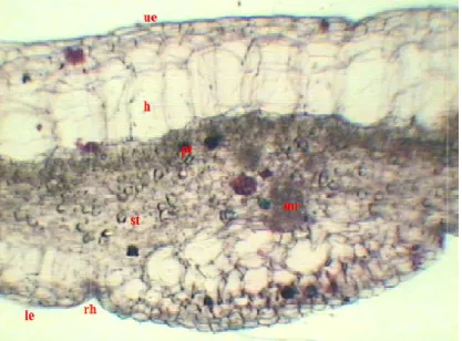
Cross section of the blade shows the usua
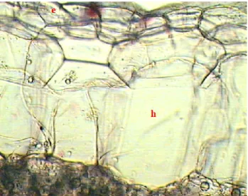
epidermis, and mesophyll. The upper epidermis consists of a layer of isodiametric cells tightly joined together, covered with a thick cuticle, supplemented with vegetable wax (Fig. 3, 4). Below the epidermis is a m
consists of palisade and a spongy tissue. The palisade tissue is poor developed, consisting of 2-3 layers of rounded assimilation cells, containing numerous chloroplasts. Small rare tannin cells and crystal
tissue consists of a 5-6 layer of large cells, with small intercellular spaces between them, and they are rounded in front of the mid rib. It is remarkable the presence of rare crystals isolated calcium oxalate (druse) (Fig. 6).
ventral surface dorsal surface
46
Begonia hydrocotylifolia Otto ex Hook. (original).
MATERIAL AND METHODS
The plant mature leaves of Begonia hydrocotylifolia were collectd from S.C. Iris International S.R.L in august, 2016. Small pieces of leaves (petiole and blade) were fixed in FAA (formalin: glacial acetic acid: alcohol 5:5:90). Cross sections of the leaf were performed by free hand made technique (Bercu and Jianu, 2003). The samples were carmine and iodine green. Anatomical observations and micrographs were performed with a BIOROM–T bright field microscope, equipped with a TOPICA
RESULTS AND DISCUSSIONS
n cross section, is circular in shape. The outer epidermis has a single layer of cells covered by a thick cuticle. Its continuity is interrupted by the presence of glandular hairs and stomata.The glandular hairs of the petiole are two-celled, formed by a short basal cell and a capitate glandular cell (Fig. 3, B). It is followed by the cortex, differentiated ternal region and an inner region. The external area is represented by an angular collenchyma tissue (5 layers of cells) and an internal one, represented by a number of parenchyma layers of cells. The vascular system is fascicular, represented by numerous closed collateral vascular bundles (25), with culinary arrangement, placed on two circles, with poor developed xylem and phloem vascular elements, separate by medullary rays
Begonia species leaves (Bercu, 2005;Li Jing Xiu,2007),
vascular system is embadded into a basic tissue of parenchyma nature with rare tannin cells, calcium oxalate crystals (isolated prismatic crystals, crystalline sand and druses) are present (Fig. 3, A).
Cross section of the blade shows the usual tissue sequence: upper epidermis, lower epidermis, and mesophyll. The upper epidermis consists of a layer of isodiametric cells tightly joined together, covered with a thick cuticle, supplemented with vegetable wax (Fig. 3, 4). Below the epidermis is a many-layered hypodermis (Fig. 4; 5). The mesophyll consists of palisade and a spongy tissue. The palisade tissue is poor developed, 3 layers of rounded assimilation cells, containing numerous chloroplasts. Small rare tannin cells and crystalline sand are present as well (Fig. 4; 5).The lacunar 6 layer of large cells, with small intercellular spaces between them, and they are rounded in front of the mid rib. It is remarkable the presence of rare crystals
oxalate (druse) (Fig. 6).
ventral surface dorsal surface
hydrocotylifolia Otto ex Hook. (original).
were collectd from S.C. Iris International S.R.L in august, 2016. Small pieces of leaves (petiole and blade) were fixed in FAA (formalin: glacial acetic acid: alcohol 5:5:90). Cross sections of the leaf were d Jianu, 2003). The samples were carmine and iodine green. Anatomical observations and micrographs T bright field microscope, equipped with a TOPICA
n cross section, is circular in shape. The outer epidermis has a single layer of Its continuity is interrupted by the presence of glandular celled, formed by a short It is followed by the cortex, differentiated The external area is represented by an angular collenchyma tissue (5 layers of cells) and an internal one, represented by a number of parenchyma layers of cells. The vascular system is fascicular, represented by numerous closed collateral vascular bundles (25), with culinary arrangement, placed on two circles, with poor developed xylem and phloem vascular elements, separate by medullary rays leaves (Bercu, 2005;Li Jing Xiu,2007), the vascular system is embadded into a basic tissue of parenchyma nature with rare tannin cells, calcium oxalate crystals (isolated prismatic crystals, crystalline sand and
Analele Universităţii din Craiova, seria Agricultură Montanology, Cadastre Series)Vol. XLVII 201
Fig. 3. Portion of a cross section of the petiole with a epidermis, cortex and a vascular bundle (A, x 156). Epidermis with glandular hair and stomata (B, x 230): co
coc- calcium oxalate crystal, cs
glandular hair, ph
Fig. 4. Cross section of the blade mid rib, rh- root hair, pt-
The vascular bundle of the mid rib is poorly developed, consisting of few elements of xylem, to the upper epidermis and some phloem vessels to the lower epidermis. In front of the primary mid rib, beneath the
collenchyma cells (Fig. 5, 6).
The lower one-layered epidermis, with cells smaller cells than those of the upper epidermis, including bi-cellular glandular hairs the same with those of the petiole.
place of formation, the lower epidermis forms an obvious recess (Fig. 6). A
Agricultură – Montanologie – Cadastru (Annals of the University of Craiova I 2017
47
Fig. 3. Portion of a cross section of the petiole with a epidermis, cortex and a vascular bundle (A, x 156). Epidermis with glandular hair and stomata (B, x 230): co
oxalate crystal, cs- crystalline sand, ct- tannin cell, e- epidermis, d glandular hair, ph- phloem, x- xylem.
Fig. 4. Cross section of the blade – ensamble (x 80): h- hypodermis, le- lower epidermis, mr pallisade tissue, sp- pongy tissue, ue- upper epidermis.
The vascular bundle of the mid rib is poorly developed, consisting of few elements of xylem, to the upper epidermis and some phloem vessels to the lower epidermis. In front of the primary mid rib, beneath the lower epidermis, there are two cell layers with slightly
layered epidermis, with cells smaller cells than those of the upper cellular glandular hairs the same with those of the petiole.
place of formation, the lower epidermis forms an obvious recess (Fig. 6).
gh
e
B
Annals of the University of Craiova - Agriculture,
Fig. 3. Portion of a cross section of the petiole with a epidermis, cortex and a vascular bundle (A, x 156). Epidermis with glandular hair and stomata (B, x 230): co – collenchyma,
epidermis, d- druse, gh-
lower epidermis, mr- upper epidermis. The vascular bundle of the mid rib is poorly developed, consisting of few elements of xylem, to the upper epidermis and some phloem vessels to the lower epidermis. In front of lower epidermis, there are two cell layers with slightly
48
Fig. 5. Cross section of the blade with epidermis and hypodermis –detail (x 235): e- epidermis, h- hypodremis.
Fig. 6. Cross section of the blade (x 235): cs- crystalline sand, d-druse, le- lowe epidermis, st- spongy tissue, vb- vascular bundle.
CONCLUSION
Analele Universităţii din Craiova, seria Agricultură – Montanologie – Cadastru (Annals of the University of Craiova - Agriculture, Montanology, Cadastre Series)Vol. XLVII 2017
49
The mesophyll is heterogenous and hipostomatic. The vascular system is represented by a number of vascular bundles withthe ussualyfoliar arrangement of the conductive tissues. The strength of the petiole and blade is, due to the collenchyma tissue, placed bellow the lower petiole epidermis.
ACKNOWLEDGEMENTS
We express our thanks to dr. ing. Elena Bavaru manager of S.C. Iris International S.R.L. for the vegetal material made available to us for this study.
BIBLIOGRAPHY
1. Ali M.S.,2013 - Genetic Architecture of Species level differences in Begonia Section Gireoudia. Institute of Cell and Molecular Biology, School of Biological Sciences, University of Edinburgh. PhD thesis.
2.Awal A., Taha R.M., Hasbullah N.A., 2008 - Induction of somatic embryogenesis and plant regeneration in Begonia x hiemalis Fotsch in vitro, J. Biol. Sci., 8: 920-924.
3.Bercu R., Jianu D.L., 2003 - Practicum de morfologiea și anatomia plantelor, Edit. “Ovidius” University Press, Constanța.
4.Bercu R., 2005 - On the leaf histoanatomy of some Begonia L. (Begoniaceae) species, Analele Universităţii „Ovidius”, Ser. Biologie-Ecologie, 9: 3-7.
5.Cruceru S., 2011 - Begoniaceae collection cultivated in the botanical garden in Craiova green houses. Analele Universităţii din Craiova, seria Agricultură – Montanologie – Cadastru, XLI (2):126-128.
6.Dehnel G.S., 1961 - Abnormal stomatal development in foliage leaves of Begoniaaridicaulis, Amer. J. Bot., 48:129-133.
7.Dulin M., 2008 - An investigation of paedomorphic secondary xylem and secondary woodiness in Xanthorhiza simplicissima, Coreopsis gigantea, and Mahonia bealei, Dissertation, The University of North Carolina at Greensboro (UNCG).
8.Forrest L.L., Hughes M., Hollingsworth P.M., 2005 - A phylogeny of Begonia using nuclear ribosomal sequence data and morphological characters, Syst. Bot. 30: 671–682.
9.Goodall-Copestake W.P., Harris D.J., Hollingsworth P.M., 2009 - The origin of a mega-diverse genus: Dating Begonia (Begoniaceae) using alternative datasets, calibrations and relaxed clock models. Bot. J. Linn. Soc., 159: 363–380
10. Hoover W.S., Karegeannes C., Wiriadinata H., Hunter J.M., 2004 - Notes on the geography of South-East Asian Begonia and species diversity in montane forests, Telopea, 10(3): 749–764.
11.Ianovici N., 2010 - Citohistologie şi morfoanatomia organelor vegetative, Edit. Mirton, Timişoara.
12.Jacques E.L., Mamede M.C.H., 2005 - Notas nomenclaturais em Begonia L. (Begoniaceae), Revista Brasil. Bot., 28 (3): 579-588.
13. Lee Y. S., 1974 - A study of stem anatomy in Begonia L., Phytologia, 27: 464-489.
14.Li Jing Xiu, Guan KaiYun, Ohmiya T., Nakata M., Godo T., 2007 - Anatomy on leaf cross sections of Begonia from Yunnan, China, Guangxi Zhiwu/Guihaia, 27(4): 543-550.
15.Lim M.Y., Lee E.J., Jana S., Sivanesan I., Jeong B.R., 2012 - Effect of Potassium Silicate on Growth and Leaf Epidermal Characteristics of Begonia and Pansy Grown in Vitro, Kor. J. Hort. Sci. Technol., 30(5):579-585.
16.Mclellan T., 2005 - Correlated evolution of leaf shape and trichomes in Begoniadregei (Begoniaceae), American Journal of Botany, 92(10): 1616–1623.
17. Niculescu, Mariana, 2009 - Morfología şi anatomia plantelor,vol. I, Edit.Sitech, Craiova.
50
SCIENTIFIC PAPERS-SERIES A-AGRONOMY Volume: 59 Pages: 116-121,
Published: 2016, ISSN- 1222-5339,
https://apps.webofknowledge.com/Search.do?product=WOS&SID=C2iDut4m8HPpdxgZPs 8&search_mode=GeneralSearch&prID=05baa3a9-21cf-4989-81bf-daf67bb9f815
19. NICULESCU, Mariana - METODE DE CERCETARE ŞI PREZENTARE A FLOREI, Ed. Sitech, Craiova, 2009, 119 p., ISBN 978 606-530-322-5
20.CRUCERU, Sonia, STAN I., NICULESCU, Mariana – Growing aspects of Begonia boweri on different types of sublayers mixtures, Bulletin of University of Agricultural Sciences and Veterinary Medicine Cluj-Napoca, Vol. 64, ISSN 1843- 5246,
Academic Press, Cluj-Napoca,
http://journsals.usamvcj.ro/agriculture/index,http://www.cabi.org/AbstractDatabases, p.
69-73
21.Peng C.-I., Wang H., Kono Y., Yang H.-A., 2014 - Begonia wui-senioris (sect. Platycentrum, Begoniaceae), a new species from Myanmar, Botanical Studies, 55 (13): 2-6.
22.Pireyre N., 1961 - Contributions to the morphological, histological and physiological study of cystoliths, Rev. of Cytology and Plant Biology, 23: 93–320.
23.Rosilah A.A., Kandasamy K.I., Faridah Q.Z., Namasivayam P., 2014 - Somatic embryogenesis and plant regeneration from leaf explants of endemic Begonia pavonina, Journal of Biology and Earth Sciences, 4 (2): B113-B119.
24.Sheue C.-R., Pao S.-H., Chien L.-F., Chesson P., Peng C.-I., 2012 - Natural foliar variegation without costs? The case of Begonia, Annals of Botany, 109: 1065–1074.
25.Stratu A., Costică N., Puşchiu O., 2011 - Histo-anatomical and physiological aspects of plant species with flowers decoration, Analele Știinţifice ale Universităţii „Al. I. Cuza” Iaşi, Tomul LVII, fasc. 1, s. II a. Biologie vegetală, 1-18.
26. Tang, M., Y.X. Hu, J.X. Lin, and X.B. Jin., 2002 - Developmental mechanism and distribution pattern of stomatal clusters in Begonia peltatifolia, Acta Bot. Sin., 44:384-390.
27.Tebbitt M.C., Lowe-Forrest L., Santoriello A., Clement W.L., Swensen S.M., 2006 - Phylogenetic Relationships of Asian Begonia, with an Emphasis on the Evolution of Rain-ballist and Animal Dispersal Mechanisms in Sections Platycentrum, Sphenanthera and Leprosae, Systematic Botany, 31(2): 327–336.
28.Zeilinga A.E., 1962 - Cytological investigation of hybrid varieties of Begoniasemperfloren,Euphytica, 11(2): 126-136.
Page Web
29.https://www.pinterest.com/pin/529806343643793049/
30.https://commons.wikimedia.org/wiki/File:Begonia_hydrocotylifolia_GotBot_2015_002.jp