Journal of Global Pharma Technology
Available Online at: www.jgpt.co.in
RESEARCH ARTICLE
Staphylococcus aureus
Enterotoxin as a Major Risk Factor for
Multiple Sclerosis Severity
Suhaib Khalid Ibrahim
1*,Thanaa Rasheed Abdulrahman
2,Nawfal Madhi
Sheaheed
31. MSc Microbiology/ College of Health and Medical Technology /Baghdad/Iraq. 2. PhD. Microbiology, Al-Nahrain University /College of Medicine/Iraq.
3. Consultant Neurologist, Baghdad Teaching Hospital, Medical City/Iraq.
*Corresponding Author:Suhaib Khalid Ibrahim
Abstract
Background: Staphylococcus aureus produce enterotoxins that function as super antigens which activate auto reactive CD4+ T-cells potentially target the basic myelin protein into the CNS. Objective: This study investigates the correlation betweenthe colonization of S. aureus harbouring enterotoxins with multiple sclerosis (MS) exacerbation. Methods: A total of 200 nasal swabs were collected from three study groups: Healthy controls as a Non-MS Subjects (n=100), relapsing remitting MS or exacerbated (n=50) and newly diagnosed MS. (n=50). S. aureus was isolated from the anterior nares of these groups following standard operating procedures. Antibiotic susceptibility test against 19 antibiotics using disk diffusion method were done. Staphylococcal super Antigen SeA, seB, seC genes were amplified using standard conventional polymerase chain reaction (PCR) technique and resolved by agarose gel electrophoresis. Results: In this study, Out of 100 MS patients and 100 healthy control groups, there were 36% and 46% males and 64% and 54% females respectively. A total of 81(81%) were colonized with S. aureus includes, 31(38.2%) newly diagnosed MS and 50(61.8%) exacerbated MS while only 12(12%) isolated from control non-MS group. All S. aureus isolates were resistant to Methicillin (100%) and sensitive to Impenem (100%). PCR results showed that, the frequency of enterotoxins (sea, seb and sec) in MS patients was (42.4%) including (40.8%) were from newly diagnosed and (43.3%) were from relapsing remitting while there is no enterotoxins was detected in control group. the prevalence of se A was significantly higher (p<0.00) in the MS exacerbation (72%) than in newly diagnosed group (64.5%). Conclusions: The frequency of S.aureus isolates and its enterotoxin seA gene is high in the MS patients and this gene serve
as an important marker in the severity of MS disease. Also S.aureus isolates were sensitive to imipenem which is considered as a better choice for nasal decolonization of S.aureus in the MS patients.
Keywords: Multiple sclerosis (MS), Staphylococcus aureus, Super antigens, Enterotoxin A, B, C.
Introduction
Multiple sclerosis (MS) is an autoimmune-inflammatory disease that occurs in the central nervous system (CNS) causing demyelination. The demyelination operation is related to infiltrating T-cells particular for major myelin proteins of the CNS, this disease has a peak start between 20 and 40 years old and influences women almost two times as often as men[1]. The most common form of disease is the relapsing-remitting MS (RR-MS), including around 85 - 90% of all cases. Although the etiology of MS is unknown; genetic and environmental factors play a role. Infectious pathogens are the possible environmental factors contributed to
the MS development or severity [2]. Pathogens related to the severity or development of MS involve bacteria; such as
Chlamydia pneumoniae , Mycoplasma
pneumonia and Staphylococcus aureus which
produced enterotoxins that function as superantigens [3]. It has been proposed that
S. aureus in nasal carriage is related to
several autoimmune diseases involving systemic lupus erythematosus, rheumatoid arthritis and Wegener’s granulomatosis syndrome through superantigens such as toxic shock syndrome toxin-1 {tsst-1} or
Superantigens are proteins produced mostly by bacteria and some viruses that potently activate autoreactive CD4+ T cells, inducing massive cell proliferation and cytokine production, predominantly IL-2 and interferon (IFN)-γ [5].Staphylococcus aureus
superantigens are capable of activating T cells due to their ability to bind to both major histocompatibility complex (MHC) class II molecules and specific Vβ regions of the T cell receptor. Furthermore, this leads to the activation of both antigen-presenting cells and T lymphocytes. Ultimately this mechanism results in the over-production of inflammatory cytokines and T cells followed by the presentation of autoimmune activated clinical symptoms [6].
The most important superantigens were the
Staphylococcus aureus enterotoxins (A, B, C,
D and E) and toxic shock syndrome toxin that participate in T cell activation [6]. More than 20 superantigens have been identified in S.
aureus strains and at least 80% of the clinical
strains of S. aureus harbor a minimum of one superantigen [7].A study in multiple sclerosis(MS) patients examining the association of bacterial superantigens with MS exacerbation has been recently carried out [8], therefore, colonization of the nose with enterotoxin A-producing Staphylococcus
aureus may play a role in triggering
autoreactive CD4+ T cell activation and Multiple Sclerosis exacerbations[9]. The role
of S. aureus superantigens in the etiology of
MS is currently still unknown, but Staphylococcal Enterotoxin A (sea), Staphylococcal Enterotoxin B (seb), Staphylococcal Enterotoxin C (sec) and Toxic Shock Syndrome Toxin-1 (tsst-1) have been demonstrated to be involved in the reactivation of a mouse model for MS ,known as experimental autoimmune encephalomyelitis (EAE) [10].
Furthermore, a previous study demonstrated that S. aureus superantigens, used at a nanogram range, were effective in inducing γδT cell activation in a panel of 16 gamma delta T cell clones isolated from MS patients and controls [11].A correlation between superantigens and patients with MS could lead to a better understanding of the etiology of this disease and possibly lead to MS treatment by antibiotic targeting the organisms that produce superantigens [9].The purpose of this study is to determine the prevalence of S. aureus colonization in
non-MS and MS patients, and examine whether there is any correlation between MS patients and S. aureus harbouring the superantigen sea, seb, and sec genes.
Materials and Methods Sample Collection
A total of 200 samples were enrolled in this study. A 100 nasal swab samples were collected from multiple sclerosis patients and 100 from healthy individuals during a period between November 2018 to January 2019 from Baghdad Teaching Hospital / Multiple Sclerosis Clinic in Medical City.
Bacterial Isolation
All specimens were cultured on 5% sheep blood agar and mannitol salt agar, incubated at 37ᴼC for 24 hours. Staphylococcus aureus
colonies were identified using phenotypic and biochemical tests like morphology of colony, catalase and coagulase tests [12].
Antimicrobial Sensitivity Testing
Antibiotic susceptibility test were perfumed for each S. aureus isolate by disc diffusion method according to the clinical and laboratory standards institute(CLSI) guidelines [13] against Methicillin (10 μg), Vancomycin (30 μg), Azithromycin(15 μg), Erythromycin (10μg), Tetracycline (10μg), Amikacin (10μg) ,Rifampin (5μg), Penicillin-G(10μg), Meropenem (10μg), Doxycycline (30μg), Sulfamethoxazole/trimethoprim (25 μg), Ciprofloxacin (10 μg), Clindamycin (10 μg), Imipenem (10 μg), Cefotaxime (30 μg),Gentamicin(10μg), Ceftriaxone (10μg), Ampicillin ( 25 μg), Chloramphenicol (10 μg).All antibiotic disks were provided from Bioanalyse Group Ltd, UK.
DNA Extraction
The DNA was extracted from pure isolates of
S. aureus after inoculation in Luria-Bertani
broth by Promega genomic purification kit USAaccording to the company instructions.
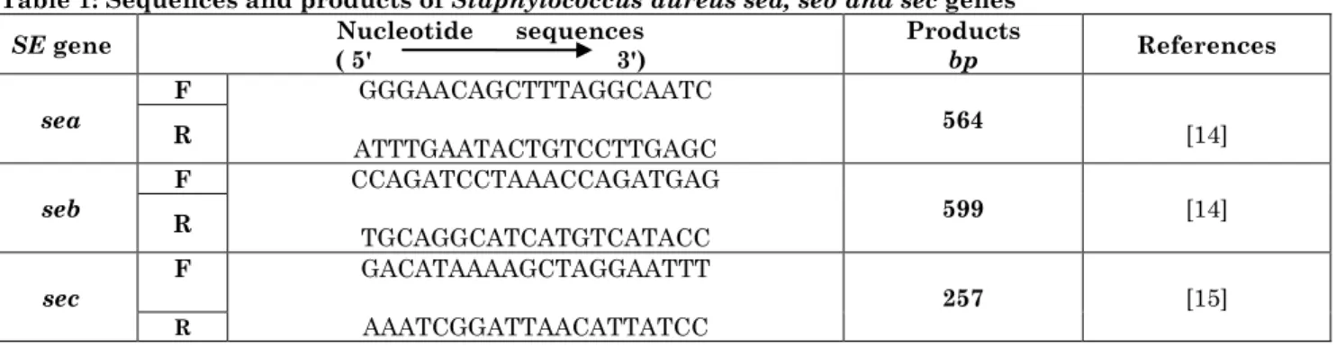
Table 1: Sequences and products of Staphylococcus aureussea, seb and sec genes
PCR amplification of enterotoxin genes was performed in the (Eppendorf, UK) Thermal Cycler under following program: 1 cycle with 94 °C 5 min. for initial denaturation, 30 cycles for denaturation, annealing and extension with 94 °C 2 min, 50 °C 2 min. & 72 °C 1min respectively and finally 72 °C 5 min for final extension [16].The PCR products were analyzed by (1%) agarose gel electrophoresis [17].
Statistical Analysis
The statistical data were analyzed by statistical package (SPSS) ver. (22.0) , Chi-square exact test was used to measure significant associations between the frequency of Staphylococcus aureus in nasal carriage of MS patients and healthy nasal carriers with enterotoxin genes distribution.
P values less than 0.05 were considered statistically significant.
Results
Staphylococcus aureus Colonization and Multiple Sclerosis
Out of 100 nasal swab samples collected from MS patients, 81 (81%) were positive for
S.aureus whereas from 100 nasal swab
samples collected from healthy controls, there were only 12(12%) positive for
S.aureus. Consequently MS patients were
most commonly colonized group in this study and there was a statistically significant differences between S. aureus colonization in healthy carriers and MS disease at (p = 0.00).On the other hand the relapsing remitting MS patients were most commonly colonized with S.aureus 50(61.8%) than newly diagnosed MS group 31(38.2%) with a p-value of (0.092), statistically non significant association between them (Table 2).
Table 2: distribution of Staphylococcus aureus in study groups
Study groups Samples NO. S.aureus positive P-value
NO. %
Non-MS group 100 12 12 0.00 (HS)*
Total MS patients 100 81 81
New diagnosed MS group 50 31 38.2
100 0.092 (NS)**
MS exacerbated group 50 50 61.8
Total groups 200 93 _
* Highly significant; ** non significant
Demographical Distribution and
S.aureus Nasal Carriage
According to gender distribution, the ratio of male to female was (1:1.7) includes 64(64%) of patients were female and 36 (36%) were male while in non-MS group there were 54(54%) female and 46(46%) male. In MS patients, a total of S.aurus positive was
81(81%) including 52(52%) female and 29(29%) male, while in control non-MS group, out of 12(12%) S.aureus positive there were 7(7%) female and 5(5%) male, statistically highly significant differences was observed between S.aureus colonization and gender distribution (P value= 0.00) as shown in (Table 3).
Table 3: Distribution S.aureus according to gender
Gender
Multiple sclerosis group Non-MS control group
S.aureus S.aureus
(+ve) (-ve) (+ve) (-ve)
Male No (%) 29(29%) 7(7%) 5(5%) 41(41%) 0.0 (HS) *
Female No (%) 52(52%) 12(12%) 7(7%) 47(47%) 0.0 (HS)
SE gene Nucleotide sequences
( 5' 3')
Products
bp References
sea
F GGGAACAGCTTTAGGCAATC
ATTTGAATACTGTCCTTGAGC 564 [14]
R
seb F CCAGATCCTAAACCAGATGAG
TGCAGGCATCATGTCATACC 599 [14]
R
sec
F GACATAAAAGCTAGGAATTT
AAATCGGATTAACATTATCC 257 [15]
Total No (%) 81(81%) 19(19%) 12(12%) 88(88%) 0.0 (HS)
100(100%) 100(100%)
* Highly significant
Antimicrobial Sensitivity Pattern
A total of 19 types of antibiotics were tested against S.aureus isolates in all study groups according to CLSI. MS patient groups were more resistant to antibiotics (53.3%) than control group (22.8%).Furthermore, within
MS group, the exacerbated MS showed more resistance (60.6%) than newly diagnosed patients (46.3%). In all study groups there is complete resistance to Methicillin (100%) where as the lowest resistance (3%) were reported against Meropenem and Imipenem as shown in (Table 4).
Table 4: Antibiotic resistance pattern of S.aureus isolates.
ANTIBIOTICS
Multiple sclerosis patients Non-MS
New Dx. MS R.R. MS. Total
No.(%) No.(%) No.(%) No.(%)
Methicillin ME 31(100%) 50(100%) 81(100%) 12(100%)
Vancomycin [VA] 3(9.6%) 5(10%) 8(10%) 0
Azithromycin[AZM] 18(58%) 43(86%) 61(75.3%) 0
Erythromycin [E] 22(71%) 45(90%) 67(82.7%) 8(66.6%)
Tetracycline [TE] 18(58%) 41(82%) 59(72.8%) 4(33.4%)
Amikacin [AK] 17(54.8%) 33(66%) 50(61.8%) 0
Rifampin[RA] 9(29%) 20(40%) 29(35.8%) 0
Penicillin-G [P] 21(67.7%) 47(94%) 68(84%) 12(100%)
Meropenem [MEM] 1(3.3%) 3(6%) 4(5%) 0
Sulfa/Trimthoprim [SXT] 20(64.5%) 45(90%) 65(80.2%) 0
Doxycycline [DOX] 8(25.8%) 26(52%) 34(42%) 0
Ciprofloxacin [CIP] 8(25.8%) 19(38%) 27(33.4%) 0
Clindamycin [DA] 9(29%) 24(48%) 33(40.8%) 0
Imipenem [IPM] 1(3.3%) 2(4%) 3(3.7%) 0
Cefotaxime [CTX] 10(32.3%) 24(48%) 34(42%) 0
Gentamicin [CN] 18(58%) 28(56%) 46(56.8%) 0
Ceftriaxone [CRO] 27(87%) 49(98%) 76(93.8%) 8(66.6%)
Ampicillin [AM] 23(74.2%) 48(96%) 71(87.7%) 8(66.6%)
Chloramphenicol [C] 9(29%) 24(48%) 33(40.8%) 0
Total 46.3% 53.3% 60.6% 53.3% 22.8%
Conventional PCR Screening for
Enterotoxin Genes
By PCR essay, the amplification of sequences of seA, seB and seC genes were
produced an amplicon of size 564 bp, 599 bp and 257 bp respectively.Figure (1, 2 and 3).
Figure 1: Gel electrophoresis of amplified PCR product (564 bp) for sea gene. Lane 1: 100 bp ladder. Lane 2: Negative control; Lanes 3– 10: Clinical isolates showing positive result. (1 % agarose, 7 v/cm2, 1
Figure 2: Gel electrophoresis of amplified PCR product (599bp) for seb gene. Lane 1: 100 bp ladder. Lanes 2 – 9 : Clinical isolates showing positive result. Lane 10: Negative control, (1% agarose, 7 v/cm2, 1
hr)
Figure 3: Gel electrophoresis of amplified PCR product (257 bp) for sec gene. Lane 1: 100 bp ladder. Lanes 2 – 4 : Clinical isolates showing positive result. Lane 5: Negative control (1 % agarose, 7 v/cm2, 1
hr)
The current study reported that, among MS patients the most commonly detected enterotoxin genes were seA 56(69.1%) followed by seB 40(49.4%) while the least prevalent enterotoxin was seC 7(8.6%). Statistically, there was a highly significant differences between the distribution of sea
and seb (P=0.00) in MS patient compared with healthy control but non-significant distribution were found in seC (P = 0.138).From overall S.aureus positive in MS groups the distribution of enterotoxins were
36(72%) of se A, 25(50%) of se B & only 4(8%) of se C in relapsing remitting group compared with newly diagnosed MS group that showed 20(64.5%), 15(48.4%) and only 3(9.7%) of se A, se B and se C respectively, whereas no enterotoxin genes were detected in non-MS control group . Significant differences were seen in the distribution of
seA , se B and se C gene between MS groups
(P=0.00002 ) compared with controls, (Table 5).
Table 5: Distribution of S.aureus enterotoxin genes
Study groups S.aureus
positive Gene distribution P-value
sea seb sec Total
No.(%) No.(%) No.(%) No.(%) No.(%)
Control 12(12) 0(0) 0(0) 0(0) 0(0)
0.00002 (HS)*
Newly MS 31(38.2) 20(64.5) 15(48.4) 3(9.7) 38(40.8)
R.R. MS 50(61.8) 36(72) 25(50) 4(8) 65(43.3)
Total 93 56(69.1) 40(49.4) 7(8.6) 103(42.4)
P-value 0.0
(HS)
0.0 (HS)
0.138 (NS)**
0.00002 (HS )
* Highly significant; ** non significant
Discussion
Multiple sclerosis is a multifactorial disease with unknown origin but environmental agents like bacterial and viral infections more participate. Staphylococcus aureus
superantigens which is considered the most
triggering agent of autoimmunity and included in several autoimmune diseases like multiple sclerosis and rheumatoid arthritis [18]. In multiple sclerosis the presence of
S.aureus enterotoxins in nasal carriage
factors for induction of autoreactive CD4+ T cells particularly in the myelin protein of CNS hence activation large number of cytokines that finally have a major role in MS severity and development [6,9]. In the present study the frequency of S.aureus nasal colonization in MS patients (81%), particularly in MS exacerbated group was high (61.8%) compared with non-MS healthy carriers which was only(12%) with highly significant differences (P=0.00 ), this may be due to more contact of these patients with healthcare settings although all MS patients were taken immune activation drugs that enhance immunity which indicates that the nasal bacterial colonization with enterotoxigenic S. aureus in MS patients was much more virulent and pathogenic than in non-MS group [8].
The current result in accordance with other study in Iran by Pakbas et al who reported that the nasal carriage rate of S.aureus in MS patiens & control group was (42%) and (23.3%) respectively [14]. Alternatively a study was conducted in Canada by Mulvey et al. revealed a lower prevalence of S.aureus in nasal carriage of MS patients (27%) and relatively higher in non-MS group (30%) [9].Regarding gender, this study observed that the frequency of S.aureus isolates in MS patients were 29% in males and 52% in females while in control group the percentage
of S.aureus was 5% males and 7% females.
Concerning MS patients the result of this study coincide with Sadeghi et al.,2019,who reported that the frequency of S.aureus
isolates in MS patients were 23% in males and 76% in females but this result disagreed with the same study [8] that reported the colonization of S.aureus in healthy nasal carriers was 56% males and 43% females. Globally S.aureus shows high resistance rate to several antibiotics.
The present study revealed that the S.aureus
isolates in MS patients was (53.3%) multi-drug resistance especially in MS exacerbated group (60.6%) than newly diagnosed (46.3%) and control group (22.8%) with highly significant differences between MS patients & non-MS group regarded to resistance pattern. In this study S.aureus isolates in all groups were resistant to methicillin (ME)(100%) indicates that MRSA strains were also distributed in nasal carriage of healthy control [19], as well as there is a high percentage of resistance to ceftriaxone
(93.8%), ampicillin (87.7%), penicillin-G (84%), erythromycin (82.7 %), sulphamethoxazole/ trimethoprim (80.2 %) and azithromycin (75.3%), while in control group in addition to methicillin resistance,
S.aureus isolates showed high resistance rate
only to penicillin-G(100%), indicating that all strains of S.aureus isolates in MS patients were resistant strains and responsible for multiple drug resistance (MDR) which leads to increase severity of multiple sclerosis(MS), while the S.aureus strain was different in control group in term of limited antibiotic resistance results and so non virulent. This result agreed with Mehrabi et al (2015) [16].
Who reported that resistant S.aureus strains in MS patients was considered as a risk factor for MS exacerbation with high level of resistance,against,tetracycline(80%),ampicilli n(72.22%),methicillin(66.66%),erythromycin( 66.66%),oxacillin(63.33%)and,sulphamethoxa zole/trimethoprim(61.11%), while more than (90 %) of isolates in all study groups were susceptible to vancomycin , meropenem & imipenem which is provided a good regimen for decolonization of S .aureus in nasal carriage of MS patients in order to prevent severity or exacerbation [20].
To the best of our knowledge, it is believed that, it is the first study in Iraq concerning three enterotoxins of S.aureus (sea, seb &sec)
identified in MS patients. The prevalence of
s.aureus enterotoxin genes (sea, seb, sec) was
higher in MS patients compared with control group that present no any enterotoxin genes. Statistically significant differences was observed between S.aureus harboring enterotoxins in MS patients and Non-MS Carriers (P= 0.00).
Among MS patients the most common prevalent enterotoxins were sea (69.1%) particularly sea in the relapsing remitting group was (72%) and in newly diagnosed MS was (64.5%). In addition the second most common prevalent enterotoxin in MS patients was seb (49.4%) with (48.4%) & (50%) in newly diagnosed & R.R. MS patients respectively ,while the least common enterotoxins distributed in MS patients was
sec (8.6%) with (9.7%) & (8%) in newly diagnosed & R.R. MS patients respectively.
significant correlation between sec & MS exacerbation (P= 0.138). Similarly other study results by Sadeghi et al. reported that
sea was the most common frequent (40%) enterotoxins in MS patients than in nasal carriers (23%) [8], all these results indicate that healthy individuals was carrying nonpathogenic strains of S.aureus in nasal passages that considered as a normal commensal flora fight by competing against pathogens , in contrast to S.aureus strains in MS patients were have many virulence factors like enterotoxin genes, more MDR particularly in a stage of relapsing remitting or exacerbated MS that responsible for causing increase sequel, severity, development and pathogenesis of this autoimmune disease.
Conclusion
The nasal carriage of S.aureus in MS patients particularly in relapsing remitting group was very high compared with healthy individuals that mean the potential risk of
S.aureus colonization in MS patients was
high. High frequency of S.aureus harboring enterotoxin genes particularly (seA) support the evidence that seA play a very significant role in the development and severity of MS. Decolonization regimen of S.aureus in nasal carriage of MS. Patients with more effective antibiotics like imipenem to prevent severity or exacerbation of the MS disease.
References
1. Chabas D, Fontaine B, Lyon-Caen O (2004) Multiple sclerosis review.Orphanet Encyclopedia, 1-6 .
2. Goldenberg MM (2012) Multiple Sclerosis Review. P T, 37(3):175-84.
3. Libbey JE, Cusick MF, Fujinami RS (2014) Role of pathogens in multiple sclerosis. International Reviews Of Immunology, 33(4) 33(4):266-83. DOI: 10.3109/08830185.2013.823422.
4. Chowdhary VR, Tilahun AY, Clark CR, Grande JP, Rajagopalan G (2012) Chronic exposure to staphylococcal superantigen elicits a systemic inflammatory disease mimicking lupus. J. Immunol., 189(4):2054-62.
5. Proft T, FRASER JD (2003) Review Bacterial superantigens. Clin Exp Immunol 2003; 133:299-306. doi: 10.1046/j.1365-2249.2003.02203.x 6. Torres BA, Kominsky S, Perrin GQ,
Hobeika AC, Johnson HM (2001) Superantigens: the good, the bad, and the ugly. Exp. Biol. Med., 226(3):164-76. 7. Xu SX, McCormick JK (2012)
Staphylococcal superantigens in colonization and disease. Front Cell Infect Microbiol, 2 (52): 1-11. DOI: 10.3389/fcimb.2012.00052
8. Sadeghi J, Alizadeh N, Oskoueia MA, Laghusi D, Oskoueid DS et al (2019) Frequency of superantigen encoding genes
of Staphylococcus aureus isolates collected
from multiple sclerosis (MS) patients and
nasal carriers, Microbial Pathogenesis, 127:316-319.
doi.org/10.1016/j.micpath.2018.12.010 9. Mulvey MR, Doupe M, Prout M, Leong C,
Hizon R, Grossberndt A, Klowak M, Gupta A, Melanson M, Gomori A, Esfahani F, Klassen L, Frost EE, Namaka M (2011) Staphylococcus aureus harbouring Enterotoxin A as a possible risk factor for multiple sclerosis exacerbations . Mult. Scler J., 17(4): 397-403. doi: 10.1177/1352458510391343
10.Schiffenbauer J, Johnson HM, Butfiloski EJ, Wegrzyn L, Soos JM (1993) Staphylococcal enterotoxins can reactivate experimental allergic encephalomyelitis. Proc. Natl. Acad Sci. U S A 90(18): 8543-6. 11.Shimonkevitz R, Colburn C, Burnham JA, Murray RS, Kotzin BL (1993) Clonal expansions of activated gamma/delta T cells in recent-onset multiple sclerosis. Proc. Natl. Acad Sci., 90(3):923-7.
12. Alwan AH, Talak MA (2015) Isolation and characterization of Staphylococcus aureus in spoiled food samples. Int. J. Curr. Microbiol App. Sci., 4(3): 645-651. 13.Hindler JA, Schuetz AN (2019) CLSI AST
News Update, 4(1):1-21.
15.Johnson W M, Tyler S D, Ewan E P, Ashton F E, Pollard D R, Rozee KR (1991) Detection of genes for enterotoxins, exfoliative toxins, and toxic shock syndrome toxin 1 in staphylococcus aureus
by the polymerase chain reaction. Journal of Clinical Microbiology, 29(3): 426-430. 16.Mehrabi F, Asgari A (2015) Resistant
Strains of Enterotoxigenic Staphylococcus aureus; Unknown Risk for Multiple Sclerosis Exacerbation. Iran Red Crescent Med J., 17(9):1-7. DOI: 10.5812/ircmj.12596
17.Oelkers P (2016) Molecular Biology Laboratory Manual.
18.Li J, Yang J, Lu YW, Wu S, Wang MR, Zhu JM (2015) Possible Role of Staphylococcal Enterotoxin B in the
Pathogenesis of Autoimmune Diseases. Viral Immunol., 28(7):354-9.
19.Dağı HT, Fındık D, Demirel G, Arslan U (2015) Detection of Methicillin Resistance and Various Virulence Factors in Staphylococcus aureus Strains Isolated from Nasal Carriers. Balkan Med. J.,
32(2):171-5. doi:
10.5152/balkanmedj.2015.150186.