Original Research Article
Comparative study between over underlay with classical underlay
techniques of tympanoplasty
Prashanth Kudure Basavaraj, Manjunatha H. Anandappa*, Veena Prabhakaran,
Nishtha Sharma, Shreyas Karkala
INTRODUCTION
Over the period of time, tympanoplasty has undergone notable changes. Overlay and underlay tympanoplasties are the two classical techniques.1 In underlay technique,
graft is placed medial to the handle of malleus and medial to the remaining ear drum.2 This technique of
tympanoplasty is ideal for the closure of small, posterior quadrant perforations; prevents lateralization and blunting, and is technically less challenging with good
success rate.2 In overlay technique, the graft is placed
lateral to the handle of malleus and lateral to the annulus after careful removal of the outer epithelial layer. It provides magnificent view of intra operative and post-operative anterior meatal angle, preserves middle ear space and is suitable for all types of perforated eardrum.
But each of these techniques has its own pitfalls. While lateralization, blunting of anterior meatal recess, and development of squamous epidermal inclusion cyst are
ABSTRACT
Background: The objective of the study was to compare the over underlay tympanoplasty technique with classical underlay tympanoplasty in terms of hearing impairment, graft acceptance and complications.
Methods: 60 patients of chronic otitis media, mucosal, inactive, aged between 16-60 years who presented to ENT OPD with small, medium, large and subtotal perforations having mild to moderate conductive hearing loss were included in the study. After taking informed consent, patients were randomly divided into 2 groups containing 30 patients each. In group A, graft was placed medial to the handle of malleus and medial to the annulus (underlay technique), while in group B, graft was placed lateral to the handle of malleus and medial to the annulus (over underlay technique). Both groups were reviewed after 6 months. Pre-operative and post-operative air bone gap were compared. Surgery was considered successful based on post-operative graft uptake, hearing improvement and maintenance of middle ear space.
Results: In group A, perforation was seen in 8 cases (26.7%) whereas only 3 cases (10%) in group B had re-perforation. Medialization was noted among 4 patients in group A (13.3%), and was absent in group B. Lateralization was absent in both the groups. Post-operative hearing threshold in group A was 6.2±4.56 dB and in group B was 11.45±7.38 dB.
Conclusions: Over underlay tympanoplasty is a safer technique as compared to classical underlay, showing lower rates of re-perforation or medialization and a significant improvement in hearing. Hence over-underlay is an effective method, having higher success rates.
Keywords: Tympanoplasty, Underlay, Over underlay
Department of ENT, JJM Medical College, Davangere, Karnataka, India
Received: 12 February 2020 Revised: 22 March 2020 Accepted: 27 March 2020
*Correspondence:
Dr. Manjunatha H. Anandappa, E-mail: [email protected]
Copyright: © the author(s), publisher and licensee Medip Academy. This is an open-access article distributed under the terms of the Creative Commons Attribution Non-Commercial License, which permits unrestricted non-commercial use, distribution, and reproduction in any medium, provided the original work is properly cited.
some of the disadvantages of overlay technique; on the other hand, adhesions, medialization and higher failure rate in large perforations are the main demerits of underlay technique.3 Additionally, classical underlay is
difficult to perform if handle of malleus is medialized.
The development of a novel method namely ‘over underlay’ reduces the pitfalls of underlay and overlay techniques.4 Here, the graft is placed lateral to the
manubrium of the malleus and under the remnant of tympanic membrane and annulus.5
Hence, we have undertaken this study to evaluate the outcomes and highlight the advantages of this new technique of ‘over underlay’ as opposed to the classical underlay approach of tympanoplasty.
Objectives
To compare two methods of tympanoplasty, i.e., over underlay and classical underlay for assessing improvement in hearing, graft acceptance, maintenance of middle ear space and incidence of medialization or lateralization post-operatively.
METHODS
A randomized prospective study was carried out from October 2018 to November 2019, which included 60 patients who presented to the OPD of Department of ENT at Chigateri District Hospital and Bapuji Hospital, the teaching hospitals affiliated to JJM Medical College, Davangere, Karnataka. Those fulfilling the inclusion and exclusion criteria were selected for the study.
Inclusion criteria
Inclusion criteria includes pateints age group between 16-60 years, patients having small, medium, large and subtotal perforations, those with COM, mucosal type and with dry ear for at least 4 weeks before operation and patients with mild and moderate conductive hearing loss.
Exclusion criteria
Exclusion criteria includes patients with age <16 and >60 years, with COM, squamosal type, those actively discharging ear and those patients’ COM with mixed hearing loss and hearing loss more than 60db and with total perforation.
After obtaining written consent and ethical committee approval, patients were randomly divided into two groups by envelope method. We did underlay technique for Group A and over-underlay for Group B. Intra-operatively, temporalis fascia was placed medial to the handle of malleus and medial to the annulus in group A. In group B, temporalis fascia was placed lateral to the manubrium of malleus and under the remnant of tympanic membrane and annulus.
Follow up was done after 6 months. Pre-operative and post-operative pure tone audiometry values i.e., air-bone (A-B) gap were compared and evaluated. Surgery was considered successful based on postoperative graft uptake, hearing improvement and maintenance of middle ear space with less complications.
The results were analyzed using chi square test, wherep value of <0.05 was considered significant.
Figure 1:Underlay technique of tympanoplasty - graft is placed medial to the handle of malleus and medial
to the remnant of tympanic membrane.
Figure 2: Over underlay technique of tympanoplasty-graft is placed medial to the handle of malleus and
lateral to the annulus or remnant of tympanic membrane.
RESULTS
The study sample consisting of sixty patients were divided randomly into two separate groups by envelope method. Each group contained 30 patients of either sex.
between 20-54 years and in case of Group B, it was between 17-54 years.
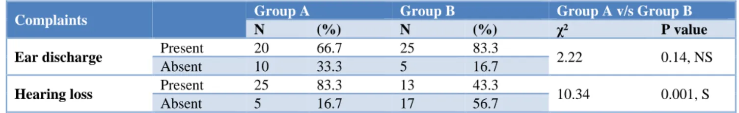
Ear discharge (Group A-66.7%, Group B-83.3%) and hearing loss (Group A-83.3%, Group B-43.3%) were the most common complaints of the patients (Table 2).
Tympanic membrane perforations were classified into four types based on the surface area of pars tensa of the tympanic membrane deficient, namely small (<25%), medium (25-50%), large (50-75%) and subtotal (>75%) (Figure 3).
Table 1: Descriptive information about the patients.
No. of cases
Group A Group B Group A
v/s Group B
30 30
Age (years)
Mean
±SD 33.3±9.6 30.9±9.7
t=0.94, p=0.35,
NS Range 20-54 yrs 17-54 yrs
Sex
Male 13 14 χ²=0.07, p=0.80,
NS
Female 17 16
Most of the patients had a large central perforation (7 from each group=23.3%). In Group A, thirteen patients (43.3%) suffered from left ear chronic otitis media, eleven (36.7%) patients had right ear chronic otitis media and six (20%) patients had bilateral chronic otitis media. In case of Group B, eleven (36.7%) patients suffered from left ear chronic mucosal disease, sixteen (53.3%) patients suffered from right chronic otitis media and 3 (10%) patients suffered from bilateral chronic mucosal infection of ear (Table 3).
Pre-operative and post-operative decibels of hearing loss were assessed. Hearing loss was classified into mild (25-40 dB) and moderate (41-55 dB) based on the pure tone audiometry values. Mean pre-operative value of hearing
loss was 35.56±6.82 dB in Group A, whereas in Group B it was 37.04±9.51 dB (Table 4). Post-operative hearing improvement (A-B gap) in both groups was assessed after 6 months. Mean A-B gap improvement in group A was noted as 6.22±4.56 dB (p≤0.001, highly significant) and was 11.45±7.38 dB in Group B (p≤0.001, highly significant).
Figure 3: Graph showing the classification of perforation based on the size of perforation.
STP=subtotal perforation, SPS=small posterior-superior perforation, SPI=small posterior-inferior perforation, SP=small posterior perforation, SAS=small anterio -superior perforation, SAI=small anterio=inferior perforation, MP=medium sized perforation in posterior quadrant, MA=medium sized perforation in anterior quadrant, MC -medium central perforation, LC=large central perforation.
Table 3: Shows the clinical diagnosis in both groups.
Com mucosal Gr A Gr B
N (%) N (%)
Left 13 43.3 11 36.7
Right 11 36.7 16 53.3
B/L 6 20.0 3 10.0
Total 30 100.0 30 100.0
χ²=2.09, p=0.35, NS.
Table 2: Describes about the complaints of the patients.
Complaints Group A Group B Group A v/s Group B
N (%) N (%) χ² P value
Ear discharge Present 20 66.7 25 83.3 2.22 0.14, NS
Absent 10 33.3 5 16.7
Hearing loss Present 25 83.3 13 43.3 10.34 0.001, S
Absent 5 16.7 17 56.7
Table 4: Comparison of pre-operative and post-operative hearing improvements in both groups.
Groups PTA (decibels) t P value
Pre-op Post-op 3M Mean difference
Gr A 35.56±6.82 29.34±6.25 6.22±4.56 7.46 <0.001, HS
Gr B 37.04±9.51 25.59±3.89 11.45±7.38 8.50 <0.001, HS
Gr A v/s Gr B
t 0.69 2.79 3.30
-
p value 0.49, NS 0.007, S 0.002, S
While comparing the complications, re-perforation (Group A=26.7% and group B=10%) and medialization (Group A=13.3%, no cases in Group B) were more in those who underwent underlay technique. Lateralization was not found in both groups (Figure 4).
Figure 4: Graph showing the post-operative complications in both the groups.
DISCUSSION
Chronic otitis media is defined as the prolonged infection of middle ear cavity which leads to perforation of tympanic membrane, persistent ear discharge, conductive hearing loss and other complications. It was considered as the most dangerous condition in ancient period. The development of antibiotics along with the improvement of knowledge about tympanic membrane helped in understanding the disease in a better way, which facilitated more effective treatment of chronic otitis media.6
In 1953, Zollner et al developed a new surgical method called tympanoplasty, which is described as a reconstructive surgery which helps to improve hearing function of the patient and to maintain a dry ear.7 It is
considered as the final surgical step for the treatment of conductive hearing loss and persistant otorrhoea caused by chronic otitis media. Various factors, such as type of graft, disease activity, eustachian tube function, surgical approach, and technique of graft placement will affect the surgical outcome.
There are several types of tympanoplasty techniques emerged over a period of time such as overlay tympanoplasty, sandwich graft tympanoplasty, loop overlay tympanoplasty, classical underlay tympanoplasty, over underlay tympanoplasty etc. out of which classical underlay and overlay are the two techniques which are used commonly. Surgeon may face many post-operative complications in overlay method, such as lateralization, formation of fibrous tissue antero-superiorly, epithelial cyst formation etc. In case of underlay technique, medial graft placement can lead to reduction of middle ear space
which leads to adhesion to the promontory resulting in graft failure.
The evolution of the new technique i.e., over underlay helps to create a movable tympanic membrane, with healthy middle ear mucosa. It also reduces the complications of the aforementioned classical techniques.
Panchal et al conducted a study to evaluate and compare the results of over underlay technique with conventional underlay myringoplasty in 40 patients aged between 15-50 years.4 Most of the patients in their study were in the
age group between 15-25 years with male predominance.4
Our study, conducted with a larger sample size of 60 patients, had a similar age predisposition, with majority of cases belonging to ages between 17-54. Although, the current study has no significant difference in the sex ratio. The predominant complaint in both studies was ear discharge along with decreased hearing.
Another study, done by Aslam et al, classified tympanic membrane defects into small, medium, large and subtotal, based on the size of the perforation.8 Most of the
perforations in both the cohort groups were medium sized.8
The present study also classified perforations similarly, with a greater number of them being large central. No significant change in the post-operative results were observed in our study based on size of perforation, a factor which was not weighed in by the above-mentioned study.
Prakash et al conducted a randomized prospective study comparing over-underlay and classical underlay techniques of tympanoplasty for a period of one year.5
They compared pre-operative A-B gap with post-operative value. Pre-post-operative A-B gap in group which underwent underlay technique was 22.63 (±9.25),while the group which had over-underlay graft placement had a mean A-B gap of 26.47 (±11.93).5 Post-operative value
after 6 months in the first group was 4.78 (±4.47), and that of the second group was 8.50 (±7.38).5 P value of
both the groups was found to be statistically significant (<0.05).5
The current study conducted by us also made similar comparisons in A-B gap, and elicited drastic pre-operative and post-pre-operative gain of hearing, which were statistical considered highly significant in both groups, with a p value of <0.001.
Yigit et al undertook a retrospective short term evaluation of over-underlay myingoplasty technique with 104 patients.6 Post-operative complications of both underlay
in over underlay technique.6 No graft lateralization or
blunting seen in this group.6
Findings of our study were in agreement to the above-mentioned study, indicating lesser incidence of re-perforation in over-underlay technique when compared to classical underlay technique of tympanoplasty. No medialization and lateralization were found in either group, with highly significant hearing improvement.
CONCLUSION
Tympanoplasty helps to improve the objective as well as subjective well-being of individuals, by improving hearing thresholds and by preventing otorrhoea. Our study concludes that over-underlay tympanoplasty is a safer alternative technique to classical underlay, showing lower rates of reperforation or medialization and a significant improvement in hearing, especially in large subtotal perforations, and where handle of malleus is medialized due to the disease process. Hence, we conclude that over-underlay is an effective method, producing higher success rate thereby rendering enhanced quality of life.
ACKNOWLEDGEMENTS
We are thankful to the department of ENT, JJM medical college, Davangere, and Chigateri district hospital, Davangere for their support.
Funding: No funding sources Conflict of interest: None declared
Ethical approval: The study was approved by the Institutional Ethics Committee(JJMMC/IEC-Sy-72-2018)
REFERENCES
1. Sergi B, Galli J, Corso DE, Parrilla C, Paludetti G. Overlay versus underlay myringoplasty: report of
outcomes considering closure of perforation and hearing function. Acta Otorhinolaryngol Ital. 2011;31(6):366-71.
2. Kartush JM, Mechaelides EM, Becvarovski Z, Rouere LMJ. Over-under tympanoplasty. Laryngoscope. 2002;112(5):802-7.
3. Indorewala S, Adedeji TO, Indorewala A, Nemade G. Tympanoplasty outcomes: a review of 789 cases. Iran J Otorhinolaryngol. 2015;27(79):101-8. 4. Panchal V, Joginder SG, Sharad H, Bhushan K,
Kaintura M. To evaluate and compare the results of over-underlay graft technique with conventional underlay myringoplasty. Ind J Otol. 2015;21:274-9. 5. Prakash MD, Viswanatha B, Kaur J, Sanyal S.
Comparative Study of the Underlay and Over-Underlay Techniques of Tympanoplasty in Perforations of the Tympanic Membrane. Res Otolaryngol. 2014;3(5):65-69.
6. Yigit, Ozgur, Alkan, Seyhan, Topuz, Ebru, et al. Short-term evaluation of over-under myringoplasty technique. European archives of oto-rhino-laryngology. Official J of the European Federation of Oto-Rhino-Laryngological Societies (EUFOS): affiliated with the German Society for Oto-Rhino-Laryngol Head Neck Surg. 2005;262:400-3. 7. Sarkar S. A review on the history of tympanoplasty.
Indian J Otolaryngol Head Neck Surg. 2013;65(3):455-60.
8. Aslam MA, Aslam MJ. Comparison of over-underlay and over-underlay techniques of myringoplasty. Pak Armed Forces Med J. 2009;3.
Cite this article as: Basavaraj PK, Anandappa MH, Prabhakaran V, Sharma N, Karkala S. Comparative study between over underlay with classical underlay techniques of tympanoplasty. Int J Otorhinolaryngol Head Neck Surg 2020;6:918-22.