O R I G I N A L A R T I C L E
Open Access
Rapid identification and detection of
Vibrio
parahaemolyticus
via different types of
modus operandi with LAMP method in vivo
Jun Li
1†, Jianfei Liu
2†, Kaifei Fu
2, Kewei Qin
2, Chenglin Wu
2, Xiaojie Yu
2, Shan Zhou
1*and Lijun Zhou
2*Abstract
Purpose:Vibrio parahaemolyticus, an easy-ignored food-borne pathogen, can cause bacterial outbreaks and human
disease during early-stage infection. In this study, we aimed to evaluate the detection efficiency of loop-mediated isothermal amplification (LAMP) as an emerging technique to directly detectV. parahaemolyticusinfection in mammalian hosts and assess its potential in clinical applications.
Methods:A LAMP assay was used for rapid identification ofV. parahaemolyticusin a variety of mouse models in which animals were infected via the digestive tract, wounds, or through general infection, and the results were compared with routine analytical methods.
Results:Our results confirmed that the LAMP assay was capable of detectingV. parahaemolyticusin different mouse organs independent of the source of bacteria, although its sensitivity depended on the route of infection and the organ affected. Foodborne-derivedV. parahaemolyticuswas the most sensitive route, with the small intestine being the most sensitive organ. The LAMP assay indicated thatV. parahaemolyticusthat spread through the blood stream had the most serious consequences during early-stage infection. Positive LAMP results were identified in all blood samples from i.v. injected mice. Furthermore, the LAMP method could directly detect trace quantities ofV. parahaemolyticusin fresh peripheral blood while conventional methods failed to do so, thereby shortening the time-to-result from days to minutes.
Conclusions:In this study, we demonstrated that the LAMP assay was effective in speeding up the detection ofV.
parahaemolyticus. Instead of being a secondary method to assist in the clinic, the LAMP assay has potential for use as the
primary technique for rapid detection ofV. parahaemolyticusin the future.
Keywords:Detection in vivo, LAMP, Mouse model,Vibrio parahaemolyticus
Background
Vibrio parahaemolyticusis a common gram-negative (G−) halophilic bacterial species that is principally distributed within sea water (DePaola et al.1990), coastal areas, and river-sea junctures (Yang et al.2017a). It can also be found in seafood (Twedt1989; Rince et al.2018). In addition to
its presence in marine environments,V. parahaemolyticus
can also be found in livestock and poultry meat,
fresh-water fish, preserved eggs, and pickles (Barker 1974).
Therefore, despite the sources of marine infection, such as seafood and marine salts, a variety of animal species can
also be carriers of V. parahaemolyticus, rendering it an
important pathogenic bacterium that is highly infectious.
Human infection with V. parahaemolyticus is an
im-portant public health problem (Baker-Austin et al. 2018). V. parahaemolyticus is the principal pathogen causing acute gastroenteritis in humans via consumption of infected seafood, for which the symptoms include
stomachache (Liu et al. 2015), diarrhea (Gong et al.
© The Author(s). 2020Open AccessThis article is licensed under a Creative Commons Attribution 4.0 International License, which permits use, sharing, adaptation, distribution and reproduction in any medium or format, as long as you give appropriate credit to the original author(s) and the source, provide a link to the Creative Commons licence, and indicate if changes were made. The images or other third party material in this article are included in the article's Creative Commons licence, unless indicated otherwise in a credit line to the material. If material is not included in the article's Creative Commons licence and your intended use is not permitted by statutory regulation or exceeds the permitted use, you will need to obtain permission directly from the copyright holder. To view a copy of this licence, visithttp://creativecommons.org/licenses/by/4.0/. * Correspondence:[email protected];[email protected]
†Jun Li and Jianfei Liu contributed equally to this work.
1Department of Medical Service, The Sixth Medical Centre, Chinese PLA (People’s Liberation Army) General Hospital, Beijing, People’s Republic of China
2018), vomiting, dehydration, chills, and fever (Raszl et al.2016). In the USA,V. parahaemolyticusis the lead-ing cause of human gastroenteritis and associated with
seafood consumption (Park et al.2018). In coastal Asian
countries, including Indonesia, V. parahaemolyticus is
the leading cause of bacterial foodborne infections (Les-mana et al.2001; Lee et al.2018). In Japan, food
poison-ing caused byV. parahaemolyticus accounts for 20~30%
of all cases of human food poisoning (Alam et al.2002).
In Europe and North America, many outbreaks of V.
parahaemolyticus food poisoning have been reported
(McLaughlin et al. 2005; Martinez-Urtaza et al. 2013;
Martinez-Urtaza et al. 2016). Coincidentally, over the
past two decades, published reports have indicated that V. parahaemolyticushas been the principal cause of bac-terial foodborne infections in China (Li et al. 2014). Ac-cording to the continuously monitoring data from 2013 mainland China from the National Foodborne Disease
Outbreaks Surveillance System (NFDOSS), V.
parahae-molyticuswas the leading factor causing 27.8% of all the microbial-driven foodborne disease outbreaks, making it the only marine bacterial species among the 5 most common pathogens, and the 5th most common species found in food (Li et al.2018).
In southern China, there is a strong seasonal pattern
in the outbreaks of human infection byV.
parahaemoly-ticus, which partially attributes to the different carriage rate of marine products throughout the year. In a study by Yang et al., investigating 504 seafood samples from 11 provinces of China, it was shown that the prevalence
of V. parahaemolyticus peaked during the summer
(33%) and reduced by half during the winter (14%) in
fish, oyster, and shrimp (Yang et al.2017b). The disease
caused by V. parahaemolyticus can spread widely but
with generally mild symptoms, which are usually
self-limiting (Rizvi and Bej2010), possibly causing people to
pay little attention to it. Due to symptoms resulting from V. parahaemolyticus, infections similar to that by other common food-borne pathogens make the prevention of V. parahaemolyticusinfection and its early-stage medical treatment easily be neglected.
With the development of technology, methods of V.
parahaemolyticusdetection have increased. To meet im-proved diagnostic accuracy, culture medium for
conven-tional phenotypes and biochemical identification
methods has undergone a process of constant improve-ment from standard alkaline peptone water (APW) to ST medium, thiosulfate citrate bile salts sucrose agar culture medium (TCBS) or Wagatsuma agar (Martinez-Urtaza et al.2006; Croci et al. 2007; Letchumanan et al.
2014). The CHROMagar Vibriomedium is gradually
be-ing applied in the clinic and significantly improves inter-pretation. Nevertheless, conventional methods still face the obstacles of time- and labor-intensive methodology.
With the advent of nucleic acid-based methods, such as multiplex PCR, real-time PCR, RAPD-PCR, REP-PCR, ERIC-PCR, GS-PCR, nucleic acid sequence-based ampli-fication (NASBA), fluorescence in situ hybridization
(FISH), and micro arrays (Di Pinto et al. 2012;
Letchu-manan et al. 2014; Nordin et al. 2016; Cho et al. 2017;
Xu et al.2018), the detection of V. parahaemolyticusis
becoming more convenient. In addition, biosensor and immunological-based methods have also become
avail-able (Kumar et al. 2011). However, these methods are
too complex for the detection of V. parahaemolyticusin
a large number of clinical samples.
Since 2000, loop-mediated isothermal amplification
(LAMP) (Notomi et al. 2000) has played a significant
role in medical diagnostics, including the diagnosis of clinical diseases (Dea-Ayuela et al.2018; Waterfield et al.
2018), qualitative detection of epidemic bacteria or
vi-ruses, and gender identification of animal embryos (Poon et al. 2004; Fujita et al. 2017; Wong et al.2018).
As an emerging method for the detection ofV.
parahae-molyticus and the rapid diagnosis of infections, LAMP has greatly boosted the capability of detection of
early-stage infections. Chen and Ge developed a toxR-based
LAMP assay in 2010 (Chen and Ge 2010), and Zhou
et al. extended the LAMP assay for V. parahaemolyticus
identification into field-based detection in 2016 (Zhou
et al. 2016). Moreover, Wang et al. optimized the assay
in 2016 by developing a multiple endonuclease
restric-tion LAMP method (Wang et al. 2016). However, few
reports or systematic studies focused on the rapid
detec-tion of early-stage V. parahaemolyticus infection in
humans using LAMP, and therefore, detailed character-istics of the LAMP assay, such as detection efficiency, sensitivity, and duration, remain unknown.
Based on the rationale described above, in the present study, the LAMP assay was optimized for rapid diagnosis of V. parahaemolyticus. Detection efficiency was
deter-mined by the detection of V. parahaemolyticus
infec-tions in a variety of mouse infection models under various conditions. By comparing this LAMP assay with current detection standards, we aimed to establish a more accurate and rapid technique for the detection of V. parahaemolyticusinfections and provided experimen-tal evidence in support of its use in such diagnostics.
Methods
Bacteria and growth conditions
In this study, a total of 71 V. parahaemolyticus strains
were used, including 69 food isolates (hereafter referred
to as strains Vp 1463~1528, Vp 4213, Vp 4215, Vp
11577), a marine isolate (Vp 1A10122), and a type strain
(Vp ATCC 17802, Table 1). V. parahaemolyticus food
isolates were kindly provided by Professor Shenghui Cui
parahaemolyticus strain 1A10122 and 17802 were pur-chased from the Marine Culture Collection of China (MCCC, Xiamen, China) and American Type Culture
Collection (ATCC, VA, USA), respectively. TheV.
para-haemolyticus strains were recovered on 2216E agar plates (BD Biosciences, NJ, USA) at 35 °C for 12 h, and routinely cultured in 2216E broth (BD Biosciences, NJ, USA) at 35 °C with shaking at 180 rpm for 12 h.
Dilu-tion or enrichment of cultured V. parahaemolyticus
strains was performed in 3% NaCl APW (Land Bridge Technology, Beijing, China).
A growth curve of V. parahaemolyticus was plotted
based on the Gompertz model usingVpATCC 17802 as
the reference strain, which calculated and indicated the
relationship between OD600 and fresh culture with an
isochronous continuous measurement method. After a 9-h culture process (data from 0 to 9 h), an equation
was established with the OD600value on the X-axis and
the CFU on the Y-axis. The growth curve was fitted
based on the optimized Gompertz equation as follows:Y
¼3:421014e−e−3:26ðx−1:53Þ. A suspension of fresh V. parahaemolyticusstrain was prepared for the animal ex-periments based on the above-mentioned equation.
PCR assay for detection ofV. parahaemolyticushemolysin
The density of the 71 V. parahaemolyticus strains was
adjusted to 3 × 10⁵CFU/mL, then cultured in 3% NaCl
APW at 35 °C with shaking at 180 rpm. After culturing for 6 h, 1 mL of the cultures was centrifuged (10510 × g) to obtain a bacterial pellet, from which genomic DNA was extracted using a DNA extraction kit (Bacterial gen-omic DNA extraction kit, DP302-02, TIANGEN, Beijing, China). Concentrations of genomic DNA were measured using a NanoDrop 8000 (Thermo Fisher, MA, USA).
Three pairs of primers targetingtdh(GenBank accession
No. M10069), trh (GenBank accession No. KP836472.1),
andtlh(GenBank accession No. AY289609) genes were
de-signed for PCR based on the DNA sequences (Table 2)
using the Primer premier 5.0 software (PREMIER Biosoft, CA, USA). Each PCR mixture consisted of a final volume
of 20μL containing 200 ng ofV. parahaemolyticusgenomic
DNA, 400 nM of each primer pair, 10 μL of 2 × GoTaq
DNA polymerase mix (Promega, WI, USA) and water. PCR
was performed as follows: initial denaturation at 94 °C for 3 min, followed by 30 cycles of denaturation at 94 °C for 40 s, annealing at 58 °C for 30 s and extension at 72 °C for 1 min, followed by a final extension at 72 °C for 5 min, then storage at 4 °C. The amplified PCR products were separated on Tris-boric acid gels in Tris-boric acid buffer to visualize the band sizes.
Mouse source and feeding conditions
Balb/C female mice (5~6 week old, 17~18 g) used in this study were purchased from Beijing Vital River Labora-tory Animal Technology Co., Ltd. (Beijing, China) and were bred under specific-pathogen-free (SPF) conditions.
Mouse infection model ofV. parahaemolyticusutilizing various routes of administration
Based on the growth curve, Vp 1474, Vp1496, Vp1513,
Vp1A10122, andVpATCC 17802 were incubated at 35
°C with shaking at 180 rpm until the culture had reached
an OD600 value of 0.35 (approximately 1.5 × 10⁶CFU).
The cell pellet was rinsed once with pre-cooled PBS, then centrifuged for 5 min at 950 × g and 4 °C, after which the supernatant was discarded. The cell pellet was
resuspended in 100 μL of pre-cooled PBS, then stored
on ice before their immediate use following the next procedure.
The V. parahaemolyticus-localized infection model consisted of the intradermal (i.d.) injection of 5 strains of V. parahaemolyticus (approximately 1.5 × 10⁶ CFU
for each strain, 100 μL) into 2 cm2of skin on the back
of each mouse adjacent to the point of attachment of the right leg to the dorsal line following sterilization. In-fected mice were then observed for 4 h for signs of
local-ized inflammation resulting from V. parahaemolyticus
infection. For theV. parahaemolyticus foodborne
infec-tion model, 5 strains of V. parahaemolyticus
(approxi-mately1.5 × 106 CFU for each strain, 100 μL) were
administered into the stomach of each mouse via intra-gastric gavage (i.g.), then infected mice were observed for 8 h to determine the extent of the intestinal
inflam-mation caused by V. parahaemolyticus. The wound
in-fection model consisted of the intravenous (i.v.) injection
of 5 strains of V. parahaemolyticus (approximately1.5 ×
106CFU for each strain, 100μL) into the caudal vein of
Table 1V. parahaemolyticusused in this study
Bacterial genus Strain ID Source Type strain
V. parahaemolyticus Vp1463–1528 (n= 66) Food samples No
V. parahaemolyticus Vp4213 Food sample No
V. parahaemolyticus Vp4215 Food sample No
V. parahaemolyticus Vp11577 Food sample No
V. parahaemolyticus Vp1A10122 Seawater isolates No
the sterilized tail of each mouse. Infected mice were
ob-served for 8 h to ascertain the response to V.
parahae-molyticus blood infection. In the V. parahaemolyticus general infection model, the left abdomen of each mouse
was sterilized under the diaphragm, and 5 strains of V.
parahaemolyticus(approximately1.5 × 106CFU for each strain, 100μL) were injected intraperitoneally (i.p.) into the abdominal cavity. Infected mice were observed for 8
h for secondary infection caused byV. parahaemolyticus
that had spread from the initial site.
Treatment of mouse peripheral blood (MPB) and major organs
After the period of observation, the mice were anesthe-tized i.p. using pentobarbital sodium (50 mg/kg, 2% PBS v/v solution). MPB was obtained from the right eye of each mouse and preserved in acid citrate-dextrose (ACD) solution. Mice were then euthanized by cervical dislocation. The whole body of each mouse was steril-ized in 75% ethyl alcohol for 5 min. The target digestive organs, including the stomach, small intestine, and liver, in addition to the kidney (urinary tract), and spleen (im-mune organ) were removed. Each organ in every group was homogenized using a tissue homogenizer (Service-bio, Wuhan, China).
Subculture ofV. parahaemolyticusand extraction of genomic DNA
Ten microliters of homogenates or 10 μL of MPB was
mixed in 10 mL of sterile 3% NaCl APW and incubated at 35°C for 12 h with shaking at 180 rpm, respectively. A
portion of each suspension (100 μL) was plated on a
TCBS plate. The plates were incubated at 35 °C for 18 h
to determineV. parahaemolyticuscolonies.
The remaining MPB was diluted with PBS to 1 mL and 1 mL of the enriched suspension from each organ used for DNA extraction using a DNA extraction kit (Genomic DNA extraction kit for blood/cell/tissue, DP304-03, TIANGEN, Beijing, China).
LAMP assay
The LAMP-based primer pairs as referred to by Yi et al. are listed in Table3(Yi et al.2014). All primers used in
this study were targeted to the tlh/ldh gene of V.
parahaemolyticus. The LAMP assay was performed ac-cording to the method of Fu et al. (Fu et al.2016), using Bst WarmStart DNA polymerase (New England Biolabs,
Herts, UK; 1 μL) mixed in 22μL buffer solution, which
included 0.2μM F3, 0.2μM B3, 1.6μM FIP, 1.6μM BIP,
0.8μM FLP, 0.8μM BIP, 1 M betaine, 8μM MgSO4, 1.6
μM dNTP, 2.5μL 10 × Thermo pol buffer (New England
Biolabs, Herts, UK), 5 μM SYTO®-9 (Invitrogen, Life
Technology, NY, USA), and ddH2O. Two microliters of
DNA template from the enriched culture and fresh MPB was used for each assay, in a total of 25μL.
Statistics
The results were analyzed using one-way analysis of
variance (ANOVA) and Dunnett’s test using IBM
Statis-tics SPSS v22 (Chicago, IL, USA). P values were
two-tailed, and the threshold for statistical significance was set at 0.05. Results are presented as the mean ± standard error of the mean (SEM) for all independent ments at each time point. All animal injection experi-ments were performed in triplicate. Three duplicate test results of each LAMP assay were used only when the triplicate results were consistent.
Results
Hemolysin detection inV. parahaemolyticusstrains
To explore the presence or absence of different
hemoly-sins inV. parahaemolyticusstrains from various sources,
expression of the tdh, trh, and tlh genes was evaluated
to determine V. parahaemolyticus hemolysin diversity.
Gel electrophoresis results indicated that all 71 strains
expressed thetlhgene, including foodborne, marine, and
type strains, apparently, a general hemolysin in V.
para-haemolyticus. However, only 4 strains possessed thetdh
Table 2Pairs of primers targeting on genetdh,trh, andtlh
Target Gene source Primer name Sequence (5′→3′) Product length
tdh GenBank accession No. M10069 tdh-F TTATTGTTGATGTTTACATT 570 bp
tdh-R ATGAAACACCAATATTTTGC
trh GenBank accession No. KP836472.1 trh-F CATTTCCGCTCTCATATGC 250 bp
trh-R GGCTCAAAATGGTTAAGCG
tlh GenBank accession No. AY289609 tlh-F GTTGTTGCTACTTTCTAGCATTTTC 1233 bp
tlh-R GATGAAAAAAACAATCACAC
Table 3LAMP primers used to detectV. parahaemolyticus
Primer number Sequence (5′→3′)
F3 GACAGCTTGTCTGATACAGG
B3 GTTCTTCGCCAGTTTTGC
FIP GCGGAAGGTTCTTCGCTTTGGCTGGTTCTTAGGTCACTTC
BIP TCTACAACTGGGCAGTTGGCCTTGATCACCAACCCCTG
LoopF GTCCACACAAAACCGTTGG
gene, all belonging to foodborneV. parahaemolyticus
iso-lates. Of the 71 V. parahaemolyticus strains, 5 strains
expressed thetrhgene, including 4 foodborne isolates and
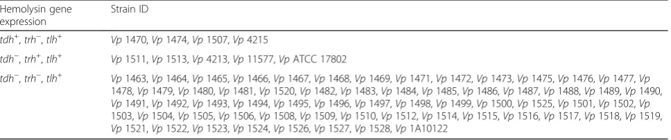
the V. parahaemolyticus type strain. The hemolysin fea-tures of the strains described above are listed in Table4.
Based on the presence of the hemolysin regulatory genes, 5 strains were selected for use in animal
experi-ments. Vp 1474 expressed TDH and TLH encoded by
the tdh and tlh genes; Vp 1496 expressed only TLH
encoded by the tlh gene; Vp 1513 expressed TRH and
TLH encoded by thetrhandtlhgenes. In comparison to
the virulence of foodborne V. parahaemolyticus strains,
Vp 1A10122 was chosen as a marine isolate, which
expressed only TLH encoded by thetlhgene.Vp ATCC
17802 was also chosen as the type strain expressing
TRH and TLH encoded bytrhandtlh.
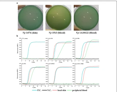
Identification ofV. parahaemolyticuscolonies on TCBS plates
Among localized infection models (Fig.1a, Table5), TCBS plates spread with skin samples from mice infected with
Vp1474 and MPB samples from mice infected with
Vp1513 andVp1A10122 developed scatteredV.
parahae-molyticuscolonies. On TCBS plates spread with skin
sam-ples from mice infected with Vp1496 and Vp1513 and
MPB samples from mice infected with Vp1474, Vp1496,
andVpATCC 17802, the presence ofV. parahaemolyticus
was ambiguous due to the presence of other bacteria on
the plates. No V. parahaemolyticus colonies were
ob-served on plates spread with skin samples from mice
in-fected withVp1A10122 andVpATCC 17802.
Among foodborne infection models (Fig. 2a, Fig. S1,
and Table 5), TCBS plates spread with stomach tissue
samples from mice infected withVp1474 andVp ATCC
17802; small intestine samples from mice infected with all experimental foodborne strains; liver tissue samples
from mice infected withVp1474 andVp1513; kidney
tis-sue samples from mice infected with Vp1474, Vp1513,
Vp1A10122, andVp ATCC 17802; spleen samples from
mice infected with Vp1513 and Vp ATCC 17802; and
MPB samples from mice infected with Vp1496 showed
recognizable V. parahaemolyticus colonies. Moreover,
on TCBS plates spread with stomach tissue samples
from mice infected with Vp1A10122, small intestine
samples from mice infected withVp1A10122, liver tissue
samples from mice infected with Vp1496 and
Vp1A10122, and MPB samples from mice infected with
Vp1513 andVp ATCC 17802, the presence of V.
para-haemolyticus was ambiguous as other bacteria
competi-tively grew on the plates. No V. parahaemolyticus
colony developed on plates spread with stomach tissue
samples from mice infected with Vp1496 and Vp1513;
small intestine and liver samples from mice infected with
Vp ATCC 17802; kidney tissue samples from mice
in-fected with Vp1496; spleen samples from mice infected
with Vp1474,Vp1496, and Vp1A10122; and MPB
sam-ples from mice infected withVp1474 andVp1A10122.
Among wound infection models (Fig. 3a, Fig. S2, and
Table 5), TCBS plates spread with stomach, small
intes-tine, liver, kidney, spleen, and MPB samples of mice
in-fected with all experimental foodborne strains and Vp
ATCC 17802 developed apparent V. parahaemolyticus
colonies, whereas on TCBS plates spread with stomach, small intestine, liver, kidney, spleen, and MPB samples
from mice infected withVp1A10122, the presence ofV.
parahaemolyticus was ambiguous due to the prolifera-tion of other bacteria on the plates.
In general infection models (Fig.4a, Fig.S3, and Table5), TCBS plates spread with stomach samples from mice
in-fected withVp1474; small intestine samples from mice
in-fected withVp1474 and Vp1496; liver, kidney, and spleen
samples from mice infected with all experimental food-borne strains; and kidney samples from mice infected with
Vp ATCC 17802 developed distinct V. parahaemolyticus
colonies. In addition, TCBS plates spread with small
intes-tine samples from mice infected with Vp1A10122, liver
samples from mice infected with Vp ATCC 17802, and
spleen samples from mice infected with Vp1A10122, the
development of V. parahaemolyticus was ambiguous as
other bacteria competitively proliferated on the plates. No V. parahaemolyticuscolony developed on the plates spread
with stomach samples from mice infected with Vp1496,
Vp1513,Vp1A10122, andVpATCC 17802; small intestine
samples from mice infected withVp 1513 and VpATCC
17802; liver and kidney samples from mice infected with
Vp 1A10122; and spleen samples from mice infected with
VpATCC 17802.
Table 4Hemolysin features ofV. parahaemolyticusstrains Hemolysin gene
expression
Strain ID
tdh+,trh−,tlh+ Vp1470,Vp1474,Vp1507,Vp4215
tdh−,trh+,tlh+ Vp1511,Vp1513,Vp4213,Vp11577,VpATCC 17802
MPB samples from mice in the wound infection model
were also directly plated for the detection ofV.
parahae-molyticus(Fig. 5a, Table5). The results indicated thatV. parahaemolyticus developed on the plates spread with
MPB samples from mice infected withVp1496,Vp1513,
Vp1A10122, andVp ATCC 17802, while MPB samples
from mice infected with Vp1474 were interfered by
other bacteria.
LAMP detection ofV. parahaemolyticus
Results of the LAMP assay demonstrated that after V.
parahaemolyticusinfection of mice through localized
in-flammation (Fig. 1b, Table 5), the skin from mice
in-fected with all experimental groups, in addition to MPB from mice infected with all experimental foodborne
strains, exhibited “S”-shaped curves, confirming the
presence ofV. parahaemolyticus.
LAMP assay results of V. parahaemolyticus digestive
system infection (Fig. 2b, Table 5) indicated that the
stomach of mice infected with Vp1496,Vp1513, andVp
ATCC 17802 exhibited “S”-shaped curves, as did the
small intestine from mice infected with all experimental
foodborne strains and Vp1A10122; the liver from mice
infected withVp1496,Vp1513, andVpATCC 17802; the
kidney and spleen from mice infected with Vp 1513, in
addition to the MPB from mice infected withVp1496.
The LAMP assay indicated that, after infection with V. parahaemolyticus through wounds (Fig. 3b,
Table 5), the MPB from mice infected with all
ex-perimental foodborne strains, the marine isolate, and
type strain exhibited “S”-shaped curves. In addition,
the stomach, small intestine, liver, kidney, and spleen samples from mice in all experimental groups were
positive for V. parahaemolyticus.
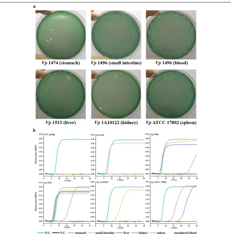
Fig. 1V. parahaemolyticusdetection in the localized infection mouse models.aThe representative ofV. parahaemolyticusproliferation identified on the specific TCBS plates from skin and MPB of localized infected mice.bThe representative ofV. parahaemolyticusdetection using rapid LAMP assay from skin and MPB of localized infected mice. White arrowhead, representative sparse colonies ofV. parahaemolyticus; P.C., positive control using plasmid containing target gene ofV. parahaemolyticusas the template of LAMP reaction; N.C., negative control using ddH2O as the
The LAMP assay indicated that the major organs from mice infected with all experimental foodborne strains
ex-hibited “S”-shaped curves when mice were infected with
V. parahaemolyticus through general infection (Fig. 4b,
Table 5), while the small intestine and spleen were the
most sensitive target organs in mice infected with the
marine strainVp1A10122 or type strainVpATCC 17802.
The LAMP assay also demonstrated that Vp1496
could be successfully identified even in fresh MPB with-out proliferation from mice in the foodborne infection
model (Fig.5b, Table5). When mice were infected with
V. parahaemolyticus in the wound model, the LAMP assay successfully detected most experimental strains,
in-cludingVp1474,Vp1496,Vp1513, andVp1A10122.
Discussion
Seafoods contaminated withV. parahaemolyticus, a
spe-cies of seafood-derived Vibrio, are common in coastal
cities. Combined with the high rates of exposure to
mar-ine or brackish environments, the abundance ofV.
para-haemolyticus results in many aquatic-derived human infections. Because its symptoms of infection are similar
to those of other bacteria, such as Escherichia coli and
Salmonellaspp. (Park et al.2018), little attention is
gen-erally paid toV. parahaemolyticus. Therefore, studies on
the epidemiology ofV. parahaemolyticus are limited,
al-though there are considerably greater numbers of vul-nerable populations than reported internationally.
Before the 1960s,V. parahaemolyticusoutbreaks were
limited to Japan (Fujino et al. 1953), but the very first
large outbreak of V. parahaemolyticus was reported in
Maryland, USA early in 1971, due to the ingestion of contaminated crab meat (Center for Food Safety and
Applied Nutrition (CFSAN) 2000). From then on, V.
parahaemolyticusinfection has become a global problem according to data from the Center for Disease Control
and Prevention (CDC) (CDC 1998), including areas
around the Pacific, Atlantic, and Indian oceans.
Virulence specificity vs.V. parahaemolyticusinfection
When V. parahaemolyticus invades a host, multiple toxins are released, and these accelerate deterioration of the site of infection. TDH, TRH, and TLH are the three
major toxins produced during V. parahaemolyticus
pro-liferation (DePaola et al. 2003). Among these, TDH and
TRH are reported to be the two main pathogenic factors
(Honda et al.1988; Nishibuchi and Kaper1995).
TDH is the primary toxin produced byV.
parahaemo-lyticusand is responsible for damage to erythrocytes and it also exhibits multi-cell type cytotoxicity. Sequence
var-iations of thetdhgene that encodes for TDH exist inV.
parahaemolyticus strains. The tdh gene family has formed as a result of phylogenetic evolution and gene
mutation (Tsunasawa et al.1987). TRH was purified and
characterized in 1988 as a V. parahaemolyticus
hemoly-sin related to TDH by Honda et al. (Honda et al. 1988)
also identified in 1989 by Nishibuchi et al. (Nishibuchi
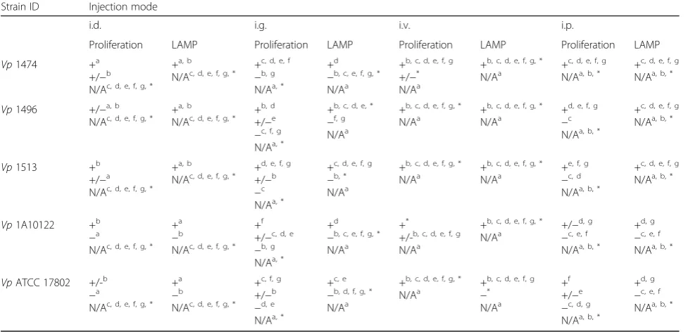
Table 5Results interpretation forV. parahaemolyticusinfection in target organs of mice
Strain ID Injection mode
i.d. i.g. i.v. i.p.
Proliferation LAMP Proliferation LAMP Proliferation LAMP Proliferation LAMP
Vp1474 +a
+/−b N/Ac, d, e, f, g, *
+a, b
N/Ac, d, e, f, g, * + c, d, e, f
−b, g N/Aa, *
+d
−b, c, e, f, g, * N/Aa
+b, c, d, e, f, g +/−* N/Aa
+b, c, d, e, f, g, *
N/Aa +
c, d, e, f, g N/Aa, b, * +
c, d, e, f, g N/Aa, b, *
Vp1496 +/−a, b
N/Ac, d, e, f, g, * + a, b
N/Ac, d, e, f, g, * + b, d +/−e
−c, f, g N/Aa, *
+b, c, d, e, *
−f, g N/Aa
+b, c, d, e, f, g, *
N/Aa +
b, c, d, e, f, g, *
N/Aa +
d, e, f, g
−c N/Aa, b, *
+c, d, e, f, g N/Aa, b, *
Vp1513 +b
+/−a N/Ac, d, e, f, g, *
+a, b N/Ac, d, e, f, g, *
+d, e, f, g +/−b
−c N/Aa, *
+c, d, e, f, g
−b, * N/Aa
+b, c, d, e, f, g, * N/Aa
+b, c, d, e, f, g, * N/Aa
+e, f, g
−c, d N/Aa, b, *
+c, d, e, f, g N/Aa, b, *
Vp1A10122 +b
−a
N/Ac, d, e, f, g, * +a
−b
N/Ac, d, e, f, g, * +f +/−c, d, e
−b, g N/Aa, *
+d
−b, c, e, f, g, * N/Aa
+*
+/-b, c, d, e, f, g N/Aa
+b, c, d, e, f, g, *
N/Aa +/−
d, g
−c, e, f N/Aa, b, *
+d, g
−c, e, f N/Aa, b, *
VpATCC 17802 +/-b
−a
N/Ac, d, e, f, g, * +a
−b
N/Ac, d, e, f, g, * +c, f, g +/−b
−d, e N/Aa, *
+c, e
−b, d, f, g, * N/Aa
+b, c, d, e, f, g, * N/Aa
+b, c, d, e, f, g
−* N/Aa
+f +/−e
−c, d, g N/Aa, b, *
+d, g
−c, e, f N/Aa, b, *
N/Anot applicable a
Local skin;b MPB;c
stomach;d
small intestine;e liver;f
kidney;g spleen;*
et al.1989). TRH is another important hemolysin and is
a recognized virulence factor. It is encoded by the trh
gene, which is closely related to thetdh gene, having up
to 68% sequence homology (Bej et al. 1999). Therefore,
TRH has the same intestinal toxicity but a different
hemocytocatheresis as TDH (Park et al.2004), also often
detected in clinical strains ofV. parahaemolyticus.
TLH is a separate virulence protein and encoded by
the tlh gene. It does not produce hemolysis on
Wagat-suma agar and is not responsible for human infection by V. parahaemolyticus tdh+ or trh+ isolates (McCarthy et al.1999). Different from thetdhandtrhgene families, tlhis a single gene and exists in all subtypes ofV. para-haemolyticus including clinical and environmental Fig. 2V. parahaemolyticusdetection in the foodborne infection mouse models.aThe representative ofV. parahaemolyticusproliferation
identified on the specific TCBS plates from the stomach, small intestine, liver, kidney, spleen, and MPB of foodborne infected mice.bThe representative ofV. parahaemolyticusdetection using rapid LAMP assay from the stomach, small intestine, liver, kidney, spleen, and MPB of foodborne-infected mice. White arrowhead, representative sparse colonies ofV. parahaemolyticus; P.C., positive control using plasmid-containing target gene ofV. parahaemolyticusas the template of LAMP reaction; N.C., negative control using ddH2O as the template of LAMP reaction; N.C.
isolates (Taniguchi et al. 1990). Therefore, TLH is a V. parahaemolyticus-specific virulence factor (Taniguchi
et al. 1985; Taniguchi et al. 1990). However, the
mech-anism of action of TLH remains unclear.
Although TDH and TRH are the two major virulence
factors ofV. parahaemolyticus, the Kanagawa reaction is
not necessarily linked to these two factors (Tada et al.
1992). Bej et al. confirmed that tdh− and trh− V.
parahaemolyticus still cause a positive Kanagawa reac-tion. Therefore, the clinical result of that specific assay (Kanagawa) is not 100% correct for the identification of V. parahaemolyticus(Ottaviani et al.2012). Thus, in our study, we detected expression of all 3 types of hemolysin
produced by V. parahaemolyticus. The hemolysin assay
indicated that all 71 V. parahaemolyticus strains
strains, 3 foodborne V. parahaemolyticus isolates had genes for the expression of TDH, TRH, and TLH. To
ensure all V. parahaemolyticus subtypes from different
sources were included in the animal experiments, 5 strains (considering both the source and virulence
fac-tors) were chosen to comprehensively representV.
para-haemolyticuslineages.
In previous studies, Tsai et al. suggested that there were
fewer than 5% prevalent V. parahaemolyticus strains in
environmental or marine food isolates (Tsai et al. 2013),
and so, it was hypothesized that the pathogenicity of V.
parahaemolyticusenvironmental isolates was weaker than clinical isolates. However, the continuously increasing
proportion of pathogenicV. parahaemolyticus has drawn
Fig. 4V. parahaemolyticusdetection in the general infection mouse models.aThe representative ofV. parahaemolyticusproliferation identified on the specific TCBS plates from the stomach, small intestine, liver, kidney, and spleen of general infected mice;bThe representative ofV. parahaemolyticusdetection using rapid LAMP assay from the stomach, small intestine, liver, kidney, and spleen of general infected mice. White arrowhead, representative sparse colonies ofV. parahaemolyticus; P.C., positive control using plasmid-containing target gene ofV.
parahaemolyticusas the template of LAMP reaction; N.C., negative control using ddH2O as the template of LAMP reaction; N.C. group, mice
attention to food safety. Up to the year 2000, a maximum
of 6% ofV. parahaemolyticusenvironmental isolates were
reported to express tdh/trh in Europe and Asia
(Letchu-manan et al. 2014), leaping to more than 8% (tdh+) and
12% (trh+) (Yang et al.2017b). In the present study, we
co-incidently found that 5.88% and 7.04% of our tested V.
parahaemolyticus strains expressedtdh/trh genes. In the future, this significantly increasing trend is very likely to be a factor influencing the outbreak of human infections
caused byV. parahaemolyticus.
Source specificity vs.V. parahaemolyticusinfection
Apart from virulence gene expression, there is a close
relationship between the source of a Vibrio isolate and
the severity of any resulting infection. Baker-Austin et al.
reviewed the differences in dissemination pathways and
infection gradients due toVibriosource diversity
(Baker-Austin et al. 2018), which also have different infection
risks. The most important source ofV. parahaemolyticus
in human infection is marine food products (Zhang
et al.2017). V. parahaemolyticusis a pathogen that
ex-ists in multiple water sources. Despite its lack of patho-genicity in fresh water, several studies have confirmed
human infection by V. parahaemolyticus isolated from
sea water. Another infection route that is easily ignored is the cross contamination of cooked foods transferred to humans through food or wounds (Brennan-Krohn et al.2016).
The correlation between the source ofVibrioand type
of infection may be reflected in genotype (Jones and Fig. 5V. parahaemolyticusdirect detection from fresh MPB of infected mice via different infection routes.aThe representative ofV.
parahaemolyticusproliferation identified on the specific TCBS plates from fresh MPB of wound-infected mice;bThe representative ofV. parahaemolyticusdetection using rapid LAMP assay from fresh MPB of localized, foodborne-derived, and wound infection mice. P.C., positive control using plasmid containing target gene ofV. parahaemolyticusas the template of LAMP reaction; N.C., negative control using ddH2O as the
Oliver 2009), which has previously been reported by
Rosche et al. for V. vulnificus (Rosche et al. 2005). In
addition, their infection data concluded that 90% of
C-genotype V. vulnificus strains was clinical isolates while
85~90% of E-genotype strains were environmental iso-lates (Rosche et al.2005). This data on strain
heterogen-eity has reinforced the correlation between Vibrio
genotype and pathogenicity. The information described
above raises a warning for the optimization of Vibrio
source identification and the updating of related data. The Vibrio source can be confirmed by whole genome
sequencing (Orata et al. 2014), therefore an
understand-ing of theVibrio source may provide an accurate
refer-ence to answer future research questions.
For V. parahaemolyticus, different sources typical of specific serotypes can lead to particular outbreaks of
hu-man infections. TheV. parahaemolyticusstrains
respon-sible for the majority of outbreaks in the USA have different serotypes from those responsible for outbreaks
in Japan. Although aV. parahaemolyticus outbreak
usu-ally happens within a limited region caused by strains with similar genotypes, a close relationship between strain heterogeneity and pathogenicity remains
(Baker-Austin et al. 2018), e.g., the pandemic O3:K6 V.
para-haemolyticus strain is generally present in the Bay of
Bangladesh region (Nair et al. 2007). However, the
mechanism by which a bacterial strain source affects a Vibriooutbreak is still not well understood.
Comparative research on pathogenicV.
parahaemolyti-cusstrains has shown that there is a diversity of
pathogen-icity when clinicalV. parahaemolyticusare isolated from
different areas of water. Considering both foodborne in-fections and regional proliferation ofV. parahaemolyticus, the V. parahaemolyticus strains were from both food-borne and seawater isolates, with ATCC type strain as the
standard for the experiments. FoodborneV.
parahaemoly-ticusstrains were all isolated from domestic consumption of seafood, including seashells, crabs, and oysters, and the seawater strain was isolated from the south China sea.
The results obtained in our study mirror theV.
parahae-molyticus infections seen in China and our LAMP assay
provides a possible solution for the rapid detection of V.
parahaemolyticusin current circumstances.
LAMP detection sensitivity
Based on Nucleic acid-PCR technology, the greatest ad-vantage of the LAMP assay lies in its high sensitivity and rapid speed of amplification. With specific pairs of primers, the LAMP assay minimized the template to 2 CFUs at most during its reaction, while other PCR
methods require 10~103-fold additional templates
com-pared with the LAMP assay (Law et al. 2015). Other
than these advantages, the LAMP assay is simple to
operate and is of low cost. Thus, the LAMP assay has fewer disadvantages than other methods.
Among the different routes of infection, V.
parahae-molyticus wound infections were the most easily de-tected by the LAMP assay. Wound infections caused by V. parahaemolyticusare not common, but they are ser-ious. Although there are few such reports, it can be life-threatening. In this study, we demonstrated that early
stages ofV. parahaemolyticusinfection can easily be
de-tected using a LAMP assay with visual interpretation of the results.
Data using the LAMP assay to detectV.
parahaemoly-ticus strains in various organs indicated that foodborne
strains were most sensitive to the assay, andV.
parahae-molyticus was detected in all organs tested in localized,
wound, and general infection models. AlthoughV.
para-haemolyticus could not be detected in all mice in the foodborne infection model, bacteria that spread to the small intestine could be detected. Our results indicated that the small intestine was a good target organ to detect
foodborne V. parahaemolyticus infection. On the other
hand, combined with different virulence values for each isolate, infectivity varied. In the foodborne infection
model, the stomach was the first organ to encounter V.
parahaemolyticus. The LAMP assay, on the contrary
showed false positive results. In all tested groups, Vp
1513 was detected with the highest efficiency, thereby demonstrating its high-level infectivity. In combination with the results of virulence gene expression, our find-ings demonstrated that the LAMP assay responded
dif-ferently to variousV. parahaemolyticusstrains.
When using the LAMP assay to detectV.
parahaemoly-ticusin different organs, the sensitivity of the LAMP assay varied in the different infection models. The results
im-plied that the infection route affected howV.
parahaemo-lyticus invaded the host, and this confirms the concern and suggestion of Baker-Austin et al., who highlighted in their research the importance of epidemiology data
up-dates, such as source ofVibrioand route of transmission
(Baker-Austin et al.2018). Combined with the LAMP
re-sults of different routes to infection, our rere-sults offer a personalized method for tracing back to the original
ex-planation for human infection byV. parahaemolyticus.
Conclusions and future perspectives
Based on previous reports, only a limited number of V.
parahaemolyticus infection cases resulted in death.
Des-pite this, V. parahaemolyticuscan still cause human
(Lee et al. 2018). This situation indirectly leads to the
excessive proliferation of V. parahaemolyticus.
There-fore, conducting early screening of potential susceptible populations plays a role in pre-warning for the limited prevention measure of its infection.
Therefore, in the early stages of V. parahaemolyticus
infection, non-invasive and rapid detection of the bac-teria is essential to prevent it from spreading further. Under this circumstance, our LAMP results
demon-strated a possible method to directly detect V.
parahae-molyticus in peripheral blood (PB). In this study, although the results showed different sensitivity when the LAMP assay was applied to specific infection models, the assay still has potential. When using the LAMP assay in the general infection model, positive results were ob-tained from all target organs, indicating that the LAMP
assay could be used forV. parahaemolyticusscreening.
Thus, the LAMP assay makes it possible to directly de-tect V. parahaemolyticus without the need for ex vivo studies. Although there are some drawbacks to the appli-cation of this method, the rapid detection speed, intuitive results, and high efficiency are promising for further inves-tigation for use in clinical cases. In addition, optimization of the LAMP assay requires further improvement.
Supplementary information
Supplementary informationaccompanies this paper athttps://doi.org/10. 1186/s13213-020-01585-6.
Additional file 1: Fig. S1.V. parahaemolyticusidentification on TCBS plates in the foodborne infection mouse models. The representative ofV. parahaemolyticusdevelopment on the specific TCBS plates from stomach, small intestine, liver, kidney and spleen of foodborne infected mice; white arrowhead, representative sparse colonies ofV. parahaemolyticus.
Additional file 2: Fig. S2.V. parahaemolyticusidentification on TCBS plates in the wound infection mouse models.The representative ofV. parahaemolyticusdevelopment on the specific TCBS plates from stomach, small intestine, liver, kidney, spleen and MPB of wound infected mice..
Additional file 3: Fig. S3.V. parahaemolyticusidentification on TCBS plates in the general infection mouse models. The representative ofV. parahaemolyticusdevelopment on the specific TCBS plates from small intestine, liver, kidney and spleen of general infected mice.
Abbreviations
G−:Gram-negative; NFDOSS: National Foodborne Disease Outbreaks Surveillance System; NASBA: Nuclear acid sequence-based amplification; FISH: Fluorescence in situ hybridization; LAMP: Loop-mediated isothermal amplification; TCBS: Thiosulfate citrate bile salts sucrose; SPF: Specific-pathogen-free; i.d.: Intradermal injection; i.g.: Intragastric gavage;
i.v.: Intravenous injection; i.p.: Intraperitoneal injection; MPB: Mouse peripheral blood; ACD: Acid citrate-dextrose; ANOVA: One-way analysis of variance; SEM: Standard error of the mean; CDC: Center for Disease Control and Prevention; PB: Peripheral blood
Acknowledgements
We thank Dr. Shenghui Cui for the generous gift ofV. parahaemolyticus
foodborne strains and for the help in their preparation. We thank Dr. Lei Shi for generously providing us with the procedure of LAMP method and the guidance.
Authors’contributions
SZ and LjZ conceived this study; KfF and LjZ designed the research; JL, JfL and KwQ performed the experiments; JfL and ClW analyzed experimental data; JL, JfL, KfF, and XjY wrote the paper. JL and JfL contributed equally to this work. All authors discussed the results, agreed on the interpretation and contributed in the finalization of the manuscript.
Funding
This work was funded by the Medical and health key project (grant no. 14J004, 12J025), the Medical Innovation Research Program (grant no. CX19027), and the Innovation Incubation Fund of the Navy General Hospital (grant no. CXPY201822).
Availability of data and materials
All data generated or analyzed during this study are included in this published article.
Ethics approval and consent to participate
All applicable international, national, and/or institutional guidelines for the care and use of animals were followed. All animal procedures complied with the institutional and national guidelines prescribed by the International Council for Laboratory Animal Science (ICLAS) from the Ministry of Health of the People’s Republic of China. All procedures performed in studies involving animals were in accordance with the ethical standards of Animal Ethics Committee of Navy General Hospital of Chinese PLA at which the studies were conducted.
Consent for publication
N/A.
Competing interests
The authors declare that they have no competing interests.
Received: 14 July 2019 Accepted: 21 February 2020
References
Alam MJ, Tomochika KI, Miyoshi SI, Shinoda S (2002) Environmental investigation of potentially pathogenicVibrio parahaemolyticusin the Seto-Inland Sea, Japan. FEMS microbiology letters 208:83–87. https://doi.org/10.1111/j.1574-6968.2002.tb11064.x
Baker-Austin C, Oliver JD, Alam M, Ali A, Waldor MK, Qadri F, Martinez-Urtaza J (2018)Vibriospp. infections. Nature reviews Disease primers 4:8.https://doi. org/10.1038/s41572-018-0005-8
Barker WHJ (1974) In: Fujino T, Sakaguchi G, Sakazaki R, Takeda Y (eds) International Symposium onVibrio parahaemolyticus. Saikon Publishing Co., Ltd., Tokyo, pp 47–52
Bej AK, Patterson DP, Brasher CW, Vickery MC, Jones DD, Kaysner CA (1999) Detection of total and hemolysin-producingVibrio parahaemolyticusin shellfish using multiplex PCR amplification oftlh,tdhandtrh. Journal of microbiological methods 36:215–225
Brennan-Krohn T, Pica N, Sandora TJ, McAdam A (2016) Closing the brief case: safe to go back in the water?Vibrio parahaemolyticuswound infection associated with brackish water. Journal of clinical microbiology 54:1672.
https://doi.org/10.1128/jcm.02661-15
CDC (1998) Outbreak ofVibrio parahaemolyticusinfections associated with eating raw oysters--Pacific Northwest, 1997. MMWR Morb Mortal Wkly Rep 47:457– 462
Center for Food Safety and Applied Nutrition (CFSAN) (2000) Draft risk assessment on the public health impact ofVibrio parahaemolyticusin raw molluscan shellfish. U. S. Food & Drug Administration. Washington, DC. Chen S, Ge B (2010) Development of atoxR-based loop-mediated isothermal
amplification assay for detectingVibrio parahaemolyticus. BMC microbiology 10:41.https://doi.org/10.1186/1471-2180-10-41
Cho Y et al (2017) Rapid identification ofVibriospecies isolated from the southern coastal regions of Korea by MALDI-TOF mass spectrometry and comparison of MALDI sample preparation methods. J Microbiol Biotechnol 27:1593–1601.https://doi.org/10.4014/jmb.1704.04056
Dea-Ayuela MA, Galiana-Rosello C, Lalatsa A, Serrano DR (2018) Applying loop-mediated isothermal amplification (LAMP) in the diagnosis of malaria, leishmaniasis and trypanosomiasis as point-of-care tests (POCTs). Curr Top Med Chem 18:1358–1374.https://doi.org/10.2174/
1568026618666181025095735
DePaola A, Hopkins LH, Peeler JT, Wentz B, McPhearson RM (1990) Incidence of
Vibrio parahaemolyticusin U.S. coastal waters and oysters. Applied and environmental microbiology 56:2299–2302
DePaola A et al (2003) Molecular, serological, and virulence characteristics of
Vibrio parahaemolyticusisolated from environmental, food, and clinical sources in North America and Asia. Applied and environmental microbiology 69:3999–4005
Di Pinto A, Terio V, Di Pinto P, Colao V, Tantillo G (2012) Detection ofVibrio parahaemolyticusin shellfish using polymerase chain reaction-enzyme-linked immunosorbent assay. Lett Appl Microbiol 54:494–498.https://doi.org/10. 1111/j.1472-765X.2012.03231.x
Fu K, Li J, Wang Y, Liu J, Yan H, Shi L, Zhou L (2016) An innovative method for rapid identification and detection ofVibrio alginolyticusin different infection models. Frontiers in microbiology 7:651.https://doi.org/10.3389/fmicb.2016.00651
Fujino T, Okuno Y, Nakada D, Aoyama A, Fukai K, Mukai T, Ueho T (1953) On the bacteriological examination of Shirasu food poisoning vol:4
Fujita N, Ayukawa Y, Fuke M, Teraoka T, Watanabe K, Arie T, Komatsu K (2017) Rapid sex identification method of spinach (Spinacia oleracea L.) in the vegetative stage using loop-mediated isothermal amplification. Planta 245: 221–226.https://doi.org/10.1007/s00425-016-2618-z
Gong XH et al (2018) Epidemiology, aetiology and seasonality of infectious diarrhoea in adult outpatients through active surveillance in Shanghai, China, 2012-2016: a cross-sectional study. BMJ open 8:e019699.https://doi.org/10. 1136/bmjopen-2017-019699
Honda T, Ni YX, Miwatani T (1988) Purification and characterization of a hemolysin produced by a clinical isolate of Kanagawa phenomenon-negative
Vibrio parahaemolyticusand related to the thermostable direct hemolysin. Infection and immunity 56:961–965
Jones MK, Oliver JD (2009)Vibrio vulnificus: disease and pathogenesis. Infection and immunity 77:1723–1733.https://doi.org/10.1128/iai.01046-08
Kumar BK, Raghunath P, Devegowda D, Deekshit VK, Venugopal MN, Karunasagar I, Karunasagar I (2011) Development of monoclonal antibody based sandwich ELISA for the rapid detection of pathogenicVibrio
parahaemolyticusin seafood. International journal of food microbiology 145: 244–249.https://doi.org/10.1016/j.ijfoodmicro.2010.12.030
Law JW, Ab Mutalib NS, Chan KG, Lee LH (2015) Rapid methods for the detection of foodborne bacterial pathogens: principles, applications, advantages and limitations. Frontiers in microbiology 5:770.https://doi.org/10.3389/fmicb. 2014.00770
Lee LH, Ab Mutalib NS, Law JW, Wong SH, Letchumanan V (2018) Discovery on antibiotic resistance patterns ofVibrio parahaemolyticusin Selangor reveals carbapenemase producingVibrio parahaemolyticusin marine and freshwater fish. Frontiers in microbiology 9:2513.https://doi.org/10.3389/fmicb.2018. 02513
Lesmana M, Subekti D, Simanjuntak CH, Tjaniadi P, Campbell JR, Oyofo BA (2001)
Vibrio parahaemolyticusassociated with cholera-like diarrhea among patients in North Jakarta, Indonesia. Diagnostic microbiology and infectious disease 39:71–75
Letchumanan V, Chan KG, Lee LH (2014)Vibrio parahaemolyticus: a review on the pathogenesis, prevalence, and advance molecular identification techniques. Frontiers in microbiology 5:705.https://doi.org/10.3389/fmicb.2014.00705
Li WW et al. (2018) Analysis of foodborne disease outbreaks in China mainland in 2013. Chinese Journal of Food Hygiene 30:293-298 doi:10.13590/j.cjfh.2018. 03.015
Li Y et al (2014)Vibrio parahaemolyticus, southern coastal region of China, 2007-2012. Emerging infectious diseases 20:685–688.https://doi.org/10.3201/ eid2004.130744
Liu Y et al (2015) A foodborne outbreak of gastroenteritis caused byVibrio parahaemolyticusand norovirus through non-seafood vehicle. PloS one 10: e0137848.https://doi.org/10.1371/journal.pone.0137848
Martinez-Urtaza J, Baker-Austin C, Jones JL, Newton AE, Gonzalez-Aviles GD, DePaola A (2013) Spread of Pacific NorthwestVibrio parahaemolyticusstrain. N Engl J Med 369:1573–1574.https://doi.org/10.1056/NEJMc1305535
Martinez-Urtaza J, Lozano-Leon A, Vina-Feas A, de Novoa J, Garcia-Martin O (2006) Differences in the API 20E biochemical patterns of clinical and
environmentalVibrio parahaemolyticusisolates. FEMS microbiology letters 255:75–81.https://doi.org/10.1111/j.1574-6968.2005.00052.x
Martinez-Urtaza J et al (2016) Epidemiological investigation of a foodborne outbreak in Spain associated with U.S. West Coast genotypes ofVibrio parahaemolyticus. SpringerPlus 5:87.https://doi.org/10.1186/s40064-016-1728-1
McCarthy SA, DePaola A, Cook DW, Kaysner CA, Hill WE (1999) Evaluation of alkaline phosphatase- and digoxigenin-labelled probes for detection of the thermolabile hemolysin (tlh) gene ofVibrio parahaemolyticus. Lett Appl Microbiol 28:66–70
McLaughlin JB et al (2005) Outbreak ofVibrio parahaemolyticusgastroenteritis associated with Alaskan oysters. N Engl J Med 353:1463–1470.https://doi. org/10.1056/NEJMoa051594
Nair GB, Ramamurthy T, Bhattacharya SK, Dutta B, Takeda Y, Sack DA (2007) Global dissemination ofVibrio parahaemolyticusserotype O3:K6 and its serovariants. Clin Microbiol Rev 20:39–48.https://doi.org/10.1128/cmr.00025-06
Nishibuchi M, Kaper JB (1995) Thermostable direct hemolysin gene ofVibrio parahaemolyticus: a virulence gene acquired by a marine bacterium. Infection and immunity 63:2093–2099
Nishibuchi M, Taniguchi T, Misawa T, Khaeomanee-Iam V, Honda T, Miwatani T (1989) Cloning and nucleotide sequence of the gene (trh) encoding the hemolysin related to the thermostable direct hemolysin ofVibrio parahaemolyticus. Infection and immunity 57:2691–2697
Nordin N, Yusof NA, Abdullah J, Radu S, Hushiarian R (2016) Sensitive detection of multiple pathogens using a single DNA probe. Biosens Bioelectron 86: 398–405.https://doi.org/10.1016/j.bios.2016.06.077
Notomi T, Okayama H, Masubuchi H, Yonekawa T, Watanabe K, Amino N, Hase T (2000) Loop-mediated isothermal amplification of DNA. Nucleic Acids Res 28:E63 Orata FD, Keim PS, Boucher Y (2014) The 2010 cholera outbreak in Haiti: how
science solved a controversy. PLoS Pathog 10:e1003967.https://doi.org/10. 1371/journal.ppat.1003967
Ottaviani D et al (2012) NontoxigenicVibrio parahaemolyticusstrains causing acute gastroenteritis. Journal of clinical microbiology 50:4141–4143.https:// doi.org/10.1128/jcm.01993-12
Park KS, Ono T, Rokuda M, Jang MH, Okada K, Iida T, Honda T (2004) Functional characterization of two type III secretion systems ofVibrio parahaemolyticus. Infection and immunity 72:6659–6665. https://doi.org/10.1128/iai.72.11.6659-6665.2004
Park MS, Park KH, Bahk GJ (2018) Interrelationships between multiple climatic factors and incidence of foodborne diseases. International journal of environmental research and public health 15.https://doi.org/10.3390/ ijerph15112482
Poon LL et al (2004) Rapid detection of the severe acute respiratory syndrome (SARS) coronavirus by a loop-mediated isothermal amplification assay. Clin Chem 50:1050–1052.https://doi.org/10.1373/clinchem.2004.032011
Twedt RM (1989)Vibrio parahaemolyticus. In: Doyle MP (ed) Foodborne bacterial pathogens. Marcel Dekker Inc., New York, pp 552-4.
Raszl SM, Froelich BA, Vieira CR, Blackwood AD, Noble RT (2016)Vibrio parahaemolyticusandVibrio vulnificusin South America: water, seafood and human infections. Journal of applied microbiology 121:1201–1222.https:// doi.org/10.1111/jam.13246
Rince A et al (2018) Occurrence of bacterial pathogens and human noroviruses in shellfish-harvesting areas and their catchments in France. Frontiers in microbiology 9:2443.https://doi.org/10.3389/fmicb.2018.02443
Rizvi AV, Bej AK (2010) Multiplexed real-time PCR amplification oftlh,tdhandtrh
genes inVibrio parahaemolyticusand its rapid detection in shellfish and Gulf of Mexico water. Antonie van Leeuwenhoek 98:279–290.https://doi.org/10. 1007/s10482-010-9436-2
Rosche TM, Yano Y, Oliver JD (2005) A rapid and simple PCR analysis indicates there are two subgroups ofVibrio vulnificuswhich correlate with clinical or environmental isolation. Microbiol Immunol 49:381–389
Tada J et al (1992) Detection of the thermostable direct hemolysin gene (tdh) and the thermostable direct hemolysin-related hemolysin gene (trh) ofVibrio parahaemolyticusby polymerase chain reaction. Mol Cell Probes 6:477–487 Taniguchi H, Kubomura S, Hirano H, Mizue K, Ogawa M, Mizuguchi Y (1990)
Cloning and characterization of a gene encoding a new thermostable hemolysin fromVibrio parahaemolyticus. FEMS microbiology letters 55: 339–345
Taniguchi H, Ohta H, Ogawa M, Mizuguchi Y (1985) Cloning and expression in
Tsai SE, Jong KJ, Tey YH, Yu WT, Chiou CS, Lee YS, Wong HC (2013) Molecular characterization of clinical and environmentalVibrio parahaemolyticusisolates in Taiwan. International journal of food microbiology 165:18–26.https://doi. org/10.1016/j.ijfoodmicro.2013.04.017
Tsunasawa S, Sugihara A, Masaki T, Sakiyama F, Takeda Y, Miwatani T, Narita K (1987) Amino acid sequence of thermostable direct hemolysin produced by
Vibrio parahaemolyticus. J Biochem 101:111–121
Wang Y, Li D, Wang Y, Li K, Ye C (2016) Rapid and sensitive detection ofVibrio parahaemolyticusandVibrio vulnificusby multiple endonuclease restriction real-time loop-mediated isothermal amplification. Technique Molecules (Basel, Switzerland) 21:E111.https://doi.org/10.3390/molecules21010111
Waterfield T, Fairley D, Lynn F, Blackwood B, Shields MD (2018) A protocol for a systematic review of the diagnostic accuracy of Loop-mediated-isothermal AMPlification (LAMP) in diagnosis of invasive meningococcal disease in children. Systematic reviews 7:86.https://doi.org/10.1186/s13643-018-0747-0
Wong YP, Othman S, Lau YL, Radu S, Chee HY (2018) Loop-mediated isothermal amplification (LAMP): a versatile technique for detection of micro-organisms. Journal of applied microbiology 124:626–643.https://doi.org/10.1111/jam. 13647
Xu D, Ji L, Wu X, Yan W, Chen L (2018) Detection and differentiation ofVibrio parahaemolyticusby multiplexed real-time PCR. Canadian journal of microbiology 64:809–815.https://doi.org/10.1139/cjm-2018-0083
Yang JH et al (2017a) Distribution and antimicrobial susceptibility ofVibrio
species associated with zooplankton in coastal area of Korea. Mar Pollut Bull 125:39–44.https://doi.org/10.1016/j.marpolbul.2017.07.054
Yang Y, Xie J, Li H, Tan S, Chen Y, Yu H (2017b) Prevalence, antibiotic
susceptibility and diversity ofVibrio parahaemolyticusisolates in seafood from South China. Frontiers in microbiology 8:2566.https://doi.org/10.3389/fmicb. 2017.02566
Yi MY et al (2014) Real time loop-mediated isothermal amplification using a portable fluorescence scanner for rapid and simple detection ofVibrio parahaemolyticus. Food Control 41:91–95.https://doi.org/10.1016/j.foodcont. 2014.01.005
Zhang Z, Lou Y, Du S, Xiao L, Niu B, Pan Y, Zhao Y (2017) Prevalence ofVibrio parahaemolyticusin seafood products from hypermarkets in Shanghai. J Sci Food Agric 97:705–710.https://doi.org/10.1002/jsfa.7715
Zhou S, Gao ZX, Zhang M, Liu DY, Zhao XP, Liu Y (2016) Development of a quadruplex loop-mediated isothermal amplification assay for field detection of fourVibriospecies associated with fish disease. SpringerPlus 5:1104.
https://doi.org/10.1186/s40064-016-2760-x
Publisher’s Note