Original Research Article
Study of prevalence and types of anemia in primary hypothyroidism
Rakesh K. Yadav
1*, Mansoor Ahmad
1, Manoj K. Mathur
1, Vishal Dhingra
2INTRODUCTION
Thyroid gland which is present in lower part of neck releases hormones that affects metabolism of all body cells including bone marrow cells thus increasing erythropoiesis, so hypothyroidism leads to anemia. Hypothyroidism is common disease with varying frequency between countries. Subclinical hypothyroidism defined as elevated serum TSH level associated with normal T3 and T4 values with minor symptoms, while in Overt/clinical hypothyroidism, TSH≥10 mIU/L and
unbound T4 level falls & with symptoms.
Hypothyroidism can cause wide variety of anemic disorders. Anemia in hypothyroidism can be normocytic normochromic, microcytic hypochromic and macrocytic hypochromic. Anemia severity is associated with hypothyroidism degree.1 Microcytic anemia is due to
malabsorption of iron and loss of iron by menorrhagia. Macrocytic anemia is caused by malabsorption of vitamin B12, folic acid and inadequate nutrition. Normocytic normochromic anemia is characterized by reticulopenia, hypoplasia of erythroid lineage, decreased level of erythropoietin, mainly erythrocyte survival. The most frequently encountered anemia type is normochromic normocytic anemia. The reason of this is bone marrow
ABSTRACT
Background: Hypothyroidism is common disease with varying frequency between countries. Anemia in hypothyroidism can be normocytic normochromic, microcytic hypochromic and macrocytic hypochromic. Anemia severity is associated with hypothyroidism degree. Objective of this study to study the association between anemia and hypothyroidism and prevalence and types of anemia in primary hypothyroidism (subclinical and overt both).
Methods: This cross sectional study was carried out at tertiary care hospital in North India. Newly diagnosed 100 primary hypothyroid patients and 100 controls with age and sex matched evaluated for anemia. Prevalence and types of anemia were studied and severity of anemia was correlated with that of hypothyroidism.
Results: Anemia was observed in 90 patients with hypothyroidism. Symptoms due to anemia were higher in cases than in controls. RBCs morphology showed normocytic normochromic in 59, microcytic hypochromic in 26 and macrocytic hypochromic in 15 cases. Serum anti-TPO positivity was present in 71.1% in cases as compared to 33.33% in controls. Anemia was severe in cases with high TSH.
Conclusions: Anemia was more prevalent in cases of hypothyroidism than in euthyroid controls. Normocytic normochromic type of anemia was most common type in this study. Serum anti-TPO positivity was 71.1% in cases. There was statistically significant negative correlation between TSH and haemoglobin. Symptoms of anemia were more in hypothyroid patients than in euthyroid anemic patients.
Keywords: Anemia, Anti-TPO, Primary hypothyroid, TSH
1Department of Medicine, 2Department of Pathology, M.L.N. Medical College (Allahabad) Prayagraj, Uttar Pradesh,
India
Received: 13 May 2019
Revised: 03 September 2019
Accepted: 07 September 2019
*Correspondence:
Dr. Rakesh K. Yadav,
E-mail: [email protected]
Copyright: © the author(s), publisher and licensee Medip Academy. This is an open-access article distributed under the terms of the Creative Commons Attribution Non-Commercial License, which permits unrestricted non-commercial use, distribution, and reproduction in any medium, provided the original work is properly cited.
repression due to thyroid hormone deficiency as well as lack of erythropoietin production arising from reduction in need of O2. Sympathetic system symptoms like
anxiety, palpitations, irregular heartbeats etc. may worsen on treatment with thyroxine if patient is iron deficient.2
Thyroid hormones also increase.2,3 DPG
(diphosphoglycerate) levels assisting in the transmission of oxygen into the tissues.3,4,5 Erythrocyte life cycle in
hypothyroidism is normal, and there is hypoproliferative erythropoiesis.6 Pernicious anemia caused by failure of
vit B12 absorption in hypothyroidism or due to associated
autoimmune conditions like celiac disease. In
hypothyroidism there is menorrhagia, reduced
iron/B12/folate absorption, are causes of anemia. According to data of WHO, anemia prevalence is 24.8% throughout the world and it is more frequent in underdeveloped countries. Prevalence of anemia in subclinical and overt hypothyroid group was 26.6% and 73.2% respectively. Thus, frequency of anemia in subclinical hypothyroidism is higher than that of general population. Aim of this study to study the prevalence and types of anemia in primary hypothyroidism (subclinical and overt both).
METHODS
Materials
The cross sectional study was conducted at Moti Lal Nehru Medical College, Prayagraj (Formerly Allahabad) and its associated hospital, Swaroop Rani Nehru Hospital from September 2017 to August 2018.
Case selection
Patients (both male and female) age >18 years attending Medicine OPD in MLN Medical College Prayagraj (Formerly Allahabad) was the source of the data.
Inclusion criteria
• All individuals aged between 18 to 60 years of either sex with newly diagnosed case of subclinical and overt primary hypothyroid patients were included in study.
Exclusion criteria
• Patients previously diagnosed with anemia or on treatment for anemia (based on history)
• Patients taking treatment for thyroid disorders (based on history)
• Pregnant patients
Methodology
The study protocol was explained to all participants and written informed consent was taken in both English and Hindi. Approval for the study was obtained from the Ethics Committee in the institution. A detailed history, a thorough clinical examination and investigations were
done for evaluation of anemia. Complete blood count (CBC) and peripheral smear examination were the basic investigations for anemia. Height and body weight were measured using a digital scale, and body mass index (BMI) was calculated as follows:
BMI = body weight in (kg)/height² in (meter).
Anemia was classified according to (WHO Classification):
• Mild - Hb 10 to 12 gm% • Moderate - Hb 8 to 10 gm% • Severe - Hb <8 gm%.
The study population was investigated for the following parameters: Complete blood count (CBC) and peripheral smear examination were the basic investigations for anemia. Based on the RBC morphology they were divided into the following groups and the specific investigations were carried out to determine the etiology of anemia.
Normocytic normochromic: coombs test (to rule out autoimmune etiology)
Microcytic hypochromic: stool for occult blood, upper
gastrointestinal endoscopy (wherever indicated),
Complete iron profile (to rule out iron deficiency)
Macrocytic hypochromic: Vitamin B12 levels and serum folate level. Test for anti-parietal cell antibodies was not done.
Estimation of serum anti-TPO antibodies in addition to the thyroid function test (T3, T4, and TSH), GBP with reticulocyte count, complete iron Profile and serum Vit. B12 & Folic Acid was carried out in both the groups.
The collected data was analyzed by applying appropriate statistical tests- chi square test, (with continuity correction for all tables (2×2) and fisher exact test (for all 2×2 tables where p-value of chi-square test is not valid due to small counts), unpaired t-test (if data passes normality test), mann-whitney test (if data fails normality tests).
RESULTS
Table 1: Base line Characteristics.
Characterstics Cases (100)
Control
(100) p-value
Mean age and SD 33±7 34±8 p=0.7916
Male patients 12(12%) 15(15%) -
Female patients 88(88%) 85(85%) -
Figure 1: Gender wise comparison between case and control.
In the cases, 89% patients had complaints of fatigue, while in the control group 12% of the participants had complaints of fatigue. On comparing it was found that fatigue occurs significantly higher in the cases than in the controls (p=.001). Dyspnea on exertion was present in 76% cases and 7% of the controls. On comparing it was observed that the dyspnea on exertion was significantly higher in the cases than in the controls (p=.001). Generalized weakness was present in the 93% cases and 5% of the controls. (Table 2).
Table 2:Symptoms.
Parameters Case (100) Control (100) p value
Fatigue Y-89(89%)
N-11(11%)
Y-12 (12%) N-88 (88%)
0.001 (<0.05) Dyspnea on
Exertion
Y- 76(76%) N- 24(24%)
Y-7(7%) N-93 (93 %)
0.02 (<0.05) Generalized
Weakness
Y-93 (93%) N-7 (7 %)
Y-05(05%) N -95(95 %)
0.001 (<0.05)
The hemoglobin level in the cases and controls was 10.13±1.09 gm/dl and 12.32±0.82 gm/dl respectively. On comparing it was observed that the hemoglobin was significantly lower in the cases as compared to the controls (p=.0001) (Table 3).
Table 3:Haemoglobin.
Parameters Case (100)
Control
(100) p value
Hb 10.13±1.09 12.32±0.82 0.0001(<0.05)
Hb is significantly lower in the cases as compared to the controls. Out of 100 cases included in the study, 59%
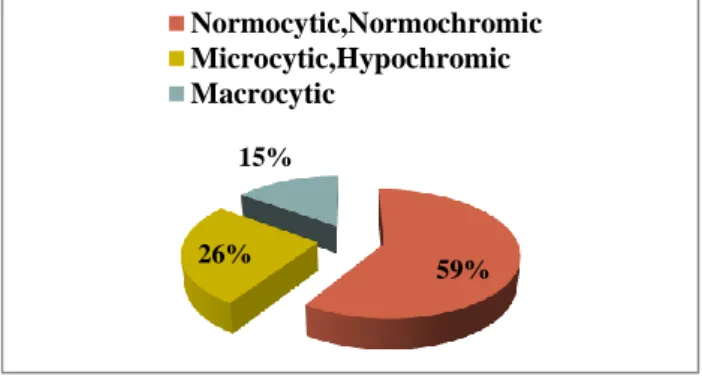
cases had normocytic normochromic RBCs morphology. Microcytic hypochromic morphology was observed in 26% cases and 15% had macrocytic morphology (Figure 2). However, in control population it was found that 95% normocytic, 2% had microcytic and 3% had macrocytic morphology (Figure 3).
Figure 2: RBC morphology in hypothyroid patients with anemia.
Figure 3: RBC morphology in control population with anemia.
It was observed that in cases 90% were found to be anemic and 10% were non anemic, while in control group 18% were found to be anemic and 82% were non anemic (Table 4, Figure 4).
Figure 4: Anemia in cases and control.
cases control
male 12 15
female 88 85
0 10 20 30 40 50 60 70 80 90 100
No
.o
f
p
a
tien
ts
59% 26%
15%
Normocytic,Normochromic Microcytic,Hypochromic Macrocytic
95%
2% 3%
Normocytic Microcytic Macrocytic
case control
Anemic 90 18
Non -anemic 10 82
0 10 20 30 40 50 60 70 80 90 100
No
.
o
f
Pa
tien
Table 4:Anemia in cases and control.
Case Anemia Non-anemic
100 90 10
Control Anemia Non-anemic
100 18 82
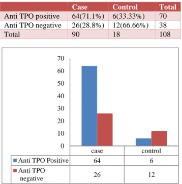
Total number of cases with hypothyroidism was 100 in our study & out of these 90 (90 %) patients had anemia and in anemic group 64 patients (71.1%) had anti-TPO positive antibodies while 26 patients (28.8%) were negative for anti-TPO antibodies (Table 5, Figure 5).
Table 5:Anti TPO in cases and control with Anemia.
Case Control Total
Anti TPO positive 64(71.1%) 6(33.33%) 70
Anti TPO negative 26(28.8%) 12(66.66%) 38
Total 90 18 108
Figure 5: Anti TPO positivity in cases and control with Anemia.
The serum T3 level (ng/dl) in the cases and controls was 2.06±0.63 and 2.44±0.61 respectively. On comparing it was observed that the serum T3 level in the two groups were comparable to each other. The serum T4 level (ng/dl) in the cases and controls was 1.26±0.42 and 1.15±0.242 respectively. On comparing the serum T4 level of the two groups were comparable to each other. The serum TSH level (mIU/mL) in the cases and controls were 28.44±9.32 and 3.47±0.62, respectively. On comparing it was found that the serum TSH level were significantly higher in the cases as compared to that of controls (p=0.0001) (Table 6).
Out of 26 microcytic patients in case 15 had iron deficiency and out of 2 microcytic patients in control group, none had iron deficiency anemia (Table 7).
Out of 15 macrocytic patients, in cases, 12 had Vit B12 deficiency, while out of 3 macrocytic patients in control group 1 patient had Vit B12 deficiency (Table 8).
Table 6:TSH of case and control.
Parameters Case (100) Control
(100) p value
TSH 28.44±9.32 3.47±0.62
0.0001(<0.05) TSH is significantly higher in the cases as compared to the control
Table 7:Serum Iron of Case and Control.
No. of patient (microcytic)
No. of patients having iron deficiency
Percentage
Case
(100) 26 15 57.69%
Control
(100) 2 0 0%
Table 8:Vit B12 level of Case and Control.
No of patient (macrocytic)
No. of patients having Vit B12
deficiency
Percentage
Cases
(100) 15 12 80 %
Control
(100) 3 1 33.33 %
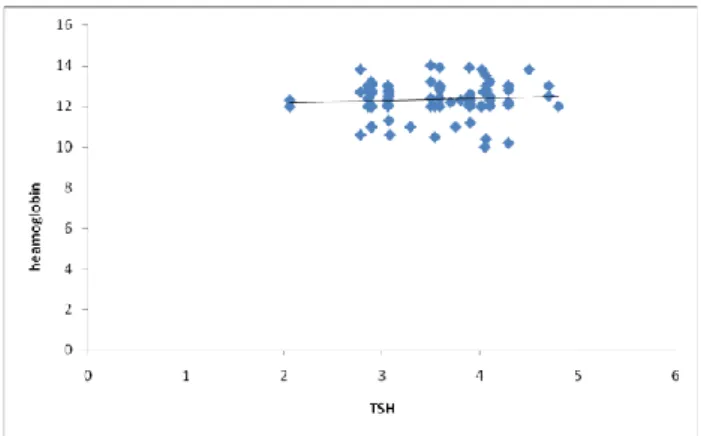
Correlation between TSH and Hb in cases
Applying Pearson’s correlation analysis between TSH and Hb level, there was a negative correlation between these two factors in cases. Pearson’s correlation coefficient was -0.45. This was statistically significant with a P value of 0.005 (Figure 6).
Figure 6: Correlation between TSH and Hb in cases.
Correlation between TSH and hemoglobin in controls
Applying Pearson’s correlation analysis between TSH and Hb level, there was no correlation between these two factors in controls. Pearson’s correlation coefficient was 0.006. This was statistically non- significant with a P value of 0.94 (Figure 7).
case control
Anti TPO Positive 64 6
Anti TPO
negative 26 12
0 10 20 30 40 50 60 70
0 2 4 6 8 10 12 14
0 10 20 30 40 50 60 70
H
em
o
g
lo
bin
Figure 7: Correlation between TSH and hemoglobin in controls.
DISCUSSION
In this study it is observed that hypothyroidism is more common in females as compared to males. These findings are similar to the Mehmet E et al., 2012 study.7 in which
sex distribution revealed that the proportion of males and females in the hypothyroid group were 12% and 88% respectively while in the control group the proportion was 16% and 84% respectively. According to the study by Das C et al.1 it was found that 70% of the subjects were
female .In accordance with the previous studies it can be concluded that the prevalence of anemia in hypothyroid patients is higher in the female population compared to males. This can be multifactorial mainly caused by monthly menstrual blood loss in the females. The complaints of fatigue, tiredness, dyspnea on exertion and generalized weakness occur more frequently in the hypothyroid patients as compared to the controls in this study.
The hemoglobin levels in the hypothyroid patients was lower (10.13 gm %) compared to the controls (12.32 gm%), in this study. In a study by Mehmet E et al.7 it was
found that the hemoglobin level in the hypothyroid subjects and control groups was 11.9 and 12.8 gm% respectively and it was lower in the hypothyroid subjects.7 The mean hemoglobin levels of the
hypothyroid and control groups was 12.2 and 13.6 gm%, in the study by Dorgalaleh A et al, 2013 study.8
The study of RBC morphology in this study revealed that the 59% of cases and 95 % of controls were having
normocytic normochromic RBCs. Microcytic
hypochromic RBCs were greater in the hypothyroid patients (26%) than in the controls (2%). Thus, study of RBC morphology revealed greater abnormalities in the hypothyroid population.
The mean serum TSH levels in our study were 28.44 mIU/ml in cases and 3.47mIU/ml in the controls. The serum TSH levels were significantly higher in the hypothyroid patients compared to the controls. These findings were comparable to findings of the study by
Mehmet E et al.7 In a study by Mehmet E et al.6 it was
found that the mean serum TSH levels in the hypothyroid group and control group was 43.1 and 1.7 mIU/mL, respectively and the serum TSH levels were higher in the hypothyroid group compared to the control group. In the study by Dorgalaleh A et al.8 the mean serum TSH levels
were 4.97 and 2.6 mIU/ml, respectively in the hypothyroid and control groups respectively. In the present study there was negative correlation between TSH levels and hemoglobin levels in cases. Such findings were also seen in a study conducted by Dorgaleleh et al.8
where a decreased level of hemoglobin was seen in hypothyroid population with raised TSH as compared to the total population. It was observed in this study that, 26 out of 100 patients in the cases were having microcytic aneamia and 56.79% of them were found to have aneamia due to iron deficiency although only 2 patients of the control population had microcytic anemia and out of which no patients were found to have iron deficiency.
In this study it was observed that 15 patients in cases were found to have macrocytic anemia out of which 80% patients were found to have Vit B12 deficiency and in the control group only 3% had macrocytic anemia and out of which 33.33% people were having Vit B12 deficiency.
The results of the direct and indirect Coomb’s test were similar in the hypothyroid and control subjects in this study. TPO antibodies were positive in the 64% of the cases and 6% of the controls and incidence in the hypothyroid cases was higher than in the controls. These findings were similar to the studies by Mehmet E et al, and Das C et al.1,7 In a study by Mehmet E et al, TPO
antibody positivity was observed in 100% hypothyroid subjects and 22.5% controls. In the study by Das C et al, TPO antibody was positive in 58.3% cases.
Thus, presence of TPO antibodies should be evaluated in
the patients with a risk for development of
hypothyroidism. It was found that higher incidence of anti-TPO antibodies was in anemic cases compared to the anemic controls. Previous studies have not shown association of TPO antibodies with that of anemia among the hypothyroid patients. This is a significant finding in this study which will help predict the risk of anemia in hypothyroid patients with anti-TPO antibodies.
CONCLUSION
this study. The study of RBC morphology in our study revealed that the 59% of cases and 95 % of controls were having normocytic normochromic RBCs. Microcytic hypochromic RBCs were greater in the hypothyroid patients (26%) than in the controls (2%). In cases, most common types of anemia is normocytic normochromic followed by microcytic hypochromic followed by macrocytic hypochromic. The mean serum TSH levels in our study were 28.44 mIU/ml in cases and 3.47mIU/ml in the controls. Twenty six out of 100 patients in the cases were having microcytic aneamia and 56.79 % of them were found to have aneamia due to iron deficiency. Only 2% patients of the control population had microcytic anemia and none had iron deficiency anemia. Fifteen (15) patients had macrocytic anemia, out of which 80% patients were found to have Vit B12 deficiency. In the control group only 3% had macrocytic anemia and out of which 33.33% people were having Vit B12 deficiency. TPO antibodies were positive in the 64% of the cases and 6% of the control and incidence in hypothyroid cases was higher than in the controls. It was found that higher incidence of anti-TPO antibodies in anemic cases (71.1%) compared to the anemic controls (33.33%).
Funding: No funding sources Conflict of interest: None declared
Ethical approval: The study was approved by the Institutional Ethics Committee
REFERENCES
1. Das C, Sahana PK, Sengupta N, Giri D, Roy M, Mukhopadhyay P. Etiology of anemia in primary hypothyroid subjects in a tertiary care center in
Eastern India. Indian J Endocr Metab. 2012;16, Suppl S2:361-3.
2. Horton L, Coburn RJ, England JM, Himsworth RL.
The hematology of hypothyroidism. Q J Med. 1976;45(177):101-23.
3. Das KC, Mukherjee M, Sarkar TK, Dash RJ,
Rastogi GK. Erythropoiesis and erythropoietin in hypoand hyperthyroidism. J Clin Endocrinol Metab. 1975;40:211-20.
4. Fein HG, Rivlin RS. Anemia in thyroid diseases. Med Clin North Am. 1975;59:1133-1145.
5. Jason WH, Stephen FH, Rajasehkar R, Govind B,
Peter HRG. Anemia in celiac disease is
multifactorial in etiology. Am J Hematol.
2007;82:996-1000.
6. Bamashmous S.A., Al-Nuzaily M.A.K. , Al-Maktari
L.A.S. , Taresh S.A.G. and Ali F.H.H. Prevalence and etiology of anemia in overt and subclinical hypothyroid women in Sana’a, Yemen. J Clin Res Letters. 2013,4(1):57-60.
7. Mehmet E, Aybike K, Ganidagli S, Mustafa K. Characteristics of anemia in subclinical and overt hypothyroid patients. Endocrine J. 2012;59(3):213-20.
8. Dorgalaleh A, Mahmoodi M, Varmaghani B, Node FK.Effect of thyroid dysfunctions on blood cell count and red blood cell indices. Iran J Ped Haematol Oncol. 2013;3(2):73-7.