A Dissertation on
“A COMPARITIVE STUDY BETWEEN APACHE II
AND RANSON SCORING SYSTEMS IN
PREDICTING THE SEVERITY OF ACUTE
PANCREATITIS"
Dissertation submitted to
THE TAMIL NADU Dr.M.G.R.MEDICAL UNIVERISTY
CHENNAI
with partial fulfilment of the regulations
for the Award of the degree
M.S. (General Surgery)
Branch – I
MADRAS MEDICAL COLLEGE ,
CHENNAI.
BONAFIDE CERTIFICATE
Certified that this dissertation is the bonafide work of Dr. G.KESAVAN on “A COMPARITIVE STUDY BETWEEN APACHE II AND RANSON SCORING SYSTEMS IN PREDICTING THE
SEVERITY OF ACUTE PANCREATITIS” during his M.S. (General
Surgery) course from July 2014 to September 2014 at the Madras Medical College and Rajiv Gandhi Government General Hospital, Chennai – 600003.
Prof.Dr.P.RAGUMANI. M.S. Director,
Institute of General Surgery,
Madras Medical College & Rajiv Gandhi Government
General Hospital, Chennai – 600 003
Prof.Dr.K.RAMASUBRAMANIAN, M.S., Professor of General Surgery,
Institute of General Surgery, Madras Medical College & Rajiv Gandhi Government General Hospital,
Chennai – 600 003.
Prof.Dr.R.VIMALA M.D, DEAN,
ACKNOWLEDGEMENT
I would like to express my deep sense of gratitude to the Dean, Madras Medical College and Prof.Dr.P.RAGUMANI M.S, Director, Institute of General Surgery , MMC & RGGGH, for allowing me to undertake this study on “A COMPARITIVE STUDY BETWEEN APACHE II AND RANSON SCORING SYSTEMS IN PREDICTING THE SEVERITY OF ACUTE PANCREATITIS”
I was able to carry out my study to my fullest satisfaction, thanks to guidance, encouragement, motivation and constant supervision extended to me, by my beloved Unit Chief Prof. Dr. K. RAMASUBRAMANIAN M.S. Hence my profuse thanks are due for him.
I am bound by ties of gratitude to my respected Assistant Professors,
Dr.A.Prabakar , Dr.S.Umarani and Dr.S.VijayaLakshmi in general, for placing and guiding me on the right track from the very beginning of my career in Surgery till this day. I would be failing in my duty if I don’t place on record my sincere thanks to those patients who inspite of their sufferings extended their fullest co-operation.
DECLARATION
I, certainly declare that this dissertation titled, “
A COMPARITIVE
STUDY BETWEEN APACHE II AND RANSON SCORING
SYSTEMS IN PREDICTING THE SEVERITY OF ACUTE
PANCREATITIS
”, represent a genuine work of mine . The contribution of any supervisors to the research are consistant with normal supervisory practice, and are acknowledged.I, also affirm that this bonafide work or part of this work was not submitted by me or any others for any award , degree or diploma to any other university board , neither in India or abroad . This is submitted to The Tamil Nadu Dr.MGR Medical University, Chennai in partial fulfilment of the rules and regulation for the award of Master of Surgery Degree Branch 1 (General Surgery).
Date :
LIST OF ABBREVATIONS USED
ACCR - Amylase Creatinine Clearance Ratio
APACHE II - Acute physiology and chronic health evaluation
AUC - Area Under Curve
CAPAP - Carboxy Peptidase Activation Peptide
CFTR - Cystic Fibrosis Transmembrane Regulator
CRAI - Continous Regional Arterial Infusion
CRP - C-reactive Protein
CTSI - Computed Tomography Severity Scoring
IL - Interleukin
NFKB - Nuclear Factor Kappa B
NPV - Negative Predictive Value
PLA2 - Phospholipase A2
PPV - Positive Predictive Value
PSTI - Pancreatic Secretory Trypsin Inhibitor
SAPS - Simplified Acute Physiology Scoring
TAP - Trypsinogen Activated Peptide
TABLE OF CONTENTS
Sl. No.
Contents
Page No
1.
INTRODUCTION1
2.
OBJECTIVES2
3.
REVIEW OF LITERATURE3
4.
METHODOLOGY81
5.
OBSERVATION ANDRESULTS
83
6.
DISCUSSION91
7.
CONCLUSION97
8.
SUMMARY98
9.
BIBLIOGRAPHY10.
ANNEXURES· FIGURES
· PROFORMA
[image:10.595.127.490.100.704.2]1
INTRODUCTION
“Acute pancreatitis includes a wide spectrum of disease, from one with mild self limiting symptoms to fulminant process with multiorgan failure and high mortality”.Given the wide spectrum of disease seen, the care of the patients with pancreatitis must be highly individualized .
The “early diagnosis and precise scoring of disease severity” are important goals in the initial evaluation and the management of pancreatitis. Pancreatitis not only must be differentiated from a myriad of other potential diagnoses, but patients also must be stratified to identify those with severe disease and to guide appropriate therapy.
2
OBJECTIVES
3
REVIEW OF LITERATURE
History:
“An early description of acute pancreatitis was given by Ambrose Pare in 1579. However, the importance of pancreas and the severity of its inflammatory disorders were not appreciated until its function as a digestive organ was delineated in the mid seventeenth century. In 1886, Nicholas Senn suggested that operative treatment for acute pancreatitis might be indicated in pancreatic gangrene or abscess formation”.
“In 1901, Opie described gall stones as a cause of pancreatitis by documenting and impacted gall stone in the ampulla of Vater during autopsy of a patient who died of pancreatitis. The first plausible explanations of the pathogenesis of acute pancreatitis were hypothesized by Halsted, Osler, and Opie, contemporaries at Johns Hopkins hospital”.
4
life- threatening, multiorgan failure including sepsis, renal failure, acute respiratory distress syndrome, and death.
Improved outcomes are clearly linked to advancements in supportive care Currently, the only effective therapeutic interventions address the complications of acute pancreatitis, most commonly biliary sepsis, pancreatic necrosis, pseudocysts, infection, and sepsis; the latter account for mortality rates in excess of 50% to 80%. The advent and integration of minimally invasive surgery, including advanced endoscopic, radiologic and interventional techniques, are changing the management of complicated, acute pancreatitis. Incorporation of these modalities has brought into question the use and timing of operative management for pancreatic necrosis, pseudocysts, and gallstones.
DEFINITIONS:
5
Mild acute pancreatitis consists of no organ dysfunction and an uneventful recovery2.
Severe pancreatitis manifests as organ failure and/or local complications such as necrosis, abscess, and pseudocyst3(Table 1).
Other acceptable markers of severe pancreatitis are 3 or more of the 11 Ranson criteria for nongallstone pancreatitis (Table 2) and the second Acute Physiology and chronic Health Evaluation (APACHE II) score higher than 8”.
Dynamic contrast – enhanced CT scans can distinguish interstitial from necrotizing Pancreatitis.
“An acute fluid collection is fluid located in or near the pancreas that lacks a definite wall and that occurs early in the course- of acute pancreatitis”
“A pseudocyst is a fluid collection that persists for 4 to 6 weeks and becomes encapsulated by a wall of fibrous or granulation tissue”.
6
[image:19.595.101.508.172.480.2]“Pancreatic necrosis is a diffuse or focal area (s) of nonviable pancreatic parenchyma, which is typically associated with peripancreatic fat necrosis”.
Table 1 : Atlanta Criteria for Severe Acute Pancreatitis
NATURAL HISTORY:
7
“There are two time peaks for morality. Most studies in the United States and Europe show that about one half the deaths occur within the first week or two, usually from multiorgan failure. 11-14 Death can be very rapid. About one fourth of all deaths in Scotland occur Within 24 hours of admission, and one third 48 hours15 After the second week of illness patients succumb to pancreatic infection associated with multiorgan failure”.
“Some studies in Europe report a very high rate of late mortality from infection.Patients who are older and have comorbid illnesses have a substantially higher rate of mortality than younger healthier patients16 . In those who survive the illness, severe pancreatic necrosis can scar the pancreas, resulting in a structure of the main pancreatic duct with subsequent obstructive chronic pancreatitis and permanent diabetes and malabsorption17”
8
PATHOLOGY:
“Most causes of acute pancreatitis (i.e., alcohol, gallstones, and drugs) involve initial injury to peripheral acinar cells, fat necrosis, and autodigestion. The peripheral cells are distant from the arterial supply of pancreatic lobules, and some parenchymal damage likely is due to abnormalities of the microcirculation”. In comparison, infections agents are directly toxic to acinar cells and cause generalized acinar cell necrosis associated with an acute inflammatory infiltrate21. In contrast, the earliest lesion produced by pancreatitis due to hypotension is ductal necrosis22.
“Pathologically, there are two main types of pancreatitis interstitial and necrotizing. Interstitial pancreatitis (also called edematous pancreatitis) is characterized by interstitial edema associated with inflammatory cells within the parenchyma. Although parenchymal necrosis may occur, it is microscopic. Small foci of fat necrosis characteristically punctuate the surface of the gland. This type of finding is usually associated with a mild clinical course”.
9
areas of necrosis, which may involve acinar cells, islet cells, and the pancreatic ductal system. Pancreatic necrosis is present mostly in the periphery of the lobules, but it may progress to involve most of the gland. Severe interstitial fat necrosis involves small veins and venules, which may be infiltrated by granulocytes, leading to thrombosis, necrosis, and rupture. Arterial thrombosis is observed infrequently”.
PATHOGENESIS
:
“The initial step in the pathogenesis of acute pancreatitis is conversion of trypsinogen to trypsin within acinar cells in sufficient quantities to overwhelm normal mechanisms to remove active trypsin8. Trypsin, in turn, catalyzes conversion of pro enzymes, including trypsinogen and inactive precursors of elastase, phospholipase and carboxypeptidase, to active enzymes9. Normally, small amounts of trypsingogen are spontaneously activated within the pancreas, but intrapancreatic mechanisms quickly remove activated trypsin10. Pancreatic secretory trypsin inhibitor (PSTI, now called SPINKI) binds and inactivates about 20% of the trypsin activity11.”
10
acute pancreatitis is unclear12. Activation of trypsinogen occurs before biochemical or morphologic injury to acinar cells, in association with colocalization of lysosomal enzymes, such as cathepsin B, and digestive enzymes, including trypsinogen with unstable vacuoles24. Thus, complete inhibition of cathepsin B either may prevent or may be a treatment for acute pancreatitis25. However, enzyme colocalization may occur without inducing significant acinar cell injury”.
“Mutations in the cystic fibrosis transmembrane conductance regulator (CFTR) gene (CTFR) have also been implicated in panceatitis. CFTR anion channel allows for secretion of chloride and bicarbonate into the ducts and, thus, flushing of the liberated enzymes and proenzymes into the duodenum. More than 1200 mutations have been described for the CFTR gene”.
“A third genetic abnormality associated with pancreatitis is a mutation of the SPINK 1 gene. As noted previously, SPINK 1 protects the pancreatic acinar cell by inhibiting prematurely activated trypsin. Mutations of the SPINKI gene presumably limit the activity of the protein, but the exact mechanism is unclear”.
11
the pancreatic duct38,39 and obstruction of the pancreatic duct at the ampulla secondary to stone(s) or to edema resulting from the passage of a stone40. Reflux of bile into the pancreatic duct could occur when the distal common bile and pancreatic ducts from a common channel and a gallstone becomes impacted in the duodenal papilla”. Alternatively, bile could reflux into the pancreatic duct from the duodenum through an incompetent sphincter of Oddi inured by recent passage of a gallstone15.”
“Experimentally, reflux of bile, particularly if infected or mixed with pancreatic enzymes, causes pancreatic injury. Mixtures of bile and pancreatic enzymes raise the permeability of the main pancreatic duct, which is associated with local parenchymal inflammation41. The common channel theory is somewhat problematic, however, because pancreatic duct pressure is invariably higher than common bile duct pressure, making bile reflux unlikely. Reflux of bile from the duodenum is also an unlikely pathogenetic factor, because pancreatitis does not occur in conditions with easily demonstrable reflux, such as after surgical sphincteroplasty or endoscopic sphincterotomy16.”
12
ductal and acinar cells. Experiments in the opossum support this theory: Ligation of the pancreatic duct causes severe necrotizing pancreatitis38, and decompression of the duct within 3 days prevents progression to acinar cell necrosis and severe inflammation40.”
PATHOPHYSIOLOGY:
“The pathophysiology of acute pancreatitis starts with local acinar injury, which if unchecked leads to local inflammatory complications, a systemic response, and sepsis. Pathophysiologic mechanisms include microcirculatory injury, leukocyte chemoattraction, release of proint amatory and anti- inflammatory cytokines, oxidative stress, leakage of pancreatic fluid into the region of the pancreas, and bacterial translocation to the pancreas and systemic circulation”.
13
14
affect acinar cell glutathione concentrations may lead to increased oxidant stree and more severe pancreatitis”.
“Meanwhile, ischemia and severe inflammation of the gland can engender disruption of the main and secondary pancreatic ducts, leading to local fluid accumulations within and surrounding the pancreas that can eventuate into pseudocysts49.”
15
“ARDS, which is secondary to microvascular thrombosis ,may be induced by active phospholipase A (lecithinase), which digests lecithin, a major component of lung surfactant. Acute renal failure has been explained on the basis of hypovolemia and hypotension. Myocardial depression and shock are likely secondary to vasoactive peptides and a myocardial depressant factor. Metabolic complications include hypocalcemia, hyperlipidemia, hyperglycemia with or without ketoacidosis, and hypoglycemia. The pathogenesis of hypocalcemia is multifactorial and include hypoalbuminemia (the most important cause), hypomagnesemia, calcium- soap formation, hormonal imbalances (e.g., parathyroid hormone, calcitonin, and glucagon), binding of calcium by free fatty acid- albumin complexes, intracellular translocation of calcium, and systemic exposure to endotoxin54.”
16
arteriovenous shunting in the gut. Indeed, in canine experimental pancreatitis, enclosing the colon in impermeable bags prevents translocation of bacteria from the colon to the pancreas58.”
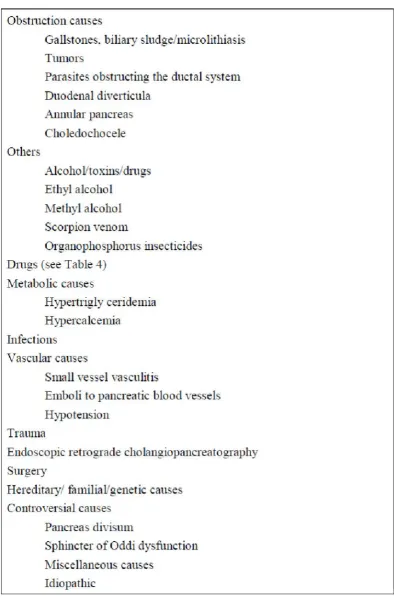
ETIOLOGY:
17
18
OBSTRUCTIVE CAUSES:
Gallstones:
“The most common obstructive process leading to pancreatitis is gallstones, which cause approximately 40% of cases of acute pancreatitis59. Cholecystectomy with clearing of the common bile duct of stones prevents recurrence, confirming the cause and effect relationship60. However, only 3% to 7% of patients with gallstones experience pancreatitis60. The risk of development of acute pancreatitis due to gallstones is relatively greater in men (relative risk, 14 to 35) than in women (relative risk, 12 to 25)60. However, more women experience gallstone pancreatitis because gallstones are more common in women60. Acute pancreatitis occurs more frequently when stones are les than 5mm in diameter (odds ratio, 4 to 5)61. Small stones are more likely than large stones to pass through the cystic duct and cause ampullary obstruction”.
Biliary sludge/microlithiasis:
19
or calcium bilirubinate granules63. On ultrasound it is a mobile, low amplitude echo that does not produce a shadow and that layers in the most dependent part of the gallbaladder”.
“Sludge occurs with functional or mechanical bile stasis. Common associations are a prolonged fast, total parenteral nutrition, and distal bile duct obstruction. In addition, the cephalosporin antibiotic ceftriaxone can complex with bile to form a sludge within the biliary system when its solubility in bile is exceeded; rarely, this sludge causes stone64 that disappear after the patient stops taking the drug. Commonly, biliary sludge occurs in acute pancreatitis with no abvious cause. However, the association between biliary sludge and acute pancreatitis is unproved. There is no prospective, randomized study documenting that removing sludge or microcrystals via cholecystectomy prevents further attacks of pancreatitis”.
TUMOURS:
20
Metastases from other primary tumors (lung, breast) to the pancreas have also caused pancreatitis70. Large adenomas of the major papilla can likewise occasionally cause obstructive pancreatitis. Other obstructive conditions that are rarely associated with acute pancreatitis are choledochoceles, duodental diverticula, annular pancreas, and space occupying parasites that obstruct the pancreaticobiliary system, such as Ascaris and Clonorchis”.
ALCOHOL TOXINS AND DRUGS
Ethyl alcohol
21
The fact that pancreatitis develops in only a small percentage of chronic alcoholics suggests underlying genetic susceptibility. To date, no strong genetic connection has been found to explain this occurrence”.
Other toxins:
“Methyl alcohol, organophosphorus insecticides, and the venom of the Trinidad scorpion have all been reported to induce pancreatitis. The mechanism of the latter two is believed to be hyperstimulation of the pancreas. Smoking raises the risk of alcoholic and idiopathic, but not gallstone, pancreatitis”.
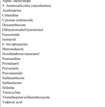
Drugs:
22
caused pancreatitis on rechallenge. The reader should notice that classes of drugs are not used but, rather, the specific drugs. Because drug induced pancreatitis is very uncommon and most cases are idiosyncratic, it would not be correct to state that a whole class of drugs (e.g., angiotensin- converting enzyme inhibitors) are potential offenders, if only one of the class (perindopril) has been documented to cause the disease”.
23
[image:36.595.135.477.198.548.2]that drugs can cause pancreatitis after years of use. Drug- induced pancreatitis tends to be mild and self- limited”.
Table 3: Drugs that Cause Pancreatitis after Rechallenge
Alpha- methyldopa
24
METABOLIC CAUSES:
Hypertriglyceridemia:
“Hypertriglyceridemia is perhaps the third most common identifiable, noniatrogenic cause of pancreatitis, after gallstones and alcoholism. Serum triglyceride concentrations exceeding 1000mg/dL (11mmol/L) may precipitate attacks of acute pancreatitis. Patients may have lactescent (milky) serum owing to increased concentrations of very-low- densitiy lipoprotein( VLDL), and at higher VLDL levels, owing to hyperchylomicronemia. The pathogenesis of hypertriglyceridemic pancreatitis is unclear, but the release of free fatty acids may damage pancreatic acinar cells or capillary endothelium”.
25
second is an alcoholic patient who is found to have hypertriglyceridemia on hospital admission. The third (15% to 20%) is a nondiabetic, nonalcoholic, non obese person who has drug or diet- induced hypertriglyceridemia. Drug induced disease is more likely to occur if there is underlying hypertriglyceridemia.Most people who abuse alcohol have moderate, but transient, elevations of serum triglycerides. This condition is likely an epiphenomenon and not the cause of pancreatitis,because alcohol raises serum triglyceride concentrations in a “dose-dependent” manner. For example, the prevalence of serum triglyceride concentrations higher than 227mg/dL (2.5mmol/L were 10%, 14% and 2091> in persons who had 3 to 5,6to 8 and 9 or more alcoholic drinks per day, respectively. Alcoholic patients with severe hyperlipidemia often have a coexisting primary genetic disorder of lipoprotein metabolism”.
Hypercalcaemia:
26
calcium) are responsible for pancreatitis, particularly because sudden infusion of high levels of calcium into rats leads to conversion of trypsinogen to trypsin, hyperamylasemia, and dose- dependent morphologic changes of acute pancreatitis such as edema and acinar cell necrosis”.
“Hypercalcemia due to hyperparathyroidism is a proposed cause of pancreatitis.However, primary hyperparathyroidism causes less than 0.5% of all cases of acute pancreatitis, and the incidence of acute pancreatitis in hyperparathyroidism varies from 0.2% to 1.5%94. Rarely, pancreatitis occurs with other causes of hypercalcemia, including metastatic. Bone disease, total paranteral nutrition, sarcoidosis, vitamin D toxicity and peri-operative infusions of calcium in high doses during cardiopulmonary bypass surgery”.
INFECTIONS:
27
biochemical evidence (more than three times elevation of serum lipase or amylase) and characteristic symptoms; and “possible pancreatitis” exists if there is only asymptomatic biochemical evidence. The definitive criterion for an infection causing pancreatitis is finding the organism in the pancreas or pancreatic duct through stain or culture. Probable criteria are culture of the organism from pancreatic juice or blood or serologic evidence combined with a characteristic clinical or epidemiologic setting. The criterion of a possible infection is culture of the organism from other body sites or serologic evidence of infection”.
28
gondii, and possibly opportunistic organisms such as Mycobacterium aviumcomplex75.”
VASCULAR DISEASE:
“Rarely, pancreatic ischemia causes pancreatitis. In most cases it is mild, but fatal necrotizing pancreatitis may occur. Ischemia may result from vasculitis (systemic lupus erythematosus, polyateritis nodosa), atheromatous embolization of cholesterol plaques from the aorta to the pancreas after transabdominal angiography, intraoperative hypotension, hemorrhagic shock, ergotamine overdose, and transcatheter arterial embolization for hepatocellular carcinoma. Acute pancreatitis has occurred in long distance runners, perhaps on an ischemic basis. Also, ischemia is one possible explanation for pancreatitis after cardiopulmonary bypass”.
TRAUMA:
29
spine, such as in an automobile accident. In blunt trauma, it is important to determine preoperatively whether there is injury to the pancreas, because the severity of pancreatic injury determines whether the pancreas must be included in the surgical plan.Even in the absence of serious injury to adjacent organs, surgery may be necessary to treat a pancreatic ductal injury.This injury cause acute duct rupture and pancreatic ascites.Serum amylase activity maybe increased in abdominal trauma whether or not the pancreas has been injured”.
Endoscopic Retrograde CholangioPancreaticography:
30
SURGERY:
“Postoperative pancreatitis can occur after abdominal or thoracic surgery. Pancreatitis occurs after 6% of liver transplantations and after 0.4% to 7.6% of cardiopulmonary bypass operations.Significant risks for pancreatitis after cardiopulmonary bypass are preoperative renal insufficiency, postoperative hypotension, and perioperative administration of calcium chloride.Mortality for postoperative pancreatitis is said to be higher (up to 35%) than for other forms of pancreatitis”.
HEREDITARY AND GENETIC CAUSES:
31
between this abnormality and pancreatitis is weak for the following reasons: (1) 1% to 4% of the general population have these mutations, (2) less than 1% of mutation carriers experience pancreatitis, and (3) the severity of pancreatitis is similar whether patients are homozygous, heterozygous, or compound heterozygous, suggesting complex genetics.PRSSI gene testing, CFTR genome testing, and SPINKI analysis are all commercially available”.
CONTROVERSIAL CAUSES:
Pancreas divisum:
32
Sphincter of oddi dysfunciton:
“Dysfunction of the sphincter of Oddi is also a controversial cause of pancreatitis.Series that study patients with recurrent acute pancreatitis report that sphincter of Oddi dysfunction (usually defined as pancreatic sphincter pressure> 40mm Hg) is the most common abnormality discovered, occurring in approximately 35% to 40%) of patients.The argument that this entity causes acute pancreatitis is based on the many experiential series reporting that endoscopic pancreatic sphincterotomy or many experiential series reporting that endoscopic pancreatic sphincterotomy or surgical sphincteroplasty reduces or eliminates the attacks of pancreatitis”.
Miscellaneous causes:
33
disease cause druginduced pancreatitis. Celiac disease has also been described in association with pancreatitis, but the relationship remains uncertain. Pancreatitis has been seen in patients who have suffered severe burns. A relationship between smoking and acute pancreatitis has been suggested”.
CLINICAL FEATURES:
“It is difficult to diagnose acute pancreatitis through history and physical examination because clinical features are similar to those of many acute abdominal illness”.
HISTORY
Abdominal pain
34
Occasionally, pain gradually increases and takes several hours to reach maximum intensity. Pain is steady and moderate to very severe in intensity. There is little pain relief with change of body position. frequently, pain is unbearable, steady, and boring, band- like radiation of the pain to the back occurs in one half of patients. Pain that lasts only a few hours and then disappears suggests another disease, such as biliary colic or peptic ulcer. Pain is absent in 5% to 10% of attacks, and a painless presentation may be a feature of serious fatal disease”.
Nausea and Vomiting:
“Ninety percent of patients with acute pancreatitis have nausea and vomiting. Vomiting may be severe, may last for hours, may be accompanied by retching, and may not alleviate pain. Vomiting may be related to severe pain or to inflammation involving the posterior gastric wall”.
Physical examination:
35
colonic ileus. Almost all patients have tenderness in the upper abdomen, which may be elicited by gently shaking the abdomen or by gentle
percussion. Guarding is more marked in the upper abdomen. Tenderness and guarding are less than expected from the intensity of
discomfort. Abdominal rigidity, as occurs in diffuse peritonitis, is unusual but can be present, and differentiating it from a perforated viscus may be impossible in these instances. Bowel sounds are reduced and may be absent. Additional abdominal findings include ecchymosis in one or both flanks (Turner’s sign) or abut the periumbilical area (Cullen’s sign), owing to extravasation of hemorrhagic pancreatic exudates to these areas. These signs occur in less than 1% of cases and are associated with a poor prognosis. Rarely, there is a brawny erythema of the flanks caused by extravasation of pancreatic exudates to the abdominal wall. During the disease a palpable epigastric mass from a pseudocyst or a large inflammatory mass may appear”.
36
days it may increase to 1010F to 1030F owing to the severe retroperitoneal inflammatory process and the release of inflammatory mediators from the pancreas.Tachypnea and shallow respirations may be present if subdiaphragmatic inflammatory exudates causes painful breathing. Dyspnea may accompany pleural effusions, atelectasis, congestive heart failure, or ARDS. Chest examination may reveal limited diaphragmatic excursion if abdominal pain causes splinting of the diaphragm, or dullness to percussion and decreased breath sounds at the lung bases if there is a pleural effusion. There may be disorientation, hallucinations, agitation, or coma, which may be due to alcohol withdrawal, hypotension, electrolyte imbalance, hypoxemia, fever, and/or toxic effects of pancreatic enzymes on the central nervous system.Icterus may be present due to choledocholithiasis (gallstone pancreatitis), bile duct obstruction from edema of the head of the pancreas, or coexistent liver disease”.
37
clinical improvement. If they occur over a joint they may be confused with arthritis”.
“Some physical findings point to a specific cause of acute pancreatitis. Hepatomegaly, spider angiomas, and thickening of palmar sheaths favor alcoholic pancreatitis. Eruptive xanthomas and lipemia retinalis suggest hyperlipidemic pancreatitis. Parotid pain and swelling are features of mumps. Band keratopathy (an infiltration on the lateral margin of the cornea) occurs with hypecalcemia”.
LABORATORY DIAGNOSIS:
38
PANCREATIC ENZYMES
Serum and Urine Amylase
39
not necessary (surgery). A limitation of serum amylase measurement is that it is not 100% sensitive or specific”.
SERUM LIPASE:
“The sensitivity of serum lipase measurements for the diagnosis of acute pancreatitis is similar to that of serum amylase measurements, between 85% and 100%.Some researchers claim a greater specificity than with serum amylase because almost all lipase originates from the pancreas (there is a small amount of gastric lipase), and the lipase value is normal when the serum amylase value is nonspecifically elevated, as in salivary gland dysfunction, tumors, gynecologic conditions, and macroamylasemia.Serum lipase content is always elevated on the first day of illness and remains elevated longer than serum amylase content. Consequently, some authorities suggest combining lipase with amylase values as a test for acute pancreatitis. However, the author and others have found that combining enzyme values does not improve diagnostic accuracy”.
OTHER PANCREATIC ENZYMES:
40
include PLAz, trypsin, carboxylester lipase, carboxypeptidase A, colipase, elastase, and ribonuclease. None, alone or in combination, is better than serum amylase or lipase, and measurements of most are not routinely available”.
Standard blood tests:
41
Other blood and urine tests:
“Many nonenzymatic proteins are over expressed in acute pancreatitis. Pancreatitis associated protein (PAP), a heat shock protein, is undetectable in the normal pancreas but its level markedly increases in acute pancreatitis. The sensitivity of PAP and pancreatic- specific protein (PSP) measurements is no better than that of conventional tests, but PAP and PSP values are as accurate as serum amylase value for the detection of acute pancreatitis”.
“The methemalbumin level rises in acute pancreatitis, but it also does so in serious intra- abdominal conditions such as intestinal infarction”.
RADIOLOGIC DIAGNOSIS:
ABDOMINAL PLAIN FILM:
42
spread and location of pancreatic exudates. Gastric abnormalities are caused by exudates in the lesser sac, which produces anterior displacement of the stomach with separation of the contour of the stomach from the transverse colon. Abnormalities of the small intestine, which are due to exudates in proximity to small bowel mesentery, include ileus of one or more loops of jejunum (the sentinel loop), of the distal ileum or caecum, or of the duodenum. Generalized ileus may occur in severe disease. Other abnormalities of the hollow GI tract may also be present”.
“Other findings on plain radiography of the abdomen may give clues to etiology or Severity, including calcified gallstones (gallstone pancratitis), pancreatic stones or calcification (chronic pancreatitis with a bout of acute inflammation), and ascites (severe pancreatitis). Gas in the retroperitoneum may suggest a pancreatic abscess”.
CHEST RADIOGRAPHY:
43
are only on the right side. During the first 7 to 10 days, there also may be signs of congestive heart failure or ARDS. Pericardial effusion is rare”.
ABDOMINAL ULTRASONOGRAPHY:
44
ENDOSCOPIC ULTRASONOGRAPHY:
“Usually, endoscopic ultrasonography (EUS) is not helpful in acute pancreatitis.However, it is more sensitive than either abdominal ultrasonography or CT to detect common duct stones. One potential use of EUS is to exclude a common duct stone in patients with severe pancreatitis and jaundice (serum bilirubin> 5mg/dL),ERCP, in this situation, may worsen pancreatitis and potentially introduce infection into necrotic areas of the pancreas. Thus, EUS might eliminate the need for urgent ERCP in severe gallstone pancreatitis”.
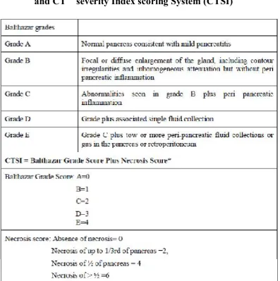
COMPUTED TOMOGRAPHY:
45
pancreatic necrosis. If there is normal perfusion of the pancreas, interstitial pancreatitis is said to be present Pancreatic necrosis (perfusion defects after intra venous contrast agent is given) may not appear until 48 to 72 hours after onset of acute pancreatitis. CT – or ultrasound- guided needle aspiration can confirm a suspected infections”.
“Contraindications for using intravenous contrast agent are a patient’s history of prior sever allergy (respiratory distress or anaphylaxis) and significant renal impairment (serum creatinine >2mg/dL). If severe renal impairment requires dialysis, intravenous contrast medium may be used. Hives or less sever allergic reactions with previous administration of iodinated contrast material are not contraindications. In a patient with such a history, however, a nonionic contrast agent should be used; also, glucocorticoids and diphenhydramine (Benadryl) should be administered before scanning”.
46
[image:59.595.112.510.324.724.2](CTSI) score (Table 4). The higher the CTSI score, the more severe the pancreatitis clinically. Although the presence of gas in the pancreas suggest pancreatic infection with a gas- forming organism, this findings can also accompany sterile necrosis with microperforation of the gut or adjacent pseudocyst into the pancreas. In the great majority of pancreatic infections, however, CT scanning shows no gas”.
Table 4: computed tomography (CT) Grading System of Balthazar
47
MAGNETIC RESONANCE IMAGING:
“Magnetic resonance imaging (MRI) provides information regarding the severity of pancreatitis similar to that given by CT. MRI is as good as CT in detecting necrosis and fluid collections and is a better method to detect choledocholithiasis and ductal disruption, especially after intravenous secretion is administered. Gadolinium, unlike intravenous contrast agents used for CT, is safe to use in renal failure. MRI, however, is less accessible and more expensive than CT”.
ENDOSCOPIC RETROGRADE CHOLANGIO PANCREATICO
GRAPHY:
“ERCP is limited to patients with severe acute pancreatitis due to gallstones with persistent common bile duct obstruction as well as to those in whom the stone could not be removed during surgery”.
DIFFERENTIAL DIAGNOSIS:
48
hours rather than several days. The pain of a perforated ulcer is sudden, becomes diffuse, and precipitates a rigid abdomen; movement aggravates pain. Nausea or infarction, the clinical setting often is an older person with cardiac arrhythmia or arteriosclerotic disease who experiences sudden pain out of proportion to physical findings, bloody diarrhea, nausea, and vomiting. Abdominal tenderness may be mild to moderate, and muscular rigidity may not be pronounced despite severe pain. In intestinal obstruction, pain is cyclical, abdominal distention is prominent, vomiting persists and may become feculent, and peristalsis is hyperactive and often audible”.
Table 5: Differential Diagnosis of Acute Pancreatitis
Biliary pain/acute cholecystitis Perforated hollow viscus
Mesenteric ischemia or infarction Closed- loop intestinal obstruction Inferior wall myocardial infarction Dissecting aortic aneurysm
49
PREDICTORS OF SEVERITY:
“Predicting severity of pancreatitis early in the course of disease is critical to maximize therapy and to prevent and minimize organ dysfunction and complications. Clinical assessment, multiple prognostic scoring lists(Ranson’s, Glasgow/Imrie Coma scales, APACHE II), peritoneal fluid analysis, organ failure scores, individual laboratory tests, and CT scanning have all been touted as helpful for this purpose”.
SCORING SYSTEMS:
Clinical signs:
50
[image:63.595.114.503.142.457.2]RANSON’S CRITERIA (TABLE 6)
Table 6:Ranson’s 11 Prognostic Criteria for Pancreatitis
“Data from Ranson JHC, Rifkind KM, Roses DF, et al: Prognostic signs and the role of operative management in acute pancreatitis. Surg Gynecol Obstet 139:69, 1974;and Ranson JHC Etiological and prognosis factors in human acute pancreatitis: A review.Am J Gastroenterol 77: 633, 1982. NA, not applicable”.
51
Table 2). Higher Ranson’s score predicts more severe disease. In mild pancreatitis (score ≤2) the mortality is 2.5%, and in severe pancreatitis (score 2:3) the mortality is 62%. Also the higher the Ranson’s score, the higher the incidence of systemic complications, necrosis, and infected necrosis. These criteria continue to remain in wide use in both the United states and Europe”.
“The Ranson criteria have several drawbacks. First, the two lists are cumbersome.Second, an accurate Ranson’s score takes 48 hours to compute, and the criteria have not been validated beyond the 48 – hours time limit. Third, not all laboratories measure all the parameters in routine blood tests (e.g., lactic dehydrogenase). Fourth, the overall sensitivity of the Ranson criteria (using 3 signs, as, the cutoff) for diagnosing severe disease is only 40%to 88% and the specificity 43% to 90%. The positive predictive value is approximately 50% and the negative predictive value around 90%”. Therefore, the best use of Ranson’s criteria is to exclude severe disease78,79,82.
THE APACHE SYSTEM:
52
APACHE I
“In the original form, APACHE contained 34 physiologic measurements and included many continuous variables. A value of 0 to 4 was assigned to each variable, according to its degree of abnormality. Shortly after its introduction apache 1 system was disfavoured, because of practical problems like collection of large number of variables. Also under the rules of APACHE system any unmeasured variable was assumed to be normal and weighted as zero. This gave rise to questions about the models general applicability. Another major criticism of original APACHE system was that the variables were chosen by a group of physicians and hence there was a potential of bias. These inaccuracies in the original APACHE system prevented its widespread use. However, it did serve as a prototype for the development of two subsequent systems”.
SAPS
53
APACHE II
“Published in 1985 by the same author this is the second version of the APACHE system and it contains refinements based on experience with the original APACHE system. APACHE II has been extensively used and has received far more attention in the literature then any of the other methodologies for ICU outcome prediction. It contains 12 continuous variables from the original APACHE system and also takes into account age of the patient, pre- morbid conditions and Glasgow coma scale”.
DEVELOPMENT OF APACHE II
54
“Some of the thresholds and weights for the physiologic variables have been changed ego Glasgow coma score, serum creatinine. Also since Alveolar – arterial O2 gradient (p [A-a] O2 is heavily dependent on
inspired O2 (F1O2) a direct weighting was given to all paO2 values when
F1O2 is less than 0.5”.
“To eliminate the problem of missing values and concerns about the assumption that an unmeasured variable was normal, measurement of all 12 variables was made mandatory for usage of APACHE II. The recorded values of the variables are based on the most deranged values during the past 24 hours”.
55
i. It has a well defined outcome (hospital death)
ii. It was derived from a large database (5815 patients from 13 hospitals)
iii. Source of bias present in its prototype was understood and corrected”.
“Short comings of APACHE II system.
Because of extensive usage, important sources of error and bias in the APACHE II system were revealed. First, APACHE II performs well overall in several ICU population but it is inaccurate when looking at specific disease categories because the data base from which it was derived, through large, did not contain many patients in major disease subsets such as cardiac surgery, oncology etc. Second APACHE II does not account for prior treatment or clinical course before the patient enters ICU, this has been labeled as lead time bias. Third, APACHE II requires determination of a single admission diagnosis, a subjective process prone to bias. Finally, despite the reduction in number of variables, measurement error from bedside data collection are still on issue”.
56
system which is now being applied widely to acute pancreatitis clinical trials”.
57
Table 7: Acute Physiology and Chronic Health Evaluation
(APACHE)- II Scoring System of Disease Severity
Glasgow Score
58
Table 8: Modified Glasgow Criteria: Within 48 Hours of Admission
Organ Failure Scores:
59
severe disease in all systems.A study from Scotland demonstrated that the Goris score was more predictive of death than the Glasgow/Imrie score. In this study of 279 patients with acute pancreatitis, there were no deaths in 189 patients with a Goris score of 0, 7 deaths (9%) in the 75 patients with a Goris score of 1 to 4, and 10 deaths (67%) in the 15 patients with a score higher than 5.Greater use of organ failure scores are likely to improve prognostication in acute pancreatitis”.
PERITONEAL LAVAGE
“Percutaneous recovery of any volume of peritoneal fluid with a dark color or recovery of at least 20mL of free intraperitoneal fluid of dark color portends a significant mortality. The sensitivity of peritoneal lavage is 36% to 72%, and the specificity is greater than 80% to 100%. An advantage is that peritoneal lavage can be used any time, but it has not gained wide acceptance because it is invasive”.
LABORATORY MARKERS:
60
amyloid A, and procalcitonin may prove valuable because their concentrations in blood or urine may serve to separate mild from severe acute pancreatitis”.
“Hematocrit value: A high hematocrit value on admission or failure of a high value to diminish after 24 hours of rehydration is believed to be a sing of hemoconcentration due to retroperitoneal fluid loss and thus a marker of severe disease. One study showed that a hematocrit higher than 44% had a sensitivity of 72% on admission and of 94% after 24 hours for detection of organ failure. The negative predictive value at 24 hours was 96%.74 However, another study from Germany found no correlation between admission hematocrit value and o rgan failure”.
“C-Reactive Protein: Measurement of CRP, an acute – phase reactant produced by the liver, is used extensively in Europe as a marker of severe pancreatitis. CRP is inexpensive to measure and readily available. The sensitivity for detecting severe disease is 60% to 100% (with cutoffs of 100-210mg/L), and the specificity is 75% to 100%”.
61
“Polymorphonuclear leukocyte Elastase: Polymorphonuclear leukocyte elastase rises very early (before CRP) in acute pancreatitis. High levels have been reported to differentiate severe from mild disease, but the test is not generally available”.
“Phospholipse A2: PLAz is involved in the synthesis of prostaglandins and degrades surfactant in the lung. It may play a role in the pulmonary dysfunction associated with acute pancreatitis. Levels of catalytic type II PLAz have been reported to differentiate between mild and severe disease within 24 hours of admission”.
62
(CAPAP) assay has also been shown to predict early severe acute pancreatitis”.
“Serum Amyloid A: Serum amyloid A is another early acute- phase reactant that is synthesized in the liver and is associated with the extent of tissue inflammation. Two studies have demonstrated that the level of this serum protein can differentiate mild from severe disease”.
“Procalcitonin: The pro peptide procalcitonin is another acute- phase reactant that has been shown to differentiate mild from severe acute pancreatitis within the first 24, hours after symptom onset. A serum strip test has been developed for this measurement that has a sensitivity of 86% and a specificity of 95% in detecting organ failure”.
COMPUTED TOMOGRAPHY:
63
scores correlate better with local complications (pseudocysts and abscesses) than with mortality. Among the 37 patients with grade D or E findings, 54% had a local complication, whereas only 2 of 51 (3.9%) with grades A through C experienced this problem. Thus, the data do not confirm that the CTSI is any more predictive than the grade A-E score. There is controversy in the literature as to whether the extent of necrosis on CT predicts organ failure. Two studies did not find any correlation between these two events. In a third study, however, there was a strong correlation”.
CHEST RADIOGRAPHY
64
TREATMENT
GENERAL CONSIDERATIONS:
“The patient with acute pancreatitis requires aggressive intravenous hydration and adequate analgesia to eliminate or markedly reduce pain. An order for no oral intake (NPO) is usually in force until nausea and vomiting have subsided. Abdominal pain is treated with analgesics, given parenterally every 3 hours. Morphine can also be used.Dosing is monitored carefully and adjusted daily according to ongoing needs. Although morphine has been reported to increase sphincter of Oddi tone and to raise serum amylase levels, its use to treat the pain of pancreatitis has not been shown to adversely affect outcome. Nasogastric intubation is not used routinely because it is not beneficial in mild pancreatitis. This modality is used only to treat gastric or intestinal ileus or intractable nausea and vomiting. Similarly, proton pump inhibitors and histamine Hz receptor blocking agents are not beneficial and are not used”.
65
measurements and oxygen supplementation are mandatory in this situation. In cannot be overly emphasized that any patient who exhibits signs of early organ dysfunction should be immediately transferred to intensive care monitoring because deterioration can be rapid and fatal. This may be one of the most important decisions the clinician must make”.
FLUID RESUSCITATION:
66
RESPIRATORY CARE:
67
CARDIOVASCULAR CARE:
“Cardiac complications of severe acute pancreatitis include congestive heart failure, myocardial infarction, cardiac arrhythmia, and cardiogenic shock. An increase in cardiac index and a decrease in total peripheral resistance may be present; they respond to infusion of crystalloids. If hypotension persists even with appropriate fluid resuscitation, intravenous dopamine may help maintain the systemic blood pressure. Unlike other vasoconstrictors, dopamine does not impair the microcirculation of the pancreas”.
METABOLIC COMPLICATIONS:
68
69
ALGORITHM FOR MANAGEMENT OF ACUTE
70
ANTIBIOTICS64
[image:83.595.112.508.363.698.2]“Antibiotics are not needed in mild pancreatitis. However, pancreatic infection(infected necrosis and, less so, abscess) and nonpancreatic sepsis (line sepsis, urosepsis or pneumonia) are major sources of morbidity and mortality in severe acute pancreatitis.Thus, it would seem logical to consider antibiotic prophylaxis to improve the outcome”.
Table 9: Prospective, Randomized Prophylactic Antibiotic Trials in
71
ENDOSCOPIC THERAPY
Urgent Removal of gallstones in gallstone pancreatitis
72
pancreatitis is unaccompanied by cholangitis, results of the German study would suggest withholding ERCP, whereas those of the English study would suggest proceeding with urgent early ERCP”.
Endoscopic Therapy of Pancreatic Duct Rupture
“Pancreatic ductal rupture leading to peri pancreatic fluid collections is common in necrotizing pancreatitis. In a prospective ERCP study of biliary pancreatitis, Uomo and colleagues SO noted a 30.5% rate of main pancreatic duct leakage. It has been proposed that early endoscopic stenting of the main pancreatic duct in patients with this problem may shorten hospital stay and reduce the need for subsequent necrosectomy. No controlled studies have yet been reported, and there is the theoretical concern that stenting may cause infection of a sterile fluid collection”.
NUTRITIONAL THERAPY
73
discretion.Twenty-one percent of patients has pain on refeeding of 250 kcal/day. Those who did so nearly doubled ‘ their stay in the hospital (33vs. 18 days) compared with those in whom pain did not reappear. A prefeeding serum lipase level more than three- fold higher than normal doubled the risk of a pain relapse with refeeding (39%, versus 16% in those with normal serum lipase). However, most patients with lipase values that high prior to refeeding did not have pain with refeeding. These observational data are insufficient to guide clinicians as to when to start refeeding. Serum lipase can remain elevated for long periods after pancreatitis, and it seems reasonable to feed the ‘patient clear’ liquids- with no or very few calories when pain and nausea have subsided without regard to the enzyme levels. The diet can then be advanced slowly as tolerated”.
74
latter group had a higher number of septic complications and a longer hospitalization”.
75
“Thus these studies demonstrate that enteral nutrition is cheaper and safer and is preferable in patients with severe acute pancreatitis. When nutrition should be initiated and for how long it must be continued, however, are still not established. Furthermore it is unclear whether nasoenteric feedings are needed or whether nasogastric or even oral feedings are similarly effective if the patient tolerates this modality. Along those lines, a UK group randomly assigned 50 patients with severe pancreatitis to either nasogastric or nasoenteric tube feedings. No difference between the groups was seen in the ability to tolerate feedings, in markers of inflammation, or in morbidity or mortality”.
SURGERY
76
severe disease). However, in the only randomized study comparing early (within 72 hours of admission) with late (>12 days after admission) necrosectomy, the mortality was higher after early operation than after later debridement (56% vs. 27%)”.
“Some investigators have reported that it is important to, differentiate sterile necrosis from infected necrosis via fine needle aspiration of the pancreas. Sterile necrosis scan be managed nonoperativley, because the mortality rate of this condition without surgery is less than 5%. Infected necrosis (as documented by fine- needle aspiration of the pancreas), however, is widely regarded as an indication for immediate surgical debridement because of the belief that infected necrosis treated medically has a nearly uniform fatal outcome. On the other hand, surgical therapy of infected pancreatic necrosis carried a substantial mortality rate, 15% to 73%. This fact has led to the recommendation that patients who are into showing improvement with maximal medical therapy or who show new signs of organ failure should undergo fine needle aspiration of the pancreas”.
77
surgical mortality. This group operated a mean of 31 days after onset of illness (considerably later than most groups) and documented only a 6.2% surgical mortality with no difference in mortality between patients with sterile and infected necrosis; these results are corroborated by another surgical team who compared early necrosectomy (a mean of 5.6 days after symptoms) with late necrosectomy (mean of 16.6 days after symptoms). Earlier operation was associated with a much higher mortality than delayed surgery (42% vs. 14%) whether infected or sterile necrosis was present. These studies suggest that delaying operation to allow for the acute inflammatory process to subside improves mortality whether or not infection is present”.
78
necrosectomy. Thus, if at all possible, surgical necrosectomy for sterile or infected necrosis should be avoided unless the patient does not experience improvement. If intervention is needed, delaying until the fourth week or later is advisable. Drainage of a pancreatic abscess by surgical, radiologic, or occasionally, endoscopic approaches is advised. Unlike pancreatic infected necrosis, a pancreatic abscess is a poorly marginated collection of pus near the pancreas that appears on CT scanning as a low density mass that may contain air bublles. The cause may be secondary liquefaction and secondary infection of an area of necrosis or infectin of a pancreatic pseudocyst. Most pancreatic abscesses occur later than infected necrosis, at least 4 weeks after the onset of acute pancreatitis. In general, the mortality of a pancreatic abscess is less than that o infected necrosis”.
OTHER APPROACHES OF QUESTIONABLE EFFICACY
79
octreotide, have failed to show convincing evidence of efficacy in the treatment of acute pancreatitis. The use of anti- inflammatory cytokines has so far not been beneficial. The largest experience has been with lexipafant, a PAF inhibitor, after initial promising reports, subsequent studies have not shown clear efficacy”.
80
COMPLICATIONS:
[image:93.595.115.498.265.748.2]“The complications of acute pancreatitis can be divided into local complications secondary to the inflammatory process in the retroperitoneum and systemic complications (table 10)”.
81
METHODOLOGY
The present study is a prospective study of 33 cases of Acute pancreatitis admitted in Rajiv Gandhi Government General Hospital, Chennai, during the study period of July 2014 to Sep 2014. 33 cases for the purpose of the study were selected on the basis of the non probability (purposive) sampling method.
Source of study :
All patients diagnosed with acute pancreatitis admitted in Rajiv Gandhi Government General Hospital, Chennai
Inclusion criteria :
All patients diagnosed with acute pancreatitis based on the clinical suspicion and elevated serum amylase
Exclusion criteria :
· hyperamylasaemia due to other causes
· chronic pancreatitis
· acute on chronic pancreatitis
82
Method of collection of data :
All patients diagnosed with acute pancreatitis based on the clinical suspicion and increased serum amylase levels admitted in Rajiv Gandhi Government General Hospital, Chennai are assessed with multiple clinical and laboratory variables of both Ranson and Apache II scoring system and the final score of the patient from both the scoring systems are assessed to know their efficacy in predicting the severity of the disease (higher the score more severe the disease).
The sensitivity, specificity, positive predictive value and negative predictive value of Ranson and Apache II scoring system in relation to the raised serum amylase level were evaluated and compared with standard published literature.
STATISTICAL METHODS APPLIED :
Data was analysed statistically using WILCOXON SIGN RANK TEST and FISHERS EXACT TEST by SPSS version 17.
83
OBSERVATION AND RESULTS
A total of 33 patients were included in the study. All had an admitting diagnosis of acute pancreatitis All 33 patients fulfilled the inclusion criteria:
· clinical suspicion of pancreatitis
· increased amylase
· features of Pancreatitis on USG ABDOMEN
Of the 33 patients, age range was 28-60 years (mean-44 years), 20(60.6 %) were men and 13(39.39%) women. The causes of acute pancreatitis included biliary stone 14(42.4%), alcoholism 10(30.3%), idiopathic 9(27.2%). 6(18.1%) patients were chronic smokers and 8(24.2%) had atleast one co-morbid disease. The common concomitant diseases were hypertension (37.5%), diabetes mellitus (25%), ischaemic heart disease (5%).
84
failure in 2(6.06%), respiratory 1(3.03%) and renal 1(3.03%) all seen in patients with severe score as per APACHE II . No death occurred and mortality was nil. Local complications occurred in 2 patients (6.06%) and both had acute fluid collection. All the complications were seen in patients with severe score as per APACHE II and none as per RANSON score.
[image:99.595.132.486.348.529.2]RANSON Scores:
Table 11: Ranson scoring system results
Score Frequency Percentage
< 3 30 90.9%
3 - 4 3 9.09%
5 - 6 Nil -
> 6 Nil -
Total 33 100%
(Score >3 suggests severe pancreatitis)
85
[image:100.595.124.492.144.318.2]APACHE II Scores:
Table 12: Apache II scoring system results
Score Frequency Percentage
0 – 5 24 72.7% 6 – 10 6 18.18%
11 - 15 3 9% >15 Nil -
Total 33 100% (Score > 8 suggest severe pancreatitis )
In our study 8 patients were diagnosed to have score more than 8 of the 33 cases, suggesting that 24.24 % had severe pancreatitis as per Apache II scoring criteria.
86
Table 13 : Analysed data of Ranson and Apache II scores
Severity of acute
Pancreatitis Scores
Median Interquartile
Range Z P
ApacheII 2 7 4.491 <0.0005 Ranson 0 1
87
Table 14 : Ranson – patient frequency
Frequency Percent
Valid Mild Severe
Total
30 3 33
90.9 9.1 100.0
Table 15 : Apache II - patient frequency
Frequency Percent
Valid Mild Severe
Total
25 8 33
[image:102.595.140.505.367.532.2]88
Table 16: Percentage table (both scoring system together)
p=0.01
Graph: 2
89
[image:104.595.117.503.146.368.2]APACHE II
Table 17: Predictive performance: Area under curve= 0.717
P<0.0005 Sensitivity= 100%
Specificity=80% ppv=62%
90
[image:105.595.114.506.146.423.2]Ranson
Table 18 : Predictive performance :- Area under curve= 0.667
P<0.0005 Sensitivity= 66.7%
Specificity= 86.7% Ppv =33%
91
DISCUSSION
“Severe acute pancreatitis ususally declares itself shortly after the onset of symptoms and delayed progression from mild to severe disease is uncommon9. Assessment of the severity of acute pancreatitis is important for early identification of patients who may benefit from additional supportive and specific therapeutic procedures.It is also important to standardize clinical data for comparision of results between centres1,10. Ideal predicting criteria should therefore be simple, non-invasive,accurate and quantitative, and the assessment tests should be readily available at the time of diagnosis.Amongst the multi factorial scorine systems, Ranson system is classical though the Apache II system appears to provide the best accuracy” .
92
“The incidence of acute severe pancreatitis in this study was 24.2% (8cases), Apache II score showed 75.8 % mild(23cases) & 24.2% (8cases) severe pancretitis and Ranson score showed 91.1% (30cases)mild & 9.09% (3cases)severe .These results were probably due to Apache II system having more number of variables and also incudes the chronic health status of the pateint than the Ranson scoring system resulting in Apache II being more accurate in predicting the severity of pancreatitis”.
[image:108.595.112.506.537.649.2]“In a study done by Yeung YP6, the results are follows. It is compared with present study.82”
Table 19: Comparision of AGE in present study with standard
literature
AGE(years ) Present study (33
cases)
Yeung .Y. p study (101
cases)
93
Table 20: Comparision of the SEX in present study with standard
literature
SEX Present study Yeung.Y.P study
Male 20 (60.6%) 43(42.6%) Female 13 (39.3 %) 58(57.4%)
Table 21:Comparision of the causative factors
CAUSE Present Study Yeung .Y.P study
Biliary stones 14(42.4%) 59(58.4%) Alcohol 10(30.3%) 3(3.0%) Idiopathic 9(27.2%) 23(22.8%) Other factors Nil 16(15.8%)
[image:109.595.122.493.312.486.2]94
and gallstones in 42.4% compared to alcohol being 92.6% and gallstones 19% in Savio G Barreto and Jude Rodrigues study and in Kimmo I. Halonena et al study alcohol accounted to 79.1% and gall stones to 13.2% showing that alcohol is most frequently the etiological factor”.71,72
“Of the 33 patients, 23(69.69%) had mild disease while 8 (24.24%) had severe disease (based on Atlanta Criteria for Severe Acute Pancreatitis; APACHE II score > 8 was considered severe,and RANSON score>3 was severe). In Savio G Barreto and Jude Rodrigues study 67% had mild disease while 33% patients had severe disease. 74.07% had mild disease and 25.93% had severe disease in Abbasi J. Akhtar et al study”
73,71.
“The overall mortality rate was nil as compared to Savio G Barreto and Jude Rodrigues study where the overall moratlity was 12% and 14% in Abbasi J. Akhtar et al study 73,71.Comparing outcomes in patient groups based on a range of APACHE II scores, it was observed that complications like acute fluid collection, major organ failure were more common when APACHE II scores exceeded 10 and patients considered severe as per RANSON score had no complications”.82
95
“Contrary to expectation, pseudocysts were observed in 2 patients whose APACHE II scores on admission were less than 5. These patients presented to the hospital later than 48 hours after the onset of symptoms by which time the severity of the attack may have subsided and the recorded scores were spuriously low”.
[image:111.595.110.510.468.659.2]“The sensitivity, specificity, positive predictive value and negative predictive value were comparable with other studies in prediction of severity. On admission APACHE II scores were very sensitive for prediction of major organ failure”.
96
97
CONCLUSION
“The study include 33 patients (M:F=20:13) with acute pancreatitis, peak incidence was in the fourth decade with alcohol accounting for 30.3% of the attacks while gall stones accounted for 40.4%”.
“An APACHE II score of ≥ 10 on admission predicted a complicated outcome in patients with acute pancreatitis with a sensitivity of 100%, specificity of 80%, positive predictive value of 62% and negative predictive value of 100%. Scores below 10 predicted an uncomplicated outcome” .
“On admission APACHE II score was a better predictor of systemic complications (sensitivity 100%) than RANSON score ( sensitivity 66.7%). Patients with APACHE II scores >10 benefitted from initial ICU care with aggressive therapy aimed at disease cure and dealing with the complications”.
98
SUMMARY
“In our study of the 33 patients, 8 (24.2%) patients suffered from severe acute pancreatitis of which all 8 were diagnosed with severe pancreatitis by APACHE II score while only 3 of these 8 patients were diagnosed with severe pancreatitis by RANSON score”.
“The complications, systemic and local were seen in patients considered to be having sev