DELINEATING MIDBRAIN CIRCUITS UNDERLYING MOTIVATED BEHAVIORS
Alice Stamatakis
A dissertation submitted to the faculty at the University of North Carolina at Chapel Hill in partial fulfillment of the requirements for the degree of Doctor of Philosophy in
the Graduate School (Neurobiology Curriculum).
Chapel Hill 2014
Approved by: Garret D. Stuber
ABSTRACT
Alice Stamatakis: Delineating midbrain circuits underlying motivated behaviors (Under the direction of Garret Stuber)
Lateral habenula (LHb) neurons convey aversive and negative reward conditions through potent indirect inhibition of ventral tegmental area (VTA)
phenotypes that were dependent on intra-LHb GABAA receptor signaling. These results suggest that noncanonical inhibitory signaling by these hybrid dopaminergic-GABAergic neurons acts to suppress LHb output under rewarding conditions. Collectively, these data demonstrate that the LHb and midbrain interact in a
ACKNOWLEDGEMENTS
TABLE OF CONTENTS
LIST OF FIGURES………..…………... viii
LIST OF ABBREVIATIONS……….……… x
CHAPTER 1: GENERAL INTRODUCTION ……….………..……. 1
History and function of dopamine……….………..………..……. 1
Composition and heterogeneity of the VTA……….………. 3
VTA circuitry………...………...… 6
Role of the lateral habenula in motivated behaviors …………...…...……… 21
Relationship between neural circuits underlying motivated behaviors and neuropsychiatric diseases………...…22
Overview of optogenetics……….………...……23
Dissertation………33
CHAPTER 2: ACTIVATION OF THE LATERAL HABENULA INPUTS TO THE VENTRAL MIDBRAIN PROMOTES BEHAVIORAL AVOIDANCE………..…………36
Introduction...……….………..… 36
Methods………..………..……….37
Results……….………...……...……43
Discussion……….……….……..……… 47
Figures………...……….…………..……… 49
CHAPTER 3: A UNIQUE POPULATION OF VENTRAL TEGMENTAL AREA NEURONS INHIBITS THE LATERAL HABENULA TO PROMOTE REWARD…….59
Introduction ………..….……….…59
Methods……….... 61
Results……….………...………… 75
Figures………...……….……….. 91
CHAPTER 4: A MAJOR GLUTAMATERGIC LATERAL HABENULA AFFERENT FROM THE LATERAL HYPOTHALAMUS NEGATIVELY CONTROLS FEEDING AND REWRAD………..………..110
Introduction ……….……… 110
Methods…………..……….……...…………...….112
Results……….………...………… 119
Discussion……….……….……… 124
Figures………...……….……… 127
CHAPTER 5: GENERAL DISCUSSION…..………...……….………. 135
LIST OF FIGURES
Figure 2.1: The LHb sends a functional glutamatergic projection to the
RMTg……...49 Figure 2.2: Acute unpredictable foot shock exposure enhances LHb-to-RMTg
glutamate release…...51 Figure 2.3: Activation of LHb inputs to the RMTg produces active and
conditioned behavioral avoidance …...53 Figure 2.4: Activation of LHb inputs to the RMTg promotes negative
reinforcement …... 55 Figure 2.5: Activation of LHb inputs to the RMTg disrupts positive
reinforcement………...…….57 Figure 3.1: THVTA neurons project to the LHb…... 91 Figure 3.2: THVTA-LHb neurons are a distinct population of neurons...94 Figure 3.3: THVTA-LHb neurons are located in the medial-anterior portion of
the VTA……...95 Figure 3.4: THVTA-LHb neurons exhibit distinct electrophysiological
characteristics ……...97 Figure 3.5: THVTA-LHb neurons express lower amounts of mRNA for
dopaminergic markers than THVTA-NAc neurons……... 99 Figure 3.6: THVTA-LHb neurons do not release detectable levels of dopamine in
the LHb……... 100 Figure 3.7: THVTA-LHb neurons release GABA in the LHb...101 Figure 3.8: GABA-negative asymmetric THVTA-LHb::ChR2 synapse...103 Figure 3.9: In vivo optical stimulation of THVTA-LHb terminals suppresses
RMTg activity……...104 Figure 3.10: In vivo optical stimulation of THVTA-LHb terminals enhances
spontaneous firing of THVTA neurons……...106 Figure 3.11: Activation of THVTA-LHb terminals produces reward-related
Figure 4.1: Genetic ablation of LH glutamatergic neurons increases
caloric intake and weight gain……...127 Figure 4.2: The anterior LH sends a glutamatergic projection to the LHb……...129 Figure 4.3: Characterization of neurotransmitter release from
VGlut2LH-LHb::ChR2 terminals ……...131 Figure 4.4: Optogenetic modulation of VGlut2LH-LHb::ChR2 circuit
LIST OF ABBREVIATIONS 3V Third ventricle
AAV Adeno-associated virus aCSF Artificial cerebral spinal fluid Arch Archaerhodopsin
BAC Bacterial artificial chromosome BLA Basolateral amygdala
BNST Bed nucleus of the stria terminalis
CamKIIα Calcium/calmodulin-dependent protein kinase II α
CART Cocaine- and amphetamine-regulated transcript CHAT Choline acetyltransferase
ChR2 Channelrhodopsin-2
DA Dopamine
DAT1 Dopamine transporter
DG Dentate gyrus
DMH Dorsal medial hypothalamus DRD2 Dopamine receptor D2 DRN Dorsal raphe nucleus EN Entopenduncular nucleus
Fx Fornix
GABA Gamma-Aminobutyric acid GAD Glutamate decarboxylase GFP Green fluorescent protein Hsyn human synapsin
Ih Hyperpolarization-activated inward rectifying current LDT Lateraldorsal thalamus
LDTg Lateraldorsal tegmental nuclei LH Lateral hypothalamus
LHb Lateral Habenula
MCH Melanin-concentrating hormone MDT Medial dorsal thalamus
MHb Medial habenula
mPFC Medial prefrontal cortex MSN Medium spiny neuron NAc Nucleus Accumbens NpHR Halorhodopsin OFC Orbital frontal cortex
PPTg Pedunculopontine tegmental nucleus PVN Paraventricular hypothalamus
TH Tyrosine hydroxylase
THVTA-LHb LHb-projecting VTA dopaminergic neurons THVTA-NAc NAc-projecting VTA dopaminergic neurons RMTg Rostromedial tegmental nucleus
CHAPTER 1. GENERAL INTRODUCTION1
HISTORY AND FUNCTION OF DOPAMINE
Animals are constantly faced with the necessity to seek out rewards, such as food and mates, while at the same time avoid dangerous and harmful situations. Therefore, motivated behaviors directed towards reward seeking and avoidance have evolved to promote survival across species (O’Connell and Hofmann, 2011). The brain reward system has been identified as an essential regulator for processing appetitive and aversive stimuli as well as generating appropriate motivated
behaviors directed towards such stimuli. Specifically, dopaminergic neurons arising from the ventral tegmental area (VTA) and substantia nigra are integral for diverse neural functions including sensorimotor functions, motor control, motivation, reward seeking, salience detection, and novelty (Bromberg-Martin et al., 2010a; Schultz, 2007; Wise, 2004).
The notion that reward seeking is driven by neurological functions was first demonstrated by Olds and Milner in a seminal 1954 study (OLDS and MILNER, 1954). Olds and Milner demonstrated that rats would press a lever for electrical stimulation of “pleasure centers” in the brain. This study fostered numerous
subsequent investigations to determine the exact neurotransmitter and neural circuits that underlie this phenomenon. Successive electrophysiological,
pharmacological, and behavioral studies identified dopamine as the neurotransmitter that was critical for reward seeking and motivated behaviors (Wise, 2008).
While it is clear that dopamine signaling contributes to reward, a debate has ensued over the precise role of dopamine to reward. Two prominent theories have emerged: reward prediction error and incentive salience. Studies supporting the role for dopamine in reward prediction have found that dopamine computes the
difference between an expected outcome and the actual outcome, and thus can act as a teaching signal (Cohen et al., 2012; Schultz et al., 1997; Wise, 2005). Incentive salience argues that dopamine functions to promote reward seeking by attributing an incentive salience to reward-predictive cues (Berridge, 2007). However, these two hypotheses should not be viewed as mutually exclusive, and it is likely that
dopaminergic neurons have multiple roles in reward behavior, and can act as both a teaching signal and to signal incentive salience.
function of select subpopulations of dopaminergic neurons, and the precise control and manipulation of specific VTA afferents and efferents.
COMPOSITION AND HETEROGENEITY OF THE VTA Anatomy and Cellular Composition of the VTA
The VTA is a midbrain structure that houses the A10 group of dopaminergic neurons (Dahlstroem and Fuxe, 1964). Cytoarchitectonic identification of the VTA is difficult, due to the lack of clear borders, and thus the VTA is often identified by it’s location to neighboring structures (Fields et al., 2007). The VTA is located posterior to the hypothalamus, anterior to the brainstem reticular formation, medial to the substantia nigra, and ventral to the red nucleus.
The VTA is comprised of a heterogeneous population of neuronal subtypes containing approximately 65% dopaminergic neurons, 30% GABAergic neurons, and 5% glutamatergic neurons (Van Bockstaele and Pickel, 1995; Carr and Sesack, 2000a; Dobi et al., 2010; Margolis et al., 2006). However, a more detailed analysis of the composition of VTA neurons reveals that it is more complicated. Recent studies have demonstrated that midbrain VTA dopaminergic neurons are capable of co-transmitting both GABA and glutamate (Stuber et al., 2010; Tecuapetla et al., 2010; Tritsch et al., 2012). Consequently, it is not completely accurate to describe the VTA as a population of discrete dopaminergic, glutamatergic, and GABAergic neurons. It should be noted, though, that it is unclear what percentage of VTA neurons are co-releasing multiple classical neurotransmitters. The cellular
axis. VTA non-dopaminergic glutamatergic neurons are located more densely in the medial and anterior portions of the VTA (Yamaguchi et al., 2007). While GABAergic neurons are located throughout the VTA, more posterior regions of the VTA tend to have a higher percentage of GABAergic neurons than the anterior regions (Jhou et al., 2009a; Kaufling et al., 2009).
Heterogeneity of VTA dopaminergic neurons
A number of recent studies have demonstrated that VTA dopaminergic neurons themselves are heterogeneous in terms of their electrophysiological
properties, their molecular profile, anatomical location, and projection field. In brain slices, dopaminergic neurons have traditionally been identified based on the
studies examining dopaminergic neurons using these electrophysiological criteria in brain slices may have excluded mPFC-projecting dopaminergic neurons.
Dopaminergic neurons, and likely VTA neurons in general, are
heterogeneous with respect to their connectivity. In general, VTA dopaminergic neurons do not collateralize to other regions of the brain, and thus separate
populations of dopamine neurons project to nonoverlapping target structures (Ford et al., 2006; Lammel et al., 2008; Swanson, 1982). Therefore, VTA dopaminergic neurons can be categorized based on their projection target. It is also likely, although less clear, that dopaminergic neurons receive unique inputs. Supporting this idea, the lateral habenula (LHb) projects predominantly onto mPFC-projecting dopaminergic neurons, while the laterodorsal tegmentum (LDT) synapses primarily onto NAc-projecting dopaminergic neurons (Lammel et al., 2012). Dopaminergic neurons are also anatomically segregated. BLA-projecting VTA dopaminergic neurons are located in the anterior-lateral portions of the VTA (Ford et al., 2006), NAc-projecting VTA dopaminergic neurons are located in the posterior-medial VTA (Ford et al., 2006; Lammel et al., 2008), and mPFC-projecting VTA dopaminergic neurons are located in the anterior-medial regions of the VTA (Nair-Roberts et al., 2008; Yamaguchi et al., 2007).
neurons projecting to the lateral shell of the NAc, and SN neurons projecting to the dorsal striatum (Lammel et al., 2008).
VTA CIRCUITRY
Interconnectivity within the VTA
Along with receiving a multitude of inputs from across the brain, which is discussed in detail below, the VTA is also locally controlled by GABAergic,
glutamatergic, and dopaminergic neurons. VTA GABAergic neurons locally inhibit VTA dopaminergic neurons, and activation of VTA GABAergic neurons decreases electrically-evoked dopamine release in the NAc (van Zessen et al., 2012),
suggesting that VTA GABAergic neurons synapse onto NAc-projecting VTA dopaminergic neurons. However, it remains unclear whether VTA GABAergic neurons inhibit other subpopulations of VTA dopaminergic neurons. In vivo electrophysiological recordings from optically-tagged VTA GABAergic neurons demonstrate that these neurons show increases in firing when a reward is expected, and in response to aversive stimuli (Cohen et al., 2012; Tan et al., 2012).
Additionally, optogenetic activation of VTA GABAergic neurons is aversive and disrupts reward consumption (Tan et al., 2012; van Zessen et al., 2012). Combined, these studies implicate a significant role for VTA GABAergic signaling in reward and aversion. However, it is unclear if these effects are due to local inhibition of VTA dopaminergic neurons, or due to inhibition of a downstream target of VTA
neurons collateralize and project to other brain structures, or if there is a population of GABAergic interneurons separate from GABAergic projection neurons.
The rostromedial tegmental nucleus (RMTg) is a GABAergic structure located immediately posterior of the VTA. Also called the tail end of the VTA, the RMTg is considered a GABAergic posterior extension of the VTA (Kaufling et al., 2009). GABAergic RMTg neurons also directly inhibit VTA dopaminergic neurons (Matsui and Williams, 2011). Similar to VTA GABAergic neurons, RMTg neurons also show increases in firing in response to noxious stimuli (Hong et al., 2011; Jhou et al., 2009b). While VTA GABAergic neurons and RMTg neurons show similar responses to aversive stimuli, the two structures appear to be distinct in terms of connectivity. The VTA sends GABAergic projections to the forebrain, while the RMTg lacks these projections (Van Bockstaele and Pickel, 1995; Carr and Sesack, 2000a; Jhou et al., 2009a; Kaufling et al., 2010; Taylor et al., 2014). Additionally, while the VTA and RMTg both receive LHb afferents, the LHb projection to the RMTg is notably more robust (Balcita-Pedicino et al., 2011).
Dopaminergic neurons also release dopamine locally via somatodendritic release (Björklund and Lindvall, 1975; Geffen et al., 1976). Dopamine receptor D2 autoreceptors are expressed on the soma and dendrites of midbrain dopaminergic neurons. These D2 receptors are coupled to G protein-coupled potassium channels (GIRK2), and thus activation of D2 receptors hyperpolarizes dopaminergic neurons (Beckstead et al., 2004). Dopaminergic neurons also release dopamine
Glutamatergic neurons in the VTA also release glutamate locally to control the activity of VTA dopaminergic and non-dopaminergic neurons (Dobi et al., 2010), although the function of this local glutamatergic release is uncertain.
Optogenetic activation of dopaminergic neurons
It has been clear for nearly 40 years that dopamine underlies aspects of reward and motivation (Wise, 2008). However, due to technical restraints, the exact causal relationship between dopamine and reward was difficult to ascertain. Within the last six years a number of studies have utilized optogenetics to selectively excite, inhibit, and record from dopaminergic neurons to help establish a causal relationship between dopamine and behavior.
Optogenetic activation of VTA dopaminergic neurons has confirmed that phasic activation of dopaminergic neurons is sufficient to produce behavioral conditioning and promote reward (Tsai et al., 2009). In addition, activation of
dopaminergic neurons is also reinforcing, as mice and rats will self-administer optical activation of VTA dopaminergic neurons, and projections to the NAc, demonstrating that reinforcement and reward can arise solely within the dopaminergic system (Kim et al., 2012; Steinberg et al., 2014; Witten et al., 2011). Finally, phasic activation of dopaminergic neurons to mimic a reward prediction error is sufficient to cause long-lasting reward-seeking behavior (Steinberg et al., 2013). Collectively, these data demonstrate a causal role for dopamine in reinforcement learning.
recent studies have begun to address this hypothesis and found that dopaminergic neurons mediate aspects of depression (Chaudhury et al., 2013; Tye et al., 2013). Interestingly, Tye et al. found that activation of VTA dopaminergic neurons could alleviate symptoms of depression, while Chaudhury et al. demonstrated that activation of dopaminergic neurons promoted aspects of depression. These
paradoxical findings could be the result of the different techniques the authors used to produce the depressive-like symptoms. While Tye et al. used a milder form of stress to induce depressive-like symptoms, Chaudhury et al. used a more intense chronic social-defeat stressor. Nonetheless, these studies provide a direct link between dopaminergic firing and depression and underscore the complexity of neuropsychiatric diseases and the circuitry underlying these diseases.
Inputs to the VTA
VTA dopaminergic neurons are phasically excited by rewards, and the cues that predict these rewards, and are phasically inhibited by the omission of rewards (Cohen et al., 2012; Schultz et al., 1997). And, as discussed above, dopaminergic neurons show heterogeneous responses to aversive events, and the cues that predict these aversive events. The VTA receives diverse afferents from throughout the brain, but the precise brain areas that provide input to VTA neurons and
communicate reward- and aversive-related information remains relatively unknown. Recent studies using optogenetics have begun to provide insight into which inputs are controlling aspects of motivated behaviors. Using an elegant and novel
the brain (Watabe-Uchida et al., 2012). Importantly, this study was able to identify monosynaptic inputs directly to VTA dopaminergic neurons. This study identified well-known inputs to VTA dopaminergic neurons, such as the bed nucleus of the stria terminalis (BSNT), lateral hypothalamus (LH), LHb, dorsal raphe (DRN), and laterodorsal tegmental nucleus (LDT). However, the study also identified novel dopaminergic afferents, such as the NAc and paraventricular hypothalamus (PVN).
Lateral hypothalamus
A variety of limbic regions project to the VTA and affect various components of motivated behaviors. One of the most well studied limbic input to the VTA arises from the LH. The LH is a heterogeneous structure that regulates aspects of
homeostasis and is critical for a variety of motivated behaviors (Sternson, 2013). Therefore, the connection between the LH and the VTA may be necessary for guiding motivated behavior directed towards maintaining homeostasis. Anatomical and electrophysiological evidence suggests that the LH sends glutamatergic, GABAergic, and peptidergic projections to the VTA, where they make connections onto both dopaminergic and non-dopaminergic VTA neurons (Edinger et al., 1977; Geisler et al., 2007; Hernandez and Hoebel, 1988; Korotkova et al., 2003; Maeda and Mogenson, 1981). Peptidergic projections from the LH include orexin,
of activation of the LH-to-VTA pathway is attenuated by blockade of endogenous neurotensin and NDMA receptor signaling (Kempadoo et al., 2013). However, given the complexity of the projections from the LH to the VTA, the precise integration and segregation of LH afferents, and their role in motivated behavior and homeostatic regulation will require further investigation.
Striatum
Although the VTA-to-NAc circuit has received much attention for it’s role in motivation and reward seeking (discussed below), the NAc also sends a reciprocal projection back to the VTA (Heimer et al., 1991; Kalivas et al., 1993; Lu et al., 1998; Nauta et al., 1978; Tripathi et al., 2010; Usuda et al., 1998). Dopamine receptor D1-expressing GABAergic medium spiny neurons (MSNs) in the NAc synapse onto GABAergic neurons in the VTA (Bocklisch et al., 2013; Xia et al., 2011). Selective activation of D1-expressing neurons in the NAc is highly reinforcing (Kravitz et al., 2012). Thus, the circuit mechanism by which activation of D1-expressing MSNs is reinforcing could be through inhibition of VTA GABAergic neurons, and thus
Amygdala and extended amygdala
The VTA also receives a dense projection from the BNST. Both
glutamatergic and GABAergic neurons in the BNST project to the VTA, where they synapse primarily onto GABAergic VTA neurons, although they also project to dopaminergic neurons as well (Jennings et al., 2013a; Kudo et al., 2012). Interestingly, activation of these parallel circuits affects behavior in an opposing fashion (Jennings et al., 2013a). Activation of the glutamatergic inputs to the VTA is anxiogenic and aversive, while activation of the GABAergic inputs to the VTA is anxiolytic, positively reinforcing, and rewarding. Since BNST neurons project primarily onto GABAergic neurons in the VTA, it is thought that these opposing behavioral responses are due to indirect inactivation (via BNST glutamatergic neurons) or activation (via BNST GABAergic neurons) of VTA dopaminergic neurons.
The BLA is also an important afferent to dopaminergic neurons. Although the BLA does not send appreciable projections directly to the VTA, glutamatergic BLA neurons project to the NAc, where they modulate presynaptic dopamine release, likely through an axo-axonic mechanism (Howland et al., 2002). Consistent with these observations, optogenetic activation of BLA glutamatergic inputs to the NAc is reinforcing, and dependent on dopamine signaling in the NAc (Stuber et al., 2011).
Lateral Habenula
LHb neurons tend to fire in a manner opposite to dopaminergic neurons. While dopaminergic neurons show excitations to reward-predictive cues, and inhibitions to cues that predict no rewards, LHb neurons show excitations to cues that predict the absence of rewards and are inhibited by reward-predictive cues (Matsumoto and Hikosaka, 2007). LHb neurons also show excitations to aversive stimuli and the cues that predict them (Matsumoto and Hikosaka, 2009a). Interestingly, the
excitation of LHb neurons to the cue that predicts the absence of a reward precedes the inhibition of dopaminergic neurons, suggesting that LHb neurons are providing dopaminergic neurons with a negative reward signal (Matsumoto and Hikosaka, 2007). Since LHb neurons are primarily glutamatergic, this reward information is likely relayed to dopaminergic neurons via GABAergic neurons. The LHb sends a robust glutamatergic projection to the RMTg, a GABAergic hindbrain structure that directly inhibits VTA dopaminergic neurons (Jhou et al., 2009b; Matsui and Williams, 2011), and thus the RMTg has been identified as the likely intermediary structure through which LHb neurons signal negative reward information to dopaminergic neurons (Hong et al., 2011). However, the behavioral and functional relevance of this circuit is unclear, and is one of the main goals of this dissertation, as discussed in the Dissertation section below.
Cortex
GABAergic neurons and PFC-projecting VTA dopaminergic neurons (Carr and Sesack, 2000b). The orbital frontal cortex sends a modest projection to the VTA (Watabe-Uchida et al., 2012). No studies to date have selectively activated this circuit; however, an inactivation study demonstrated that both the VTA and OFC are necessary for learning unexpected outcomes. Although it is possible that this could be mediated by an intermediary structure, it is clear that either direct or indirect connectivity between the VTA and OFC are necessary for reward prediction error (Takahashi et al., 2009).
Hindbrain
Burst firing of dopaminergic neurons results in phasic dopamine release in the NAc (Gonon, 1988; Overton and Clark, 1992), and is thought to be essential for reward prediction error. Several studies have demonstrated that burst firing in dopaminergic neurons is likely driven by inputs from the mesopontine tegmentum. Nuclei of the mesopontine tegmentum that project to the VTA include the
projection to NAc-projecting VTA dopaminergic neurons, and optogenetic activation of this projection elicits conditioned place preference, and promotes a rewarding phenotype that is dependent on dopamine signaling in the NAc (Lammel et al., 2012). Collectively, these data demonstrate that hindbrain afferents, in particular from the mesopontine tegmentum are a likely candidate for providing dopaminergic neurons with reward prediction error.
Unexplored circuits
The VTA also receives several other afferents that have been yet to be functionally or behaviorally dissected. These include GABAergic projections from the ventral pallidum, preoptic area of the hypothalamus projections, inputs from the superior colliculus, periaqueductal gray, and projections from several nuclei in the pontine, cerebellum and medulla (Geisler and Zahm, 2005). It will be important for future studies to continue to tease apart how each of these inputs onto VTA
dopaminergic neurons contribute to appetitive and aversive processing of stimuli, as well as the subsequent generation of motivated behavioral responses.
Outputs of the VTA
In general, regions of the brain that receive input from the VTA, send
projections back to the VTA (Geisler and Zahm, 2005; Taylor et al., 2014; Watabe-Uchida et al., 2012). Dopaminergic neurons in the VTA send widespread projections throughout the forebrain, including the mPFC, NAc, LHb BLA, and hippocampus (Swanson, 1982). VTA GABAergic and glutamatergic neurons also send extensive connections to both overlapping VTA dopaminergic target regions, as well as distinct regions of the brain that receive little to no dopaminergic input. Since dopaminergic neurons project to largely non overlapping regions (Ford et al., 2006; Lammel et al., 2008; Swanson, 1982), and since dopamine appears to be involved in a variety of aspects of motivated behavior, it is likely that the precise role of dopamine in various behaviors is a function of projection target.
Striatum
The most well studied dopaminergic output is to the NAc. Along with dopaminergic afferents, the NAc also receives glutamatergic afferents from limbic regions such as the BLA, prefrontal cortex, and hippocampus. The NAc projects to motor areas such as the VTA and ventral pallidum, and is thus situated to interface and process motivation and reward information to then promote goal directed behaviors (Mogenson et al., 1980).
The study of dopamine release in the NAc using electrochemistry, and in particular fast scan cyclic voltammetry (FSCV), has allowed for the quantification of dopamine release with unparalleled temporal and spatial resolution (Phillips et al., 2003a). In addition, these studies have complemented previous electrophysiological, genetic, and pharmacological studies implicating dopamine signaling in the NAc in appetitive and aversive behaviors (Wanat et al., 2009). Supporting the role of dopamine as a teaching signal, FSCV studies have demonstrated that as a rodent learns to associate a reward-predictive cue with a reward, dopamine transients in the NAc shift from occurring during the onset of the reward, to the onset of the cue (Day et al., 2007). Electrochemical studies have also demonstrated dopamine release in the NAc in response to novel stimuli, aversive stimuli, during sexual behavior, and in response to cues predicting drugs of abuse such as cocaine
(Badrinarayan et al., 2012; Phillips et al., 2003b; Rebec et al., 1997; Robinson et al., 2001; Stuber et al., 2005; Young et al., 1993).
studies demonstrating that dopaminergic neurons can co-release both GABA and glutamate in the striatum (Stuber et al., 2010; Tecuapetla et al., 2010; Tritsch et al., 2012), results from the aforementioned studies may not be exclusively attributed to the effects of dopamine release. While many studies have established a central role for dopamine in reinforcement (Berridge and Robinson, 1998; Ikemoto and
Panksepp, 1999; Wise, 2004), it remains unclear if the co-release of glutamate and GABA from dopaminergic neurons mediates aspects of reinforcement.
GABAergic neurons in the VTA also project to the striatum (Van Bockstaele and Pickel, 1995; Brown et al., 2012; van Zessen et al., 2012). Surprisingly, these neurons project specifically to cholinergic interneurons, and activation of these inputs decreases the spontaneous firing rate of postsynaptic cholinergic neurons, similar to the pause seen during reinforcement training (Brown et al., 2012). When the authors mimicked this pause in vivo, by activating the VTA GABAergic inputs to the striatum, mice were better able to discriminate a salient stimulus that predicted an aversive event. These data demonstrate that GABAergic afferents to cholinergic interneurons are important in modifying behavioral responses to conditioned stimuli. Finally, non-dopaminergic glutamatergic VTA neurons also project to the NAc
(Hnasko et al., 2012), but the precise function of this circuit is yet to be determined.
Cortex
PFC-projecting dopaminergic neurons and NAc-PFC-projecting GABAergic neurons (Carr and Sesack, 2000b). Recent studies have suggested that the VTA-to-mPFC circuit may processes aversive and stressful stimuli. mPFC-projecting VTA dopaminergic neurons show increases in AMPA/NMDA ratio following exposure to an aversive stimulus (Lammel et al., 2011). Therefore, mPFC-projecting VTA dopaminergic neurons may be the neurons that are showing phasic excitations to aversive stimuli (Brischoux et al., 2009; Matsumoto and Hikosaka, 2009b). Interestingly, optical inhibition of the VTA-to-mPFC circuit in mice with a history of social defeat
decreased social interaction, suggesting that inputs to the mPFC arising from the VTA may be involved in promoting aspects of depression (Chaudhury et al., 2013).
Limbic regions
VTA dopaminergic neurons also project to the hippocampus, where they preferentially target the subiculum, hilus, and the CA1 region (Lisman and Grace, 2005). The VTA-to-hippocampus circuit is thought to signal novelty and enhance learning. Exposure to a novel environment increases dopamine release in the hippocampus (Ihalainen et al., 1999) and enhances D1-dependent long term potentiation in the CA1 (Li et al., 2003). In addition, dopamine agonists in the
and extended amygdala, including the BLA, central amygdala, and BNST (Swanson, 1982).
While these studies demonstrate a functional circuit from the VTA to limbic regions, future studies are clearly needed to determine the causal role of the dopaminergic projection to these areas.
Unexplored Circuits
While optogenetic techniques have been employed to investigate the behavioral and functional role of the VTA-to-NAc and VTA-to-mPFC circuits, a number of dopaminergic circuits have yet to be fully defined. Importantly, although the existence of a mesohabenular pathway has been identified (Gruber et al., 2007; Phillipson and Griffith, 1980; Skagerberg et al., 1984; Swanson, 1982), the
ROLE OF THE LATERAL HABENULA IN MOTIVATED BEHAVIORS
The habenula is a bilateral epithalamic structure located on both sides of the third ventricle. The habenula is subdivided into the lateral (LHb) and medial (MHb) portions, which are anatomically, genetically, morphologically, and functionally distinct (Klemm, 2004; Nair et al., 2012). The LHb receives converging inputs from the cortex, basal ganglia, and limbic systems (Geisler and Trimble, 2008). While the LHb sends weak projections back to a few of these regions, the primary output of the LHb is to midbrain monoamanergic nuclei, including the VTA and DRN (Araki et al., 1988; Geisler and Trimble, 2008). Electrical stimulation of the LHb inhibits midbrain dopaminergic neurons, while lesions of the LHb result in activation of midbrain dopaminergic neurons (Lisoprawski et al., 1980; Nishikawa et al., 1986). Similarly, inhibition of the LHb increases dopamine release in the striatum (Lecourtier et al., 2008).
Seminal studies conducted in monkeys revealed that LHb neurons encode negative reward prediction errors (Matsumoto and Hikosaka, 2007). While midbrain dopaminergic neurons show excitations to reward predictive cues and inhibitions to cues that predict reward omissions, LHb neurons show excitations to cues that predict no reward and inhibitions to reward predictive cues (Matsumoto and Hikosaka, 2007). The excitation to the reward omission in the LHb neurons
The LHb is comprised of almost entirely glutamatergic neurons (Geisler and Trimble, 2008), and sends projections to GABAergic neurons in the RMTg (Balcita-Pedicino et al., 2011; Hikosaka et al., 2008), a discrete population of neurons posterior to the VTA, which inhibits VTA dopaminergic neurons (Matsui and
Williams, 2011). VTA-projecting RMTg neurons show Fos induction after exposure to aversive stimuli, such as foot shock and food deprivation. Collectively, these data suggest that the LHb negatively modulates midbrain dopaminergic neurons during the presentation of aversive stimuli or omission of rewards through connections with the RMTg.
The LHb has also been recently implicated as a key brain nuclei in the pathophysiology of depression. Neuroimaging studies have revealed increased activity in the habenula in models of depression in humans and rodents (Caldecott-Hazard et al., 1988; Morris et al., 1999; Shumake et al., 2003). A recent study aimed at uncovering the molecular mechanisms in the LHb mediating core symptoms of depression, found that increasing beta-CamKII increased synaptic efficacy and output of the LHb and was sufficient in producing depressive-like symptoms (Li et al., 2013).
RELATIONSHIP BETWEEN NEURAL CIRCUITS UNDERLYING MOTIVATED BEHAVIORS AND NEUROPSYCHIATRIC DISEASES
demonstrated that nearly every drug of abuse either directly or indirectly increases dopamine neurotransmission (Di Chiara and Imperato, 1988; Nestler, 2005). Further, both short term and long term exposures to drugs of abuse can have long lasting changes on dopaminergic function and afferent and efferent plasticity (Lüscher and Malenka, 2011). In addition to the role of midbrain function and connectivity on drug addiction, recent optogenetic investigations have also
implicated the VTA in mediating aspects of depression, as discussed above in the VTA Circuitry section.
Recent studies have implicated the LHb in neuropsychiatric diseases such as depression (as discussed above) and drug addiction. Drugs of abuse have both rewarding and aversive components, and it is thought that both the negative and positive reinforcing properties of drug taking contribute to drug addiction (Koob, 2013). In vivo electrophysiology in rats have demonstrated that exposure to cocaine produces biphasic responses in aversive-responding LHb neurons (Jhou et al., 2013). LHb neurons showed initial inhibition to intravenous cocaine, followed by delayed excitation, mirroring the shift from the rewarding to aversive aspects of cocaine. Lesions of LHb efferents, lesions of the RMTg, and optogenetic inhibition of the RMTg all abolished cocaine-induced avoidance behaviors (Jhou et al., 2013).
OVERVIEW OF OPTOGENETICS
neuropsychiatric diseases, and thus I utilized a novel technique called optogenetics to investigate the functional and behavioral significance of the reciprocal connectivity between the LHb and VTA. Because this technique is used throughout all of the subsequent chapters, below I have outlined the technical aspects of optogenetics, as well as in vitro and in vivo applications. Further, at the end of this dissertation I discuss a few of the important limitations and considerations associated with optogenetic experiments.
The functional complexity and genetic heterogeneity of the brain has
historically prevented researches from investigating the causal link between neural circuits and behavior. Relationships between function and behavior have
traditionally been accomplished by tissue lesioning techniques, electrical stimulation, or pharmacological activation or inactivation. Whereas these methods have
many of the technical limitations associated with electrical, lesioning, and pharmacological manipulations. Finally, combining optogenetics with slice electrophysiology and in vivo behavioral paradigms allows for an unprecedented insight into the neural circuitry involved in motivated behaviors.
Opsins and hardware to control specific neuronal pathways with light
For a full description of the different opsins currently used to study neural circuits see Yizhar et al. (Yizhar et al., 2011a). The most commonly used opsin to activate neural circuits is channelrhodopsin (ChR2). ChR2 is a light-gated cation channel that was originally isolated from blue-green algae (Nagel et al., 2003). ChR2 is maximally activated by a blue, 450-490 nm light. When activated, absorbed
photons cause a light-induced isomerization of the all-trans retinal protein, which opens the channel allowing sodium and other cations to flow through the cell. When expressed in a neuron, this influx of cations causes depolarization of the cell
membrane at resting membrane potentials, which will lead to the opening of endogenously expressed voltage-gated sodium channels to initiate an action potential.
Optogenetic inactivation of neural circuits is most commonly accomplished using the light gated chloride pump, halorhodopsin (NpHR), which was first
discovered in arachabacteria (Matsuno-Yagi and Mukohata, 1977). Introduction of wildtype NpHR into neurons demonstrated that while photoinhibition was possible, exogenous NpHR protein was not initially sufficiently expressed at neuronal
modification of NpHR, with an added endoplasmic reticulum export signal and membrane trafficking peptide sequence, results in robust expression at neuronal membranes, which facilitated its use in vivo for neuronal circuit element inhibition (Gradinaru et al., 2010). NpHR is maximally activated by a yellow/orange, ~590 nm wavelength of light, but can response to a broad wavelength range from ~520 – 620 nm. When activated, NpHR pumps chloride from the extracellular space into the cytoplasm of the cell. When expressed in a neuron, this results in hyperpolarization of the cell membrane, and can decrease neuronal firing rates (Fenno et al., 2011). Optical inhibition can also be achieved by the use of outward proton pumps, such as archaerhodopsin (Arch) (Fenno et al., 2011). Arch is maximally activated by a 560 nm wavelength of light, and activation of Arch has been shown to result in robust currents at relatively low light outputs (Chow et al., 2010). Although proton pumps such as Arch show robust inhibition of neuronal membranes, it remains
undetermined the deleterious effects these proteins have in neuronal tissues and if they show any non cell-type specific effects (Fenno et al., 2011).
Expressing opsin proteins under the control of cell type specific promoters is one method of targeted manipulations of genetically defined neuronal subtypes. Using this technique, optogenetic manipulation of glutamatergic BLA neurons to the NAc have been investigated (Stuber et al., 2011). Calcium-calmodulin-dependent protein kinase IIα (CamKIIα) is preferentially expressed in glutamatergic projection
the BLA results in ChR2 positive neurons constrained to glutamatergic projection neurons within the BLA. As discussed in detail below, implantation of an optical fiber into the NAc, allows for precise control over excitatory BLA inputs into the NAc. Other studies using the CamKIIα promoter have investigated BLA afferents to other
regions of interest such as the central amygdala (Tye et al., 2011) and to study cortical pyramidal neurons (Aravanis et al., 2007; Sohal et al., 2009; Yizhar et al., 2011b).
A transgenic approach is also a common method to achieve targeted manipulation of genetically defined cells. There now exist a number of transgenic mouse lines that selectively express ChR2 or NpHR in specific subtypes of neurons (Arenkiel et al., 2007; Zhao et al., 2011). Whereas this method ensures that virtually all neurons of a specific genetically defined population will express opsin proteins, it oftentimes do not provide anatomical specificity of expression to a discrete brain region of interest. Thus, to reliably target neuronal populations within specific brain nuclei, cre recombinase-inducible expression systems have been used in
orientation, allowing the opsin to be transcribed. Thus, delivery of these cre-inducible opsins into a specific brain region results in opsin expression in only the genetically identified cell-type in the brain region of interest. Cholinergic
interneurons in the NAc have been targeted using this method (Witten et al., 2010). Here, bacterial artificial chromosome (BAC) transgenic Choline Acetyltransferase (ChAT)::Cre mice are injected with a cre-inducible double floxed recombinant AAV vector coding for ChR2 or NpHR3.0 into the NAc. Dopaminergic neurons in the VTA have also been targeted using a transgenic approach in which TH-cre (Tsai et al., 2009) in mice or rats (Witten et al., 2011) or DAT-cre mice (Cohen et al., 2012; Stuber et al., 2010) are injected with a double floxed cre-inducible opsin vector. The use of cre-mice paired with double-floxed opsins, or the use of cell-type promoters, allows for precise control over genetically defined populations of neurons.
Different hardware setups have been utilized to deliver light in vitro and in vivo. The most common in vitro light delivery systems include filtered light from mercury arc lamps (Boyden et al., 2005; Gunaydin et al., 2010), lasers, (Cardin et al., 2010; Kravitz et al., 2012), and LEDs (Adesnik and Scanziani, 2010; Wang and Morales, 2009). In vivo, lasers coupled to optical fibers are most commonly used to deliver light into the brain (Cardin et al., 2010; Stuber et al., 2011; Tye et al., 2011). For a more in depth description of in vivo light delivery to the brain, see the section entitled In Vivo Optogenetic Strategies.
conditioning (Adamantidis et al., 2011) and Pavlovian conditioning (Stuber et al., 2011). Combining these paradigms with optogenetics allows for sub second precision control of neural circuitry time locked to discrete behavioral events. Behavioral paradigms associated with other neuropsychiatric diseases, such as open field test and elevated plus maze, have also been interfaced with in vivo
optogenetics (Tye et al., 2011). This can be achieved using real-time video tracking hardware and software to restrict optical stimulation when the animal enters a
specific area of a behavioral arena.
Slice electrophysiology paired with optogenetics to parcel out local circuits
Anatomical tracing studies and electrophysiological techniques using
electrical stimulation are often used to study the synaptic connectivity within neural circuits. However, there are significant limitations associated with both of these techniques. Anatomical tracing studies often fail to address the strength and functionality of the synaptic connections. Electrophysiological studies using electrical stimulation can address functionality, but they often fail to determine cell-type specific projections. Because of the heterogeneity of most neural tissues, electrical stimulation will typically non-specifically activated all afferents to a given neuron. Patch clamp electrophysiology paired with optogenetics circumvents the limitations associated with both of these methods because it allows for cell-type specific activation and assessment of the strength and functionality of these
immunohistochemistry), while optically stimulating site-specific or genetically defined afferents that are expressing ChR2. These techniques have been successful in parsing out neural circuits involved in addiction. In one example of this application, Chuhma et al (2011) used optogenetics in NAc brain slices to define the functional connectivity of MSNs. By conditionally expressing ChR2 in MSNs, these authors were able to investigate connections within the striatum and projections to the globus pallidus and substantia nigra (Chuhma et al., 2011), as well as examine how striatal cholinergic interneurons can regulate function of other populations of striatal neurons.
Optogenetics paired with slice electrophysiology has also been used to examine the possibility of neurotransmitter co-release. Dopamine and glutamate coincident signaling is crucial for a variety of motivated behaviors including responding to motivationally significant stimuli. A subset of TH positive
dopaminergic neurons in the VTA also express vesicular glutamate transporter-2 (VGluT2), indicating that these dopaminergic neurons are capable of packaging glutamate into synaptic vesicles (Hnasko et al., 2010). Furthermore,
confirmed that other neurons that release neuromodulators, such as acetylcholine, are also capable of glutamate corelease, such as projection neurons in the MHb (Ren et al., 2011). Utilizing optogenetic approaches to study neurotransmitter release will likely yield a plethora of novel information on the intraneuronal signaling dynamics of defined neural circuits.
In vivo optogenetic strategies
In vivo optogenetic approaches can be used for a variety of different
experiments from targeted manipulations of genetically defined cells to manipulation of specific neural pathways on a physiologically relevant time-scale. Furthermore, utilizing these in vivo optogenetic approaches in awake and behaving animals allows for precise control over neural circuitry time locked to discrete events, necessary for determining a causal relationship between structure and cue or reward-related behavior.
paradigms that involve weeks of training and testing. Chronic fibers, on the other hand, are cemented into the skull during stereotaxic surgery and allow for multiple testing sessions over an extended time period with minimal light loss (Sparta et al., 2012).
Chronic or acute optical fibers can be placed in the same brain area as the virus injection to examine the effects of optical stimulation or inhibition on genetically targeted cell bodies of interest. For example, optical activation of D2 positive
and inactivation of BLA terminals in the NAc demonstrated that this circuitry is both necessary and sufficient for cue-driven motivated behavior (Stuber et al., 2011).
Optogenetic manipulations of the neural circuitry involved in motivated behaviors have aided in supporting and refuting many hypotheses that were previously untestable as a result of technical limitations associated with traditional techniques. Many of the optogenetic studies to date investigating these circuits have used optogenetic stimulation of neurons, but optogenetic inhibition is likely to prove to be an even more powerful tool to determine both necessity and sufficiency of neural circuits for mediating reward-related behaviors. In addition, combining optogenetics with in vivo monitoring techniques such as in vivo electrophysiology, and neurochemical techniques such as microdyalisis and voltammetry, allows for actuation of neural circuits, while simultaneously measuring the neurophysiological output. The ever-increasing methods for targeted genetic manipulations of neurons as well as the continued development and refinement of optogenetic methods are unprecedented.
DISSERTATION
Neurons in the LHb are excited by aversive stimuli and are thought to inhibit midbrain dopaminergic neurons during the presentation of an aversive stimulus or the omission of a reward through synapses onto RMTg neurons. However, little is known about the electrophysiological properties of LHb excitatory inputs onto
consequences of LHb-to-RMTg activation remain unknown. To address this, I use ex vivo and in vivo optogenetic strategies to investigate how aversive stimuli alter LHb-to-RMTg glutamatergic transmission and how direct manipulation of this pathway affects behavior.
Following thorough investigation of the LHb-to-midbrain circuit I next
investigated various LHb afferents. Inputs to the LHb arise from forebrain regions including the LH, entopenduncular nucleus (EN) and prefrontal cortex (Kim and Lee, 2012; Poller et al., 2013; Shabel et al., 2012; Warden et al., 2012). Although the majority of LHb afferents arise from the forebrain, the LHb also receives a
substantial projection from the VTA, with an estimated 30-50% of LHb-projecting VTA neurons being dopaminergic (Gruber et al., 2007; Phillipson and Griffith, 1980; Skagerberg et al., 1984). Electrical stimulation of the midbrain decreases the firing rate of LHb neurons (Shen et al., 2012), but the functional and behavioral
significance of synaptic inputs to the LHb arising from VTA dopaminergic neurons remains unknown. I utilized a combination of optogenetic manipulations,
electrophysiology, and viral tracing techniques to investigate the molecular, anatomical, and genetic profiles of LHb-projecting VTA dopaminergic neurons. I next used electrophysiology combined with electrochemical techniques to determine which neurotransmitters these dopaminergic neurons release in the LHb, and how this connection affects downstream reward circuitry. Finally, I combined
reward-seeking and feeding. The LHb receives a robust glutamatergic projection from the LH (Poller et al., 2013), and thus may serve as a prominent connection between homeostatic and reward circuits. However, the precise function of this circuit is unclear. To investigate this, I utilized optogenetics in combination with a genetic ablation method to determine the behavioral and functional relevance of this circuit.
CHAPTER 2: ACTIVATION OF LATERAL HABENULA INPUTS TO THE
VENTRAL MIDBRAIN PROMOTES BEHAVIORAL AVOIDANCE2
INTRODUCTION
The neural circuitry that mediates behavioral responses to rewarding and aversive stimuli become disrupted in neuropsychiatric diseases such as drug addiction, anxiety disorders, and depression (Koob and Volkow, 2010; Shin and Liberzon, 2010). Ventral tegmental area (VTA) dopaminergic neurons show changes in firing patterns in response to both rewarding and aversive associated stimuli (Brischoux et al., 2009; Schultz et al., 1997). While dopaminergic neurons encode salient stimuli and predictive cues, the neural circuit elements that provide dopamine neurons with reward- and aversive-related information are not well defined.
The lateral habenula (LHb) has been shown to signal punishment and prediction errors, as LHb neurons are excited by aversive stimuli and inhibited by rewarding stimuli (Bromberg-Martin and Hikosaka, 2011). In addition, excitatory inputs to LHb neurons are potentiated in a learned helplessness model of
depression (Li et al., 2011). The LHb sends excitatory projections to GABAergic neurons in midbrain limbic structures such as the VTA and rostromedial tegmental nucleus (RMTg) (Jhou et al., 2009b; Matsui and Williams, 2011; Perrotti et al.,
2This chapter previously appeared as an article in the journal Nature Neuroscience.
2005), which act to inhibit dopaminergic neuron output (Ji and Shepard, 2007; van Zessen et al., 2012). While correlative evidence suggests that the LHb neurons convey negative reward-related information, whether selective activation of LHb efferents to the midbrain has behaviorally relevant consequences remains elusive. Here, we used ex vivo and in vivo optogenetic strategies to investigate whether neurotransmission at LHb-to-RMTg glutamatergic synapses is altered by acute exposure to unpredictable aversive stimuli, and how direct manipulation of this pathway affects behavior.
METHODS
Experimental subjects and stereotaxic surgery
We grouped housed adult (25–30 g) male C57BL/6J mice (Jackson
Laboratory) until surgery. We anesthetized the mice with ketamine (150 mg per kg of
body weight) and xylazine (50 mg per kg) and placed the mice in a stereotaxic frame
(Kopf Instruments). We bilaterally microinjected 0.4 µl of purified and concentrated
adeno-associated virus (AAV, ~1012 infections units per ml, packaged and titered by
the UNC Vector Core Facility) into the LHb (coordinates from bregma: −1.7
anterior/posterior, ±0.48 medial/lateral, −3.34 dorsal/ventral). LHb neurons were
transduced with virus encoding ChR2-EYFP or EYFP under the control of the human
synapsin (SYN1) promoter. Following surgery, we individually housed the mice. For
behavioral experiments, we also implanted mice with a unilateral chronic fiber
directed above the RMTg (coordinates from bregma: −3.9 AP, ±0.3 ML, −4.8 DV).
procedures in accordance with the Guide for the Care and Use of Laboratory
Animals, as adopted by the US National Institutes of Health, and with approval of the
UNC Institutional Animal Care and Use Committees.
Histology, immunohistochemistry and microscopy
We anesthetized mice with pentobarbital and killed them by perfusion with
buffered saline followed by 4% paraformaldehyde (wt/vol) in
phosphate-buffered saline. We subjected 40-µm brain sections to immunohistochemical staining
for neuronal cell bodies and/or tyrosine hydroxylase (Pel Freeze, made in sheep;
Neurotrace: Invitrogen, 640-nm excitation/660-nm emission or 435-nm
excitation/455-nm emission) as previously described (van Zessen et al., 2012). We
mounted sections and captured z stack and tiled images on a Zeiss LSM Z10
confocal microscope using a 20× or 63× objective. For determination of optical fiber
placements, we imaged tissue at 10× on an upright fluorescent microscope. We
recorded optical stimulation sites as the location in tissue where visible optical fiber
tracks terminated.
Slice preparation for patch-clamp electrophysiology
We prepared brain slices for patch-clamp electrophysiology as previously
described (Stuber et al., 2011; van Zessen et al., 2012). Briefly, we anesthetized
mice with pentobarbital and perfused transcardially with modified artificial
cerebrospinal fluid. We then rapidly removed the brains and placed them in the
same solution that we used for perfusion at ~0° C. We cut sagittal midbrain slices
RMTg (200 µm) on a vibratome (VT-1200, Leica Microsystems), placed the slices in
a holding chamber and allowed them to recover for at least 30 min before recording.
Patch-clamp electrophysiology
We made whole-cell voltage-clamp recordings of RMTg neurons as
previously described (Stuber et al., 2011). Briefly, we back-filled patch electrodes
(3.0–5.0 MΩ) for current-clamp recordings, with a potassium-gluconate internal
solution (van Zessen et al., 2012). For voltage-clamp recordings, we back-filled
patch electrodes with a cesium methanesulfonic acid internal solution (Stuber et al.,
2010). For optical stimulation of EPSCs, we used light pulses from an LED coupled
to a 40× microscope objective (1-ms pulses of 1–2 mW, 473 nm) to evoke
presynaptic glutamate release from LHb projections to RMTg. For mEPSCs and
optically evoked EPSCs, we voltage-clamped RMTg neurons at −70 mV. For AMPA
and NMDA receptor experiments, the holding potential was +40 mV. We added
picrotoxin (100 µM) to the external solution to block GABAA receptor–mediated
inhibitory postsynaptic currents for all experiments. For mEPSCs, we added
tetrodotoxin (500 nM) to the external solution to suppress action potential driven
release. We calculated the AMPA/NMDA ratio and paired pulse ratio as previously
described18. We averaged six sweeps together to calculate both the AMPA/NMDA
ratio and the paired pulse ratio. We collected mEPSCs for 5 min or until 300
mEPSCs were collected. To determine where, anterior-posterior, midbrain neurons
were light responsive, we injected TH-IRES-GFP mice with SYN1-ChR2-EYFP into
categorized the cells as light-responsive if a light pulse resulted in an average
evoked current across six sweeps of >20 pA.
Shock procedure for patch-clamp electrophysiology
We placed mice expressing ChR2-EYFP in the LHb-to-RMTg pathway into
standard mouse behavioral chambers (Med Associates) equipped with a metal grid
floor capable of delivery foot shocks for 20 min. Mice received either 19 or 0
unpredictable foot shocks (0.75 mA, 500 ms). We presented shocks with a
pseudo-random interstimulus interval of 30, 60 or 90 s. We anesthetized mice for
patch-clamp electrophysiology 1 h after the session ended (described above).
In vivo optogenetic excitation
For all behavioral experiments, we injected mice with a ChR2-EYFP or EYFP
virus and implanted them with a chronic unilateral custom-made optical fiber
targeted to the RMTg as described previously (Sparta et al., 2012). We connected
mice to a 'dummy' optical patch cable 3 d before the experiment each day for 30–60
min to habituate them to the tethering procedure. Following the tethering procedure,
we then ran mice in the behavioral procedures (see below). We used a 10-mW laser
with a stimulation frequency of 60 Hz and a 5-ms light pulse duration for all
behavioral experiments.
Real-time place preference
We placed mice in a custom-made behavioral arena (50 × 50 × 25 cm black
stimulation side. We placed the mouse in the nonstimulated side at the onset of the
experiment and delivered a 60-Hz constant laser stimulation each time the mouse
crossed to the stimulation side of the chamber until the mouse crossed back into the
nonstimulation side. We recorded behavioral data via a CCD camera interfaced with
Ethovision software (Noldus Information Technologies). We defined an escape
attempt as each time a mouse attempted to climb out of the apparatus. We only
scored an attempt if no paws were on the ground.
Conditioned place preference
The conditioned place preference apparatus (Med Associates) consisted of a
rectangular cage with a left black chamber (17 cm × 12.5 cm) with a vertical metal
bar floor, a center gray chamber (15 cm × 9 cm) with a smooth gray floor and a right
white chamber (17 cm × 12.5 cm) with a wire mesh floor grid. We monitored mouse
location in the chamber using a computerized photo-beam system. The conditioned
place preference test consisted of 4 d. Day 1 consisted of a preconditioning test that
ensured that mice did not have a preference for one particular side20. On days 2
and 3, we placed the mice into either the black or white side of the chamber
(counterbalanced across all mice) and delivered either 0.5 s of 60-Hz stimulation
with an interstimulus interval of 1 s for 20 min, or no stimulation. Approximately 4 h
later, we placed the mice into the other side of the chamber and the mice received
the other treatment. We placed the mice back into the chamber 24 h after the last
conditioning session with all three chambers accessible to assess preference for the
stimulation and nonstimulation paired chambers. To assess long-term associations
later.
Negative and positive reinforcement procedures
Behavioral training and testing occurred in mouse operant chambers
interfaced with optogenetic stimulation equipment as described previously (Sparta et
al., 2012). For the negative reinforcement procedure, we placed mice into the
chamber and delivered 500 ms of 60-Hz optical stimulation with an interstimulus
interval of 1 s. We trained mice on a fixed ratio (FR1) training schedule, in which
each nose poke resulted in 1 20-s period in which the laser was shut off and the
LHb-to-RMTg pathway was not optogenetically activated. In addition, a tone and
houselight cue turned on for the entire 20 s and turned off when the laser stimulation
returned. For the positive reinforcement procedure, we food restricted a separate
group of mice to 90% of their free-feeding bodyweight. We then trained mice for one
session per day for 1 h in the operant chambers on a FR1 schedule (in which each
nose poke resulted in 20 µl of a 15% sucrose solution, wt/vol). In addition, a tone
and houselight cue turned on for 2 s. Once the mice reached stable behavioral
responding (as determined by 3 d of over 100 active nose pokes that did not vary by
more than 20% from the first of the 3 d), mice received 2 s of 60-Hz optical
stimulation time-locked to the cue following each active nose poke. For both
behaviors, we recorded inactive nose pokes, but these had no programmed
consequences. In addition, we collected and time-stamped the number of active and
Data analysis
We used t tests and one- or two-way analyses of variance to analyze all
behavioral and electrophysiological data when applicable. When we obtained
significant main effects, we performed Tukey's HSD post hoc tests for group
comparison. For all behavioral experiments, we analyzed the data in Ethovision,
Matlab, Excel and Prism. We used six mice per group for the real-time place
preference and negative reinforcement experiments and eight mice per group for the
conditioned place preference and positive reinforcement experiments. We used no
more than two neurons from a given animal for patch-clamp electrophysiology in the
aversive stimuli exposure experiments.
RESULTS
Optogenetic Targeting of LHb Neurons and Innervation to the RMTg
To selectively activate LHb efferents to the RMTg, we introduced
channelrhodopsin-2 fused to an enhanced yellow fluorescent protein (ChR2-eYFP) in the LHb of mice using viral methods (Figure 2.1 A-C). We observed LHb terminal expression of ChR2-eYFP in midbrain structures, including the VTA and RMTg (Figure 2.1 D-F). Whole cell recordings from RMTg neurons in brain slices revealed that light pulses, to selectively stimulate LHb ChR2-expressing efferent fibers,
resulted in inward currents that were blocked by the glutamatergic receptor antagonist DNQX (Figure 2.1 G,H).
neurons following optical stimulation of LHb efferents in th-ires-GFP transgenic mice. Fibers originating from the LHb were predominantly localized to the posterior VTA and RMTg and the majority of light-responsive neurons were non-dopaminergic neurons located in the RMTg and posterior VTA (Figure 2.1 E,I,J).
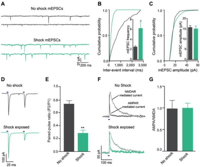
Acute unpredictable foot shock exposure enhances LHb-to-RMTg glutamate release
Since neurotransmission by LHb neurons may encode information related to aversive stimuli processing (Matsumoto and Hikosaka, 2009a), we explored whether exposure to an aversive stimulus altered excitatory neurotransmission at LHb-to-RMTg synapses. We exposed mice expressing ChR2-eYFP in LHb-to-LHb-to-RMTg fibers to either 0 or 19 unpredictable foot shocks in a single 20-min session. One hour later, we performed whole-cell recordings from RMTg neurons in close proximity to LHb-to-RMTg ChR2-eYFP-positive fibers. Voltage clamp recordings from RMTg neurons from foot shock-exposed mice displayed an increase in the frequency of miniature excitatory postsynaptic currents (mEPSCs) compared to non-shocked controls (Figure 2.2 A-C). Furthermore, LHb-to-RMTg glutamate release probability was significantly enhanced following shock exposure, as indexed by a reduction in the optically-evoked paired pulse ratio (Figure 2.2 D,E). We observed no
differences in mEPSC amplitude or optically-evoked AMPA/NMDA ratios,
Activation of LHb inputs to the RMTg produces active, passive, and conditioned behavioral avoidance
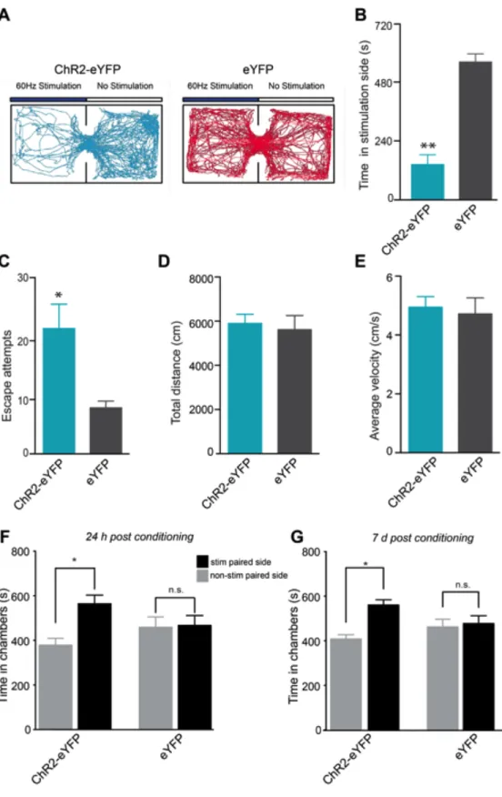
To determine whether optogenetic stimulation of LHb-to-RMTg fibers has behavioral consequences, we optogenetically stimulated this pathway in behaving mice at 60-Hz as this was the mean light-evoked firing rate of LHb neurons in brain slices (Figure 2.1 B,C). To determine if optogenetic stimulation of LHb-to-RMTg fibers resulted in passive avoidance behavior, we tested mice in a real-time place preference chamber. When an experimental mouse crossed over into a counter-balanced stimulated-designated, contextually indistinct side of an open field, light stimulation was constantly pulsed until the mouse crossed back into the
non-stimulated designated side. Mice expressing eYFP spent equal times on both sides of the chamber, whereas mice expressing ChR2-eYFP spent significantly less time on the stimulated side (Figure 2.3 A,B) and made significantly more escape
attempts (Figure 2.3 C). There were no differences in total distance traveled or average velocity between ChR2-eYFP and eYFP mice across the entire session (Figure 2.3 D,E). These data suggest that acute activation of LHb-to-RMTg fibers promotes location-specific passive avoidance behavior.
was maintained in the ChR2-eYFP-expressing mice 7 days following the last
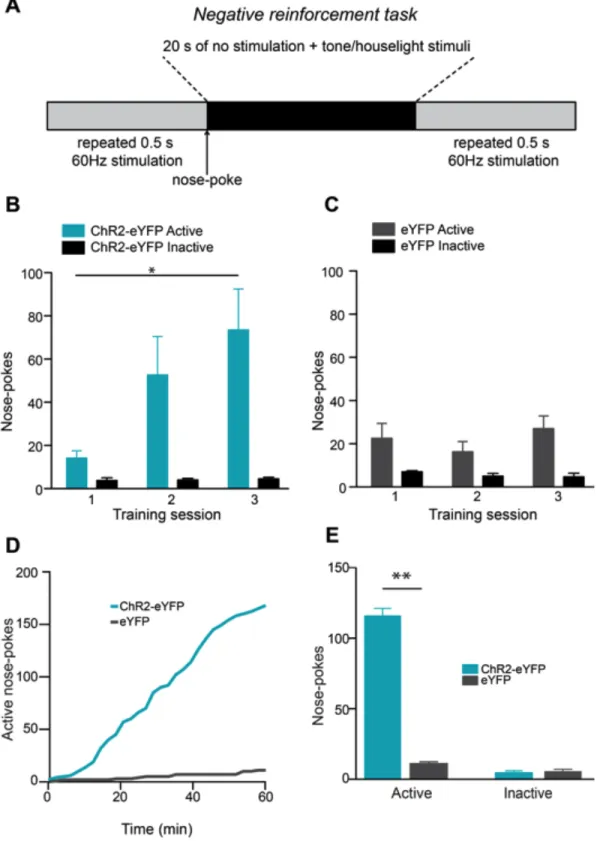
conditioning session (Figure 2.3 G) demonstrating that activity in this pathway also promotes conditioned avoidance. To determine if mice would perform an operant response to actively avoid activation of LHb-to-RMTg fibers, ChR2-eYFP or eYFP expressing mice were placed in chambers where they could nose-poke to terminate optogenetic stimulation of LHb-to-RMTg fibers (Figure 2.4 A).
ChR2-eYFP-expressing mice learned to nose-poke to terminate laser stimulation over 3 daily training sessions (Figure 4 B,C). Following training, ChR2-eYFP-expressing mice made significantly more active nose-pokes to terminate LHb-to-RMTg activation compared to eYFP-expressing mice (Figure 4 D,E), resulting in a significant increase in the percentage of time the stimulation was off (percent time stimulation was off: ChR2-eYFP: 47.5 ± 7.1 %; eYFP: 2.8 ± 0.9 %; t(10) = 6.28, p < 0.0001). These data demonstrate that LHb-to-RMTg activity can negatively reinforce behavioral responding.
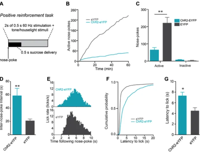
Next, we examined whether LHb-to-RMTg activation disrupted positive reinforcement. We trained a separate group of mice to nose-poke to earn liquid sucrose rewards. Following stable responding, nosepokes to earn sucrose in subsequent test sessions where paired with a 2s, 60-Hz LHb-to-RMTg stimulation (Figure 2.5 A). ChR2-eYFP-expressing mice receiving stimulations made
= 1.64, p = 0.12), suggesting that stimulation of this pathway time-locked to an operant response served as a punishment.
DISCUSSION
We found that activation of LHb terminals in the RMTg promotes active, passive, and conditioned behavioral avoidance, suggesting that endogenous activity of LHb glutamatergic inputs to the RMTg conveys information related to aversion. The data presented here suggest that the LHb’s connection with midbrain GABA neurons is crucial for promoting these behaviors. Consistent with this, direct
excitation of VTA GABA neurons disrupts reward-related behaviors (van Zessen et al., 2012) and stimulation of VTA GABA neurons or inhibition of VTA dopamine neurons promotes aversion (Tan et al., 2012). Importantly, optogenetic stimulation of LHb terminals in the RMTg suppressed positive reinforcement and supported
LHb projections are phylogenetically well-conserved (Stephenson-Jones et al., 2012), neurotransmission in this pathway is likely essential for survival by promoting learning and subsequent behavior to avoid stimuli associated with negative
FIGURES
the LHb. Neurons were counter-stained using a red fluorescent Nissl stain. D, Dorsal; V, Ventral; M, Medial; L, Lateral. (B,C) Activation of ChR2 expressed in LHb cell bodies in brain slices resulted in sustained high frequency activation during the 500 ms stimulation (n=7 cells). (D) Sagittal confocal image showing expression of ChR2-eYFP in the LHb-to-midbrain pathway via the fasciculus retroflexus fiber bundle following injection of the viral construct into the LHb. Midbrain TH+
dopaminergic neurons are shown in blue. A, Anterior; P, Posterior. (E) Horizontal confocal image showing the distribution of LHb terminals in the midbrain. (F) Confocal compressed z-stack showing that ChR2-eYFP is expressed in LHb projection fibers in the RMTg after virus injection into the LHb. (G) Postsynaptic optically-evoked EPSCs recorded from RMTg neurons were significantly attenuated following bath application of 10µM DNQX (t6 = 3.94, p = 0.07, n = 4 cells). (H) LHb efferents to the RMTg were stimulated at 60 Hz for all behavioral tasks. Optically-evoked EPSCs at this frequency for 500 ms show a significant reduction in
Figure 2.2: Acute unpredictable foot shock exposure enhances LHb-to-RMTg glutamate release. (A) Representative mEPSC traces recorded from neurons from mice immediately following either 0 or 19 unpredictable foot shocks. (B)
synapses were significantly depressed from mice that received foot shocks (t14 = 3.56, p = 0.003, n = 8 cells/group). (F) Representative optically evoked