Research Paper
Determinants of host susceptibility to murine respiratory syncytial virus
(RSV) disease identify a role for the innate immunity scavenger receptor
MARCO gene in human infants
Monica High
a,1, Hye-Youn Cho
a,1, Jacqui Marzec
a,1, Tim Wiltshire
b, Kirsten C. Verhein
a, Mauricio T. Caballero
c,
Patricio L. Acosta
c,d, Jonathan Ciencewicki
a, Zackary R. McCaw
a, Lester Kobzik
e, Laura Miller-DeGraff
a,
Wes Gladwell
a, David B. Peden
f, M. Elina Serra
c, Min Shi
g, Clarice Weinberg
g, Oscar Suzuki
b, Xuting Wang
h,
Douglas A. Bell
h, Fernando P. Polack
c,i,⁎
, Steven R. Kleeberger
a,⁎⁎
a
Immunity, Inflammation, and Diseases Laboratory, National Institute of Environmental Health Sciences, Research Triangle Park, NC, USA
b
Division of Pharmacotherapy and Experimental Therapeutics, Eshelman School of Pharmacy, University of North Carolina, Chapel Hill, NC, USA
cFundación INFANT, Buenos Aires, Argentina
dConsejo Nacional de Investigaciones Cientificas y Tecnicas (CONICET), Buenos Aires, Argentina e
Department of Environmental Health, Harvard University School of Public Health, Boston, MA, USA
f
Center for Environmental Medicine, Asthma and Lung Biology, School of Medicine, University of North Carolina at Chapel Hill, Chapel Hill, NC, USA
g
Biostatistics and Computational Biology Branch, National Institute of Environmental Health Sciences, Research Triangle Park, NC, USA
h
Genome Integrity & Structural Biology Laboratory, National Institute of Environmental Health Sciences, Research Triangle Park, NC, USA
iDepartment of Pediatrics, Vanderbilt University, Nashville, TN, USA
a b s t r a c t
a r t i c l e i n f o
Article history:
Received 15 October 2015
Received in revised form 2 August 2016 Accepted 5 August 2016
Available online 6 August 2016
Background:Respiratory syncytial virus (RSV) is the global leading cause of lower respiratory tract infection in infants. Nearly 30% of all infected infants develop severe disease including bronchiolitis, but susceptibility mech-anisms remain unclear.
Methods:We infected a panel of 30 inbred strains of mice with RSV and measured changes in lung disease param-eters 1 and 5 days post-infection and they were used in genome-wide association (GWA) studies to identify quantitative trait loci (QTL) and susceptibility gene candidates.
Findings:GWA identified QTLs for RSV disease phenotypes, and the innate immunity scavenger receptorMarco was a candidate susceptibility gene; targeted deletion ofMarcoworsened murine RSV disease. We characterized a humanMARCOpromoter SNP that caused loss of gene expression, increased in vitro cellular response to RSV infection, and associated with increased risk of disease severity in two independent populations of children in-fected with RSV.
Interpretation:Translational integration of a genetic animal model and in vitro human studies identified a role for MARCOin human RSV disease severity. Because no RSV vaccines are approved for clinical use, genetic studies have implications for diagnosing individuals who are at risk for severe RSV disease, and disease prevention strat-egies (e.g. RSV antibodies).
Published by Elsevier B.V. This is an open access article under the CC BY-NC-ND license (http://creativecommons.org/licenses/by-nc-nd/4.0/).
Keywords:
Infectious disease Innate immunity Lung
Single nucleotide polymorphism SNP
Promoter Haplotype
1. Introduction
RSV is the primary cause for hospitalization during thefirst year of life in infants worldwide, and it is the leading cause of lower respiratory tract infection leading to bronchiolitis, pneumonia, and respiratory fail-ure (Lozano et al., 2012). RSV is also a significant cause of respiratory ill-ness in immunocompromised adults and the elderly (Falsey et al., 2005). While the majority of infected subjects present symptoms, the nature and severity of the symptoms vary among individuals (Ciencewicki et al., 2014; Caballero et al., 2015). For some, infection in-duces cold-like symptoms; others require hospitalization, and a small ⁎ Correspondence to: F.P. Polack, Vanderbilt University, Department of Pediatrics,
B-3307 MCN, 1161 21st Ave South, Nashville, TN 37232-2007, USA.
⁎⁎ Corresponding author at: Immunity, Inflammation, and Diseases Laboratory, National Institute of Environmental Health Sciences (NIEHS), 111 T.W. Alexander Drive, Building 101, MD D-201, Research Triangle Park, NC 27709, USA.
E-mail addresses:[email protected](F.P. Polack),
[email protected](S.R. Kleeberger). 1
These authors contributed equally to this work.
http://dx.doi.org/10.1016/j.ebiom.2016.08.011
2352-3964/Published by Elsevier B.V. This is an open access article under the CC BY-NC-ND license (http://creativecommons.org/licenses/by-nc-nd/4.0/).
Contents lists available atScienceDirect
EBioMedicine
percentage of cases require intensive care and may result in death. The wide variation in response to RSV infection suggests that susceptibility and disease are influenced by multiple host intrinsic factors (Stark et al., 2002; Miyairi and DeVincenzo, 2008; Gelfand, 2012; Feldman et al., 2015).
Previous in vivo mouse studies suggest that RSV infectivity is a mul-tigenic trait, but did not identify the genes responsible for infection (Prince et al., 1979; Stark et al., 2002; Stark et al., 2010). The innate and acquired immune systems have been implicated in the pathogene-sis of RSV infection, including surfactant association proteins, viral re-sponse receptors and proteins, and inflammatory cells and their products such as chemokines, cytokines, and soluble inflammatory me-diators [e.g. (Mukherjee and Lukacs, 2013; Varga and Braciale, 2013)]. However, roles for each of these components in the pathogenesis of RSV disease are unclear.
RSV disease is complex and, while candidate gene single nucleotide polymorphisms (SNPs) have associated with RSV disease severity in infants [e.g. (Miyairi and DeVincenzo, 2008; Daley et al., 2012; Ciencewicki et al., 2014; Caballero et al., 2015)], the mechanisms of dif-ferential disease susceptibility remain unclear. To better understand the genetic basis of RSV disease, we performed a genome-wide association (GWA) study of disease following infection in inbred strains of mice to identify candidate quantitative trait loci (QTL) and genes that associate with specific disease phenotypes. We identified 7 significant and 8 sug-gestive QTLs and candidate susceptibility genes including macrophage receptor with collagenous structure (Marco), an innate immunity scav-enger receptor. Targeted deletion of mouseMarcoworsened RSV dis-ease phenotypes, consistent with a protective role in disdis-ease pathogenesis. We then identified and characterized in vitro a human
MARCOpromoter SNP that diminished mRNA expression basally and after RSV infection. The SNP is located in an antioxidant response ele-ment (ARE) of theMARCOpromoter and is a binding site for the tran-scription factor nuclear factor (erythroid derived)-2 like 2 (NFE2L2), also known as nuclear factor erythroid 2 (NF-E2)-related factor 2 (NRF2). Deletion of the ARE also reducedMARCOmRNA expression fol-lowing RSV infection in airway epithelial cells. Moreover, compared to the wild-type C allele, we found that theMARCOpromoter rs1318645 SNP G allele associated with increased disease severity in populations of RSV-infected infants. Our modeling of disease after RSV infection in mice thus identified a host susceptibility gene candidate that has impli-cations for better understanding human disease.
2. Materials and Methods
2.1. Mice and Treatments
Mice from 30 inbred strains (male, 6–10 weeks) for the strain screen were purchased from Jackson Laboratory (Bar Harbor, ME) and housed singly with hardwood bedding (PJ Murphy Forest Products Company, Montville, NJ) in pathogen-free facilities at NIEHS. A breeding colony ofMarcodeficient mice (Marco−/−) was developed and maintained at NIEHS (Arredouani et al., 2004; Cho et al., 2012). Age- and sex-matched C57BL/6 wild-type (WT) mice were purchased from Charles River Lab-oratories (Wilmington, MA) and used as controls for experiments with theMarco−/−mice. All mice were provided free access to water and pelleted open-formula rodent diet NIH-07 (Zeigler Brothers, Gardners, PA.). Constant air temperature (72 ± 3° F), relative humidity (50 ± 15%), and light/dark cycle (12 h on/12 h off) were maintained during the experiments. Mice of each strain were randomly assigned to either RSV infection or vehicle control groups. Mice were infected in the morn-ing with 106plaque-forming units (PFU) of human RSV-19 strain in 50μl Hank's balanced salt solution (HBSS) by intranasal instillation, and returned to their cages. HBSS containing HEp-2 cell lysates were in-tranasally instilled for vehicle controls. On post-instillation days 1 and 5, mice (6–12 per group) were killed by overdose with Nembutal (sodium pentobarbital; 104 mg/kg body weight). Phenotyping for response to
RSV infection was performed in at least two separate experiments (n≥3 per group), and power calculations based on previous studies were used to determine sample sizes. All animal use was approved by the NIEHS Animal Care and Use Committee. Animals were treated hu-manely and with regard to alleviation of suffering.
2.2. Lung Bronchoalveolar Lavage Fluid (BALF) and Cell Preparation
The right lung of each mouse was lavaged in situ consecutively four times with HBSS (35 ml/kg body weight, pH 7.2–7.4) and BALF was an-alyzed for cell differentials using standard morphological and cytologi-cal criteria as described previously (Saltini et al., 1984; Cho et al., 2001). Total protein content (a marker of lung permeability) was mea-sured with the Bradford assay as previously described (Cho et al., 2001; Cho et al., 2007). Investigators were blinded to treatment for all lavage and histopathology samples (see below). Time points chosen for evalu-ation of inflammation and injury were based on a previous investigation of phenotype kinetics in lungs of a panel of 5 inbred strains of mice in-fected with RSV (data not shown). We found that the peaks of infl am-mation and injury phenotypes are time-dependent and usually occur either 1 or 5 days after infection as described previously (Cho et al., 2009).
2.3. Lung Histopathology
Left lung lobes were inflated intratracheally in situ andfixed with 10% neutral buffered zinc formalin for 24 h following procedures de-scribed previously (Cho et al., 2009). Fixed lungs were then placed in 70% ethanol until proximal and distal intrapulmonary axial tissues were excised and embedded in paraffin and sectioned (5μm). Tissue sections were stained histochemically with hematoxylin and eosin (H&E) for histopathological analyses and alcian blue-periodic acid shift (AB/PAS) double staining to identify acidic and neutral mucosubstances (Cho et al., 2009).
2.4. Measurement of Stored Intraepithelial Mucus (Volume Density, VS)
Images of histological sections were captured using an Axioskop 40 microscope, camera, and Axiovision software. Black and white images were taken with a 10 × objective lens and analyzed with Scion image analysis software. The mucus-containing area and the perimeter of basal lamina from large airways, visually judged to be third generation bronchus, were used for analysis. The volume of mucus per airway was calculated as described previously (Harkema et al., 1987).
VS values (nl/mm2
basal lamina) were calculated for each lung sec-tion and averaged for vehicle control and RSV infected groups. VS calcu-lations were averaged for each treatment group and are reported as the mean ± SEM for each strain (n = 3/infection/strain).
2.5. Quantitative Real-time Polymerase Chain Reaction (qRT-PCR)
Total RNA was isolated from non-lavaged left lung pieces using RNeasy Mini Kits (Qiagen, Valencia, CA) following manufacturer proce-dures and as described previously (Cho et al., 2009). Quantification of gene expression was determined byΔCTvalues obtained by subtracting fluorescence detected number of cycles to threshold (CT) of 18s mRNA from those of target gene RNA in the same sample.
2.6. Heritability and Correlation of Phenotypes
response [1 or 5 days post-infection (PI)] for each phenotype was used for all analyses.
2.7. Genome-wide Association Mapping
GWA mapping was performed using the mean value for each pheno-type for each strain. Data for each strain were distributed normally (no outliers removed), and no transformations were applied. We also gen-erated a RSV disease index by performing a principal components (PC) analysis of each quantitated phenotype and then applied thefirst 3 PCs for each strain to haplotype association mapping. Two different haplotype-based approaches were used: SNPster (Genomics Institute, Novartis Research Foundation, San Diego, CA) and FastMap (University of North Carolina, Chapel Hill, NC). The algorithms (Pletcher et al., 2004; Gatti et al., 2009) and rationale for using these approaches have been described elsewhere (McClurg et al., 2006; Nichols et al., 2014). Exper-imental informative SNP genotypes for GWA mapping were selected as described previously (Benton et al., 2012). The SNPs used for the 30 strains of mice were fully genotyped (i.e. not imputed).−Log10P values were considered significant if they exceeded 6.60 (genome-wide signif-icance at Pb0.05) and suggestive if they exceeded 6.06 (genome-wide suggestive at Pb0.20). Only QTLs that were identified using both algo-rithms were considered further for candidate gene analyses. Candidate gene SNP and haplotype data were obtained from the Mouse Phenome Database (MPD; Jackson Laboratory;http://phenome.jax.org; dbSNP 138 annotation). Non-informative SNPs (i.e. no heterozygosity between strains or the minor allele was found inb10% of the strains) were re-moved from further haplotype and SNP analyses.Marcohaplotype data were obtained from the MPD, and hierarchical clustering was per-formed as described previously (Cho et al., 2015).
2.8. Sequencing of MARCO Promoter and Functional Assessment
We used traditional walk-down sequencing to identify regions of
MARCOthat were polymorphic, and confirmed these sites by screening an ethnically diverse panel of individuals for allelic variation. Regions with SNPs at 5% or higher frequency were used in promoter deletion ex-periments to test for effects on transcriptional activity. Constructs were generated to encompass the full length of theMARCOpromoter (−1388 to +98), and polymorphic regions were sequentially deleted to assess differential activity.
Initial promoter deletion assays identified a polymorphic region of the MARCO promoter with high activity (−300 and + 98) that contained a putative NRF2-binding ARE. To determine whether the SNP region influenced transcript levels, the−300 to + 98 region of
MARCOcontaining the rs1318645,−156 C/G SNP (−157) was cloned upstream of the luciferase reporter gene and its expression analyzed by transient transfection using reporter assays. The−300 to−71 pro-moter region was further investigated by generating two constructs [−300 to +98 bp, (300-Luc) and−71 to +98 bp (71-Luc, lacking the SNP)] and comparing their promoter activity.
To examine effects of the promoter SNP on transcriptional activity, we amplified the 5′-flanking region of genomicMARCOpromoter to generate polymorphic variants of the 300-Luc sequence [−156 C-Luc and−156 G-Luc]. All constructs were verified by sequencing, and tran-siently transfected using Fugene HD (Promega, Madison, WI) into BEAS-2B cells (The American Type Culture Collection, Manassas, VA), and an-alyzed for luciferase activity as described previously (Cho et al., 2015).
For RSV infection we used the pGL4.20 vector, and Beas2B cells were transfected withMARCOpromoter constructs (−156 C-Luc,−156 G-Luc or−71-Luc) overnight and were serum starved for 1 h during RSV infection (MOI of 4). Cells were harvested 3, 6, 24, 48 and 72 h post-infection. Luciferase activity was assessed in three independent samples and all experiments were repeated in triplicate.
2.9. Electrophoretic Mobility Shift Assay (EMSA)
EMSA allows visualization of transcription factor binding affinity to regulatory promoter sequences in the presence of variant alleles. DNA-protein binding specificity was further assessed using antibodies specific to the binding factor of interest. An aliquot (5μg) of human lung nuclear protein (Active Motif, Carlsbad, CA) was incubated on ice with binding buffer for 15 min, followed by addition of 3 × 10⁴cpm [γ32
P] ATP (Amersham Biosciences, Piscataway, NJ) end-labeled wild type or variant probes (End-labeled probes: rs1318645−156 C (wt) FORWARD 5′-CCT GGG AGT GAC GGG TGC ATT CAG AG-3′, REVERSE 5′-CT CTG AAT GCA CCC GTC ACT CCC AGG-3′; rs1318645−156G (var-iant) FORWARD 5′-CCT GGG AGT GAG GGG TGC ATT CAG AG-3′, RE-VERSE 5′-CT CTG AAT GCA CCC CTC ACT CCC AGG-3′) and incubated for 30 min at room temperature. We used short (26 base pairs) frag-ment sequences for the EMSA experifrag-ments that overlap the consensus sequence forNRF2. We used polyclonal rabbit anti-NRF2 antiserum (2μl) or control IgG (2μl) as controls and processed for EMSA as de-scribed previously (Marzec et al., 2007). Samples were electrophoresed and autoradiographed with an intensifying screen at−70 °C. Genotyp-ing was done without knowledge of disease status (i.e. blinded to case or control). We tested for statistically significant differences in DNA-NRF2-antibody complex formation between wild-type, heterozygous, and variant genotypes using one-way ANOVA with SNK a posteriori comparisons of means.
2.10. Human RSV Disease Population
Two independent prospective case-control studies were conducted in Buenos Aires, Argentina from 2003 to 2006 (population 1) and 2010–2013 (population 2); these studies, with inclusion and exclusion criteria, were described previously (Caballero et al., 2015). Study partic-ipants were previously healthy full-term infants younger than 1 year of age and born after 15 September of the previous year (i.e. approximate-ly 15 days after the end of RSV season in Buenos Aires). Participants had bronchiolitis symptoms for thefirst time in their lives, and clinical signs included wheezing with or without cough, rales, dyspnea, and increased respiratory rate and retractions of the respiratory muscles. Diagnosis was performed by trained pediatricians. Infants were tested for RSV, rhinoviruses, hPIV1–4, influenza virus A and B, and human metapneumovirus, and all participating subjects were infected only with RSV. Recruited cases were infants with bronchiolitis whose oxygen saturation upon enrollment wasb93% in room air (the primary out-come). Oxygen saturation is a clinically relevant phenotype for discrim-inating between mild and severe RSV disease (Caballero et al., 2015). Infants with bronchiolitis were recruited as controls if oxygen satura-tion upon enrollment was≥93% while breathing room air. Genotyping of infants was performed using whole blood samples (population 1) or nasal aspirates (population 2). Population sizes were based on calcu-lations to provide sufficient statistical power (~80%) tofind significant differences between mild and severe disease with a minor allele fre-quency of ~0.2. The Institutional Review Boards (IRB) of the participat-ing hospitals in Buenos Aires, as well as the Johns Hopkins University IRB (Baltimore, MD, USA), approved the protocol. We obtained in-formed consent from parents of each infant enrolled in the studies. The study conformed to standards indicated by the Declaration of Helsinki.
2.11. Allelic Discrimination Genotyping for MARCO rs1318645
Genomic DNA was isolated and characterized for purity and concen-tration as described previously (Caballero et al., 2015). TheMARCO
assay mix (C_9566534_20). The 20 × mix consisted of 18μM forward and reverse primers and 8μM of each allele-specificfluorescently la-beled (VIC or FAM) TaqMan MGB probe. Standard PCR cycling condi-tions were used as described previously (Caballero et al., 2015).
Allele-specific PCR products were detected and verified as described previously (Caballero et al., 2015). Five percent of the samples were re-peated for quality control, and we found 100% concordance.
2.12. Statistical Analysis
Lavage and tissue parameters were analyzed by 3-way ANOVA (fac-tors: exposure, time, strain) and Student-Newman-Keuls (SNK) com-parisons of means procedures. Strain distribution patterns andMarco
haplotype groups were analyzed by 1-way ANOVA and SNK compari-sons of means. Statistical associations between candidate gene
a
c
e
b
d
0.855
0.549
0.544
0.610
genotypes and phenotypes within mouse strains were calculated using the two-tailed Student'st-test. Only the significance of murine non-synonymous coding SNPs were tested in this study; if more than one non-synonymous coding SNP was found within a gene multiple com-parisons were performed. Because of potential bias effects of admixture in our human study populations, we followed methods described previ-ously to test for evidence of bias due to population stratification (Epstein et al., 2007) using 33 ancestral informative markers (AIMs; Suppl. Table S1) that were evaluated in 300 infants with mild or severe RSV disease. Genotyping was performed on the Sequenom iPlex plat-form by BioServe (Beltsville, MD, USA). In the two infant populations, we usedχ2
analyses (dominant and recessive modeling; SigmaPlot 13.0) to test for associations betweenMARCOgenotypes and RSV dis-ease severity. We assessed potential confounding of demographic vari-ables in the relationships betweenMARCOgenotype and RSV disease severity using an additive model for multiple logistic regressions. Statis-tical significance in these and all other comparisons was accepted at Pb0.05.
3. Results
3.1. Phenotypic Characterization of RSV Disease in Inbred Mice
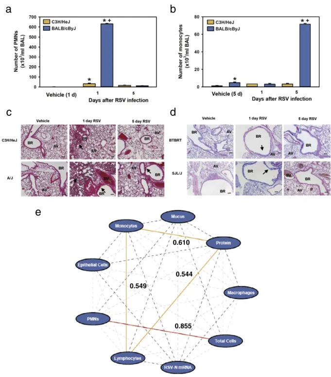
We developed a genetic model of RSV disease by infecting intra-nasally 30 strains of mice. Inflammation was assessed in BALF by measuring total protein concentration and counting inflammatory and epithelial cells 1 and 5 days PI. The time courses of inflammation and injury phenotypes indicated early (acute) and late phase re-sponses in all of the strains tested. Relative to vehicle controls, mean numbers of total cells, polymorphonuclear leukocytes (PMNs), epithelial cells, and total protein in BALF of infected mice were significantly increased 1 day PI and declined 5 days PI in the majority (N50%) of strains (representative strains,Fig. 1a; all strains, Suppl. Table S2). Numbers of BALF macrophages, lymphocytes, and monocytes (representative strains,Fig. 1b; all strains, Suppl. Table S2) peaked at 5 days PI in the majority of strains. The differences in kinetics may reflect the sequential contributions of the innate and acquired immune systems in response to infection as others have found [e.g. (Kok et al., 2012; Stoppelenburg et al., 2013)]. Qualitative inspection of lung histopathology sections was consistent with BALF inflammation phenotypes (Fig. 1c). Mucus cell metaplasia is a clini-cal feature of RSV disease, and we found the mean amount of intraepithelial cell mucus (Fig. 1d; Suppl. Table S2) and lung RSV-N mRNA expression (Suppl. Table S2) were greatest 1 day PI in 59% and 70% of the strains, respectively.
We also found statistically significant inter-strain variation in the peak of each RSV disease phenotype. That is, significantly smaller num-bers of inflammatory cells and other indicators of injury (mild disease) were found in a subset of strains (e.g. C3H/HeJ) compared to others with severe disease (e.g. BALB/cByJ) (Fig. 2a–d; Suppl. Table S2; Suppl. Fig. S1). Heritability estimates of RSV disease phenotypes ranged from 47.6% (BALF protein, 1 day PI) to 85.7% (intraepithelial mucus, 1 day PI) (Suppl. Table S2). To better understand mechanisms of response to RSV infection, we tested for correlation of disease phenotypes. Pearson's correlation analyses of RSV disease phenotypes among 30 strains found that responses 1 day PI did not correlate with responses 5 days PI (data not shown). When maximum responses for each parameter were com-pared, few were significantly correlated (Fig. 1e;Fig. 2). Interestingly, RSV-N mRNA expression was not correlated with any response pheno-type, afinding consistent with lack of correlation we found between RSV disease severity and RSV titers in infected children (Caballero et al., 2015). Large inter-strain variation and relatively high heritability of distinct disease phenotypes suggests the mouse model of response to RSV infection is multi-genic and mimics the variability of RSV disease severity found in human populations.
3.2. Identification of QTLs and Gene Candidates for RSV Disease Phenotypes
Because the phenotypic responses to RSV infection were largely not correlated, we reasoned that the phenotypes may have some QTLs in common, but likely also have unique QTLs. Therefore, we used GWA mapping to identify QTLs for each phenotype across the entire inbred strain data set. Using the maximum mean response phenotypes for each strain, mapping with SNPster identified significant (−Log10 PN6.6) QTLs for RSV-induced changes in BAL monocytes (chromo-somes 1 and 17;Fig. 3a,Table 1) and PMNs (chromosomes 3 and 8; Table 1), and thefirst PC (chromosomes 4, 11, and 13;Table 1). Sugges-tive QTLs (−Log10PN6.0) were also found for intraepithelial mucus content (chromosomes 7 and 15), BALF total protein concentration (chromosomes 9 and 19), and lung RSV-N mRNA expression (chromo-somes 1 and 11) (Table 1). Importantly, all QTLs were also identified using FastMap (data not shown). GWA mapping of other RSV response phenotypes with SNPster did not identify significant or suggestive QTLS, or were not confirmed with FastMap.
To further investigate genetic contributions to RSV disease pheno-types we searched the MPD for informative SNPs in susceptibility gene candidates within significant and suggestive QTLs, and whether they
d
c
b
a
associated with respective disease phenotypes. Genes were chosen for investigation if they had biological plausibility for a role in RSV disease and if the minor allele of candidate SNPs were present in≥10% of the
thus supporting a potential role for these genes in murine RSV disease severity.
We focused initially on the chromosome 1 QTL for monocyte infl am-mation to validate gene candidates because this QTL (~1.299 Mb) had the greatest GWA−Log10P value (Table 2). The scavenger receptor geneMarcois located in the QTL (Fig. 3b) and is in linkage disequilibri-um (LD) with the SNP that has the highest−Log P value in the QTL, and humanMARCOis upregulated in children infected with RSV [i.e. biolog-ical plausibility (Fjaerli et al., 2006)]. Hierarchical clustering ofMarco
SNPs identified 3 major haplotypes in the strain panel (Fig. 3c; Suppl. Table S3). Mean numbers of RSV-induced BAL monocytes in strains fromMarcohaplotypes 2 and 3 were significantly greater than those in strains from wild-type haplotype 1 (Fig. 3d). Potentially functional, informative SNPs are located inMarco, including the non-synonymous coding SNP rs30741725 (A/T) that causes a threonine to serine substitu-tion at the 476 amino acid residue in exon 17. Significantly increased numbers of BAL monocytes and lymphocytes 5 days PI were found in strains of mice with the minor T allele for this SNP compared with strains that have the major A allele (Table 2; Suppl. Fig. S2A and S2B). The majority of strains with the T allele haveMarcohaplotypes 2 or 3. The rs30741725 A/T SNP is in LD with other SNPs in theMarco haplo-type, so a definitive role for this SNP in differential RSV responsiveness requires further investigation. There are 5 other protein-coding genes in the QTL withMarco, but none have known biological plausibility for the RSV response phenotypes or do not meet criteria for further study (stated above).
3.3. Genetic Deletion of Marco Modifies RSV-induced Pulmonary Infl amma-tory Response
We further investigated the role ofMarcoon RSV disease severity by infectingMarco+/+andMarco−/−mice with RSV. Relative to vehicle controls, RSV increased BALF monocytes inMarco+/+andMarco−/− mice, and numbers of monocytes in Marco−/−mice were slightly lower compared toMarco+/+mice 1 day PI, during the development of the monocyte response. However, numbers were significantly greater inMarco−/−mice compared to those inMarco+/+mice at the peak of the response (5 days PI;Fig. 3e, left). Mean numbers of PMNs were also significantly increased inMarco+/+andMarco−/−mice 1 day PI, but were significantly greater in Marco−/− mice compared to
Marco+/+ mice, and remained significantly elevated at 5 days PI (Fig. 3e, right). BALF inflammation phenotypes were consistent with lung histopathology sections from Marco+/+ and Marco−/− mice (Fig. 3f). No significant genotype effects were found for other RSV BALF phenotypes (data not shown). Lack of resolution of the PMN response inMarco−/−mice is consistent with the hypothesis that inflammatory cell clearance is impaired in these mice.
3.4. Functional Assessment of the Human MARCO Promoter
Based on our GWA investigation and identification ofMarcoas a gene candidate for differential RSV disease severity in the mouse, we queried dbSNP (build 129, 2008) to identify potentially relevant SNPs in humanMARCObased on functional annotation. A missense SNP
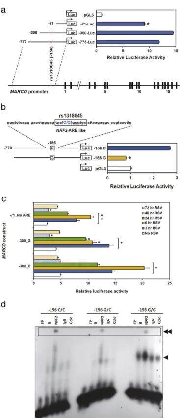
(rs61732822, Asp511Glu) in the scavenger receptor cysteine rich (SRCR) domain of exon 17 (similar to that found in mouseMarco), is predicted by SIFT (sift.jcvi.org) to have deleterious consequences, but the 1000 genomes project phase 1 (www.1000genomes.org) indicates the alternate allele frequency of 0.055 is too low to study in our popula-tions of children with RSV-induced bronchiolitis. We then asked wheth-er theMARCOpromoter contains polymorphic regions that might alter transcriptional efficiency. We consecutively deleted sections of the pro-moter from−1388 to−71 and probed for transcriptional activity using luciferase reporter constructs. We identified a region close to the tran-scription start site (−300 to−71) that, when deleted, significantly re-duced transcriptional activity (Fig. 4a). We then screened commercially available DNA samples (n = 40; Coriell Institute, Camden, NJ) for SNPs in theMARCOpromoter region. Forty subjects (20 European, 20 African-American) were chosen to provide a detection rate of 98% for SNPs present in as little as 5% of the population. The screen identified a SNP (rs1318645, −156 C/G; Suppl. Fig. S3) that contained a putative antioxidant response element for NRF2 [C al-lele, position weight matrix (PWM) score = 10.9; G alal-lele, PWM score = 7.5]. Mutation of the−156 site from C (wild type) to G completely reduced luciferase activity to that of the pGL3 basic vec-tor (Fig. 4b).
Polymorphic and ARE deletion constructs cloned in BEAS-2B epithe-lial cells were also assessed for transcriptional activity following RSV in-fection. RobustMARCOactivation was found 3, 6 and 24 h PI, and mutation from C to G at the−156 site reduced activity approximately 50% at 6 h PI, and constructs with ARE deletion caused similar reduction in activity (Fig. 4c). At 24 h PI, luciferase activity in constructs with ARE deletion was reduced by 50% compared to that of wild type−300C
Fig. 3.Genome-wide haplotype association map of BALF monocytes in 30 mouse strains infected with RSV andMarcohaplotype clustering and monocytic inflammatory response to RSV infection. (a) Manhattan plot for maximum mean BALF monocytes in 30 strains of mice. X axis, chromosome number and cumulative genomic position; Y axis,−Log10P values. The rectangle on chromosome 1 identifies a locus significantly associated with RSV-induced monocyte infiltration. (b) Detailed view of the chromosome 1 QTL for monocyte response to infection. The geneMarcois indicated within the locus. (c) Hierarchical clustering of 29 mouse strains based on 87MarcoSNPs (http://phenome.jax.org/SNP). Numbers represent uncertainty in genotype changes after clustering (Φ), whereΦ= 1−cluster entropy / crude entropy. If, after clustering, all strains in the cluster have the same genotype thenΦ= 1; ifΦ= 0, the genotype distribution within the cluster is as variable as the genotype distribution across all strains. (d) Box plots of numbers of BALF monocytes following RSV infection in strains withMarcohaplotypes 1, 2, and 3. The boundary of the box closest to zero indicates the 25th percentile, a line within the box marks the median, and the boundary of the box farthest from zero indicates the 75th percentile. Error bars above and below the box indicate the 90th and 10th percentiles. Outlying points are shown for haplotype 1. *Pb0.05 versusMarcohaplotype 1 (1-way ANOVA and Student-Newman-Keuls comparisons of means). (e) Differential pulmonary disease phenotypes after RSV infection inMarco+/+and Marco−/−mice. Means (±SEM) of monocytes (left) and PMNs (right) recovered in BALF after vehicle or RSV infection. *Pb0.05 versus vehicle;+
Pb0.05 versusMarco+/+
mice (3-way ANOVA with Student-Newman-Keuls comparisons of means). (f) Pulmonary pathology 1 and 5 days following vehicle control or RSV infection inMarco+/+
andMarco−/−mice. Arrows indicate areas of increased airway inflammation and bronchial epithelial proliferation (hyperplasia). AV = Alveoli, BR = bronchus or bronchiole, BV = blood vessel. Bar = 100μm.
Table 1
Quantitative trait loci (QTL) type, genomic location, and maximum−Log10P value iden-tified by haplotype association mapping using SNPster and FastMap in 30 inbred strains of mice.
Phenotype Chromosome QTL type Genomic location (Mb) Max
−Log10P Monocytes 1 Significant 122.292051–123.591461 8.0764
17 Significant 63.717121–63.851568 6.2074 Lymphocytes 1 Suggestive 52.516061–52.626962 4.2831
4 Suggestive 154.164390–154.306615a 4.5528 11 Suggestive 32.132950–33.743190a
4.6383 PMNs 3 Significant 90.189278–92.304415 7.0324 8 Significant 116.222735–121.343307 6.1851 Mucus 7 Suggestive 130.318155–130.535201 4.2492 15 Suggestive 10.421905–11.278506 4.3576 Protein 9 Suggestive 99.381216–99.610367 5.8803 19 Suggestive 42.164991–42.683021 5.2600 RSV N 1 Suggestive 92.939552–93.424360 4.5088 1 Suggestive 151.202928–152.311735 4.8599 11 Suggestive 116.303373–116.468685 4.5465 PC 1 4 Significant 153.237964–154.148432a 6.6645
11 Significant 31.520635–32.654798a 6.8655 13 Significant 60.210819–60.772421 6.7123
constructs (Fig. 4c). Transcription activity was close to basal levels 48 and 72 h PI (data not shown).
Gel shift analysis showed reduced NRF2 specific binding in the pres-ence of the variant G allele in the potential ARE (Fig. 4d), suggesting binding efficiency is altered at this polymorphic site. Overall binding was increased with the G allele, suggesting additional factors may bind this regulatory locus.
3.5. Role of MARCO rs1318645 in RSV Disease Severity
Next we asked whether theMARCOSNP rs1318645 that caused re-duced MARCO gene expression in vitro (see above) associated with in-creased risk of severe RSV disease in populations of infected infants from our prospective case-control study (Ciencewicki et al., 2014; Caballero et al., 2015). Demographic characteristics are described else-where (Caballero et al., 2015). Genotyped (Fig. 5a) infants with mild and severe infection had combined allelic frequencies (Suppl. Table S4) similar to those published previously (http://www.1000genomes. org/). We were unable to successfully genotype 39 infants in thefirst population and 25 in the second population, and there was no associa-tion of unsuccessful genotyping between cases and controls. Allelic fre-quencies satisfied Hardy-Weinberg equilibrium (P = 0.369) and, although admixture was present we found no evidence of bias due to population stratification (i.e. stratification was not predictive of differ-ential RSV disease severity).χ2
analysis of RSV disease severity found the rs1318645 C/G polymorphism was associated with increased risk of severe RSV disease (Fig. 5b;χ2= 5.15, P = 0.053). The association was largely attributed to GG risk effect (recessive model) where infants homozygous for the G allele were at significant risk of developing severe RSV disease compared to CC and CG genotypes (Suppl. Table S5). In a second, independent population, we found theMARCOrs1318645 C/G allele was again associated with RSV disease severity in infected infants (Fig. 5b;χ2= 8.15, P = 0.017) and similar to thefirst population, the as-sociation was driven by GG genotype (Suppl. Table S5). Asas-sociation of theMARCOrs1318645 C/G allele with RSV disease severity in the com-bined population was significantly strengthened compared to either population alone (Fig. 5b;χ2= 10.34, P = 0.003), and the homozygous GG genotype conferred approximately 62% greater risk of severe RSV disease compared to CC and CG genotypes (Suppl. Table S5). Using mul-tiple logistic regression we found theMARCOrs1318645 C/G allele was associated with increased risk of severe RSV disease (unadjusted OR: 1.654, 95% CI: 0.98, 2.58). After adjusting for gender, socioeconomic sta-tus, and breastfeeding the SNP remained associated with severe RSV disease (adjusted OR: 1.684, 95% CI: 1.03, 3.15). Similar outcomes were found in the second population (unadjusted OR: 1.744, 95% CI: 1.10, 2.44; adjusted OR: 1.82, 95% CI: 1.13, 2.56) and in the combined
populations (unadjusted OR: 1.615, 95% CI: 1.24, 2.35; adjusted OR: 1.676, 95% CI: 1.28, 2.45).
4. Discussion
The impact of RSV disease, particularly in vulnerable infants, remains a global public health concern (Lozano et al., 2012). Unfortunately, no RSV vaccines are approved for clinical use. Therefore, it is critical to identify host risk factors that lead to development of prevention and in-tervention strategies to reduce risk of RSV disease morbidity and mor-tality, as well as asthma incidence because RSV disease has been identified as a predisposing risk factor (Dakhama et al., 2005; Edwards et al., 2012; Gelfand, 2012; Feldman et al., 2015). Our study used ge-nome-wide association analyses of RSV disease parameters that model human disease to identify biologically plausible susceptibility gene can-didates, includingMarco. Moreover, a missense loss of function poly-morphism in the humanMARCOhomolog also associated with severe RSV disease in two populations of infected infants, and illustrates the utility of a genetic mouse model for understanding human disease. Im-portantly, this study provides additional genetic insight that may lead to a means to identify individuals at risk for severe RSV disease.
Considerable evidence has suggested that genetic background is an important host determinant of RSV disease severity, but full characteri-zation of susceptibility genes for this complex disease remains unclear. Previous genetic investigations of human RSV disease have questioned whether polymorphisms in gene candidates associate with innate and acquired immune responses to infection and have provided some understanding of susceptibility factors. Gene candidates in these studies were chosen because of prior knowledge about biological plausibility. We comprehensively characterized RSV disease pheno-types in a large panel of inbred strains representing genetic diversity that is present in the mouse genome. We found significant variation and robust heritability in each RSV disease phenotype between the strains. However, relatedness between maximum response phenotypes was minimal, suggesting multiple genetic pathways likely contribute to the responses.
Anh and Desmecht (2006)reported that pneumonia virus of mice (PMV, the murine form of RSV) infection in a panel of 6 strains caused strain-specific histopathologic inflammatory effects, decreased body weight, and changes in pulmonary function particularly 5–7 days PI. The studies are not directly comparable because mice were infected with different viruses and late phase PI times investigated, but the inter-strain variation in phenotypes is consistent with ourfindings. Others (Stark et al., 2002) compared RSV infection in 8 inbred strains and found significant inter-strain variation in infectivity at 4 days PI, but histopathologic changes were minimal and cellular effects were Table 2
Associations of candidate gene functional single nucleotide polymorphisms (SNPs) with RSV-induced lung inflammation phenotypes.
Phenotype QTL chromosome Gene symbol SNP rs ID Location (Mb) SNP function Major allele, mean ± SEM, N Minor allele, mean ± SEM, N P-value
Monocytes 1 Marco rs30741725 120.474787 CNSyn, Thr476Ser A, 38.4 ± 8.8; 19 T, 14.5 ± 2.7; 10 b0.001 Lymphocytes 11 Mpg rs26841220 32.231754 CNSyn, Arg254Gln G, 28.9 ± 6.1; 24 A, 80.6 ± 22.6; 6 0.004 PMNs 3 Lor rs31130301 91.885023 CNSyn, Gly293Arg G, 134.3 ± 18.8, 16 C, 275.2.5 ± 50.4; 12 0.005 8 Adamts18 rs37229383 116.233617 CNSyn, Gly957Arg G, 226.1 ± 34.1; 21 C, 116.2 ± 24.3; 7 0.046 Mucus 7 Aqp8 rs32822168 123.462409 5′UTR A, 1.13 ± 0.3; 24 C, 0.6 ± 0.2; 3 NS Protein 19 Sfrp5 S19-42272711 42.272711 3′UTR C, 342.1 ± 32.3; 19 T, 222.2 ± 13.4; 7 0.042 RSV-N 1 Ramp1 rs47959958 91.206647 3′UTR A, 5.6 ± 0.3; 14 G, 2.3 ± 0.3; 12 b0.001
Espnl rs48467891 91.320153 CNSyn, Gly406Ser G, 4.8 ± 0.5; 19 A, 2.8 ± 0.4; 11 0.011
Per2 rs32164384 91.445550 CNSyn, Ser172Thr G, 4.7 ± 0.4; 21 C, 2.4 ± 0.4; 9 0.003 1 Ptgs2 rs32559831 151.948631 Intronic A, 5.1 ± 0.5; 14 T, 2.5 ± 0.4; 10 b0.001 11 Rhbdf2 rs29403288 116.471777 3′UTR C, 4.9 ± 0.4; 20 T, 2.4 ± 0.5; 7 0.005 PC1 11 Mpg rs26841220 32.231754 CNSyn, Arg254Gln G,−0.50 ± 0.3; 24 A, 1.99 ± 0.1; 6 b0.001
discordant with the magnitude of infection likely due to the time point when acute virus effects on histopathology were no longer present.
We leveraged extensive genetic diversity among the 30 strains of mice to perform GWA of multiple RSV disease phenotypes and thus were not guided a priori by hypotheses about the role of specific genes. Significant and suggestive QTLs were identified for inflammatory cells, intraepithelial cell mucus, and RSV N mRNA expression. Several genes that may contribute to viral response, pulmonary inflammation,
and cytokine trafficking were found within the identified QTLs [e.g.
Ptgs2(Obata et al., 2013),Ramp1(Li et al., 2014),Rhbdf2(Issuree et al., 2013)], and putative functional SNPs in the genes associated with differential RSV disease phenotypes among the strains. SNPs in other QTL genes strongly associated with disease phenotypes and, while little is known about their function in infectious disease, they may be impor-tant targets for future investigation of their roles in RSV disease progres-sion. We also found significant QTLs using thefirst PC as an RSV disease index. Two of these QTLs overlapped those found for lymphocytes, and suggests that further investigation of genes within the QTLs could pro-vide greater understanding of disease susceptibility. Future GWA inves-tigations using PC analyses may provide additional insight to disease pathogenesis beyond using single phenotypes. Interestingly, genome-wide linkage analyses of RSV infectivity in back-cross and F2 popula-tions derived from C57BL/6J and AKR/J mice byStark et al. (2010) iden-tified a QTL on chromosome 6 that associated with increased susceptibility to infection, though disease phenotypes were not pre-sented. While GWA in the present investigation identified suggestive QTLs on chromosomes 1 and 11 for RSV-N mRNA expression, none were detected on chromosome 6. Differences in study design (e.g. hap-lotype mapping with many strains versus linkage analysis with 2 strains, PI time differences) may have contributed to this discrepancy.
We focused on the gene candidateMarco, which was identified by GWA for monocytes, and has been shown previously to have protective effects in the lung following inhalation of particulate matter, exposure to ozone, and bacterial infection (Arredouani et al., 2005; Dahl et al., 2007; Thakur et al., 2008; Thakur et al., 2009; Ghosh et al., 2011).
MARCOgene expression was also upregulated in children infected with RSV (Fjaerli et al., 2006). Our hypothesis thatMarcocontributes to defense against RSV-induced pulmonary inflammation was support-ed by two lines of evidence. First, strains of mice with aMarcohaplotype containing the T allele of missense coding SNP rs30741725 were at greater risk of developing more severe RSV disease compared to strains with the wild type A allele. Second, numbers of BALF monocytes and PMNs were significantly increased inMarco−/−mice compared to
Marco+/+mice. The mechanism through whichMarcomodulates the inflammatory response to RSV is not completely understood. We initial-ly hypothesized that Marco has a direct role in clearing RSV from the air-ways, perhaps binding the virus for internal degradation by macrophages. To test this hypothesis, we infected with RSV lung macro-phages isolated fromMarco+/+andMarco−/−mice but found no differ-ences in infectivity, with and without co-culturing with epithelial cells (data not shown). Marco also has an important role in clearance of ap-optotic cells (Rogers et al., 2009; Getts et al., 2014), and recent studies have targeted Marco receptors with immune-modifying particles to re-solve monocytic inflammation (Getts et al., 2014). It is plausible that
Fig. 4.Effect of a genetic polymorphism on in vitroMARCOpromoter activity. (a) Deletion analysis of theMARCOpromoter compared to pGL3 basic vector. *Pb0.05 versus− 300-Luc; group sizes = 4. (b) Mutagenesis analysis of theMARCOrs1318645 variant (−156G) compared to pGL3 basic vector. Bars (means ± SEM) represent luciferase activity in basic vector, control−156 C allele, and mutant−156 G constructs. Flanking sequence forMARCOrs1318645 is shown at top with the NRF2 response element boxed. *Pb0.05 versus−156 C (P = 0.001); group sizes = 4. Statistical analyses, one-way ANOVA and Student-Newman-Keuls comparisons of means. (c) Luciferase activity of RSV infectedMARCOreporter constructs.MARCOpromoter constructs were transiently transfected and cells infected with RSV (MOI of 4). Bars (means ± SEM) represent luciferase responses after no RSV (control) or 3–72 h post-infection (PI). Cells were harvested 3–72 h PI and relative luciferase activity assessed. *Pb0.05 versus RSV-infected−300_C (WT);+
loss of function mutations inMarcocompromise clearance of monocytes and apoptotic cells which leads to accumulation of these cells in the air-ways, further contributing to RSV disease pathology. Impaired resolu-tion of the inflammatory response to RSV infection and more severe disease inMarco−/−mice compared toMarco+/+mice is consistent with this hypothesis.
It is important to note thatMarcodoes not account for all of the monocyte inflammation caused by RSV infection. Targeted deletion of
Marcosignificantly increased susceptibility to some RSV-induced phe-notypes but did not account for all of the genetic variation identified by the strain screen. The background strain (C57BL/6 in this model, a moderate responder to RSV) likely contributes to part of this difference as the choice of background can influence the effect of targeted deletion in mice [e.g. (Baleato et al., 2008; Leontyev et al., 2012)]. Perhaps more importantly, other QTLs were also identified by the GWA and gene can-didates within them also contribute to the genetic component of the phenotype. Further investigation of these QTLs should provide addition-al insight to the genetic contribution to RSV disease susceptibility.
BecauseMarcohad an important role in modulating the infl ammato-ry response elicited by RSV, we sought to determine whether human
MARCOwas also important in RSV disease. We initially queried whether theMARCOrs1318645−156 C/G SNP caused loss of function because it was located in an ARE proximal to the transcription start site. Others had found that the transcription factor NRF2, which binds to AREs to in-crease gene transcription, had a role inMarcoexpression in mice
(Harvey et al., 2011; Bonilla et al., 2013). We showed previously that murineMarcobears a potentially functional ARE for Nrf2 binding (−940 region, PWM score = 8.9) in a model of neonate acute lung in-jury (Cho et al., 2012). In the present study, we found that deletion of the ARE and mutating the−156 site in an ARE in humanMARCOalso significantly attenuated gene expression as well asMARCO:NRF2 specif-ic binding. Moreover, deletion of the ARE also significantly reduced ex-pression ofMARCO in cells infected with RSV thus suggesting an important role for the ARE and the rs1318645 −156 C/G SNP in
MARCOfunction, basally and in response to viral infection. This is sup-ported by our previous study in which increased lung injury and infl am-mation were found inNrf2deficient mice compared to wild type mice after RSV infection (Cho et al., 2009).
We further investigated the role ofMARCOin human RSV disease by asking whether the missenseMARCOrs1318645 G allele was more prevalent in infected children with severe disease. In two independent populations, infected children homozygous for the rs1318645 G allele were more likely to have severe RSV disease than children who were heterozygous or wild-type for the C allele. The significance of the risk ef-fect was increased when the populations were combined. TheMARCO
promoter SNP increased the risk for severe RSV disease by approximate-ly 62%. A previous investigation tested whether 9 non-synonymous cod-ing variants or tag SNPs inMARCOassociated with chronic obstructive pulmonary disease and found no association, although one of the SNPs (rs41279766) associated with the risk of sepsis (Thomsen et al., 2013). Another study found tag SNPs associated with increased risk of pulmonary tuberculosis, while another was associated with resistance (Bowdish et al., 2013), thus the role ofMARCOin this disease is not clear. A caveat for this investigation is that, although we tested for a num-ber of potential co-factors in our studies, it is possible that other con-founders or unmeasured environmental factors may contribute to the differences in disease severity that associated with the MARCO
rs1318645 G allele. For example, gender has been found to be important in susceptibility to some lung diseases in mouse models (Carey et al., 2007; Cabello et al., 2015). In the present study, we used only male mice. Although we are not aware of sexual dimorphism effects in RSV disease, male and female mice respond differentially to influenza A in-fection (Larcombe et al., 2011). Another important RSV disease pheno-type is airways hyperreactivity (Yoo et al., 2013). We chose to focus the present studies on the inflammation and injury responses to RSV in-fection, but investigations into the genetic basis of susceptibility to RSV-induced airway hyperreactivity may also identify important susceptibil-ity genes. It is also feasible that different strain distribution patterns may be found with a different line of RSV.Lukacs et al. (2006)found that dif-ferent strains of RSV of the same antigenic subgroup may cause difdif-ferent RSV-induced phenotypes in BALB/cJ mice, and it is possible that other strains also respond differently to multiple RSV lines. Finally, it is impor-tant to emphasize that, while the rs1318645 SNP associated signifi cant-ly with RSV disease in these populations, other gene pocant-lymorphisms also contribute to susceptibility, including myxovirus (influenza virus) resistance 1 (MX1) (Ciencewicki et al., 2014) and toll-like receptor 4 (TLR4) (Caballero et al., 2015), and interactions between these and other, unknown, genetic contributions remain to be clarified. It is likely that continued investigation of gene candidates identified with GWA for all of the phenotypes in the mouse model will add to our understanding of RSV disease susceptibility.
In conclusion, this translational investigation identified gene candi-dates, includingMarco, for host susceptibility to multiple phenotypes of RSV disease in mice that closely mimic human disease, and a poly-morphism in humanMARCOassociated with increased risk of RSV dis-ease severity in infants. Continued investigation of the genetic basis of differential susceptibility to RSV disease may lead to a panel of informa-tive SNPs to identify children who are at risk for disease severity. In the absence of an RSV vaccine, these individuals could be treated prophylac-tically with an RSV monoclonal antibody to prevent infection and se-quelae associated with severe disease such as childhood asthma. Fig. 5.Effect of theMARCOrs1318645 C/G polymorphism on human RSV disease severity.
(a) Representative electropherograms of theMARCOpromoter and putative NRF2 binding site with the rs1318645 C/G polymorphism. (b). Role ofMARCOrs1318645 C/G in disease severity among two populations of infants infected with RSV. Infants with mild or severe RSV disease were genotyped for the MARCO SNP and determined to be homozygous for the major (C) or minor (G) allele, or heterozygous.χ2
Funding Sources
Research related to the manuscript was supported by the Intramural Research Program of the National Institutes of Health, NIEHS (Z01 ES 100557).
Conflict of Interest Statement
The authors declare no conflicts of interest or competingfinancial interests.
Author Contributions
S.R.K., M.H., H.C., J.M., and F.P.P. designed the study. M.G.C., P.L.A., M.E.S., and F.P.P. coordinated the infant population studies. M.H., L.M.-D., and W.G. bred and maintained the mice and performed the experi-ments. M.H., T.W., K.C.V., Z.R.M., L.K., D.B.P., M.S., C.W., O.S., X.W., D.A.B., F.P.P., and S.R.K. contributed to data analysis and interpretation. M.H., H.C., and S.R.K. drafted the manuscript. All authors edited the manuscript.
Acknowledgments
Drs. Michael Fessler and Donald Cook at the NIEHS provided excel-lent critical review of the manuscript.
Appendix A. Supplementary Data
Supplementary data to this article can be found online athttp://dx. doi.org/10.1016/j.ebiom.2016.08.011.
References
Anh, D.B.F.P., Desmecht, J.M., 2006.Differential resistance/susceptibility patterns to pneumovirus infection among inbred mouse strains. Am. J. Phys. Lung Cell. Mol. Phys. 291, 426–435.
Arredouani, M., Yang, Z., Ning, Y., Qin, G., Soininen, R., Tryggvason, K., Kobzik, L., 2004.The scavenger receptor MARCO is required for lung defense against pneumococcal pneu-monia and inhaled particles. J. Exp. Med. 200, 267–272.
Arredouani, M.S., Palecanda, A., Koziel, H., Huang, Y.C., Imrich, A., Sulahian, T.H., Ning, Y.Y., Yang, Z., Pikkarainen, T., Sankala, M., et al., 2005.MARCO is the major binding recep-tor for unopsonized particles and bacteria on human alveolar macrophages. J. Immunol. 175, 6058–6064.
Baleato, R.M., Guthrie, P.L., Gubler, M.C., Ashman, L.K., Roselli, S., 2008.Deletion of CD151 results in a strain-dependent glomerular disease due to severe alterations of the glo-merular basement membrane. Am. J. Pathol. 173, 927–937.
Benton, C.S., Miller, B.H., Skwerer, S., Suzuki, O., Schultz, L.E., Cameron, M.D., Marron, J.S., Pletcher, M.T., Wiltshire, T., 2012.Evaluating genetic markers and neurobiochemical analytes forfluoxetine response using a panel of mouse inbred strains. Psychophar-macology 221, 297–315.
Bonilla, D.L., Bhattacharya, A., Sha, Y., Xu, Y., Xiang, Q., Kan, A., Jagannath, C., Komatsu, M., Eissa, N.T., 2013.Autophagy regulates phagocytosis by modulating the expression of scavenger receptors. Immunity 39, 537–547.
Bowdish, D.M., Sakamoto, K., Lack, N.A., Hill, P.C., Sirugo, G., Newport, M.J., Gordon, S., Hill, A.V., Vannberg, F.O., 2013.Genetic variants of MARCO are associated with susceptibil-ity to pulmonary tuberculosis in a Gambian population. BMC Med. Genet. 14, 47.
Caballero, M.T., Serra, M.E., Acosta, P.L., Marzec, J., Gibbons, L., Salim, M., Rodriguez, A., Reynaldi, A., Garcia, A., Bado, D., et al., 2015.TLR4 genotype and environmental LPS mediate RSV bronchiolitis through Th2 polarization. J. Clin. Invest. 125, 571–582.
Cabello, N., Mishra, V., Sinha, U., DiAngelo, S.L., Chroneos, Z.C., Ekpa, N.A., Cooper, T.K., Caruso, C.R., Silveyra, P., 2015.Sex differences in the expression of lung inflammatory mediators in response to ozone. Am. J. Phys. Lung Cell. Mol. Phys. 309, L1150–L1163.
Carey, M.A., Card, J.W., Voltz, J.W., Germolec, D.R., Korach, K.S., Zeldin, D.C., 2007.The im-pact of sex and sex hormones on lung physiology and disease: lessons from animal studies. Am. J. Phys. Lung Cell. Mol. Phys. 293, L272–L278.
Cho, H.Y., Zhang, L.Y., Kleeberger, S.R., 2001.Ozone-induced lung inflammation and hy-perreactivity are mediated via tumor necrosis factor-alpha receptors. Am. J. Phys. Lung Cell. Mol. Phys. 280, L537–L546.
Cho, H.Y., Morgan, D.L., Bauer, A.K., Kleeberger, S.R., 2007.Signal transduction pathways of tumor necrosis factor—mediated lung injury induced by ozone in mice. Am. J. Respir. Crit. Care Med. 175, 829–839.
Cho, H.Y., Imani, F., Miller-DeGraff, L., Walters, D., Melendi, G.A., Yamamoto, M., Polack, F.P., Kleeberger, S.R., 2009.Antiviral activity of Nrf2 in a murine model of respiratory syncytial virus disease. Am. J. Respir. Crit. Care Med. 179, 138–150.
Cho, H.Y., van Houten, B., Wang, X., Miller-DeGraff, L., Fostel, J., Gladwell, W., Perrow, L., Panduri, V., Kobzik, L., Yamamoto, M., et al., 2012.Targeted deletion of nrf2 impairs
lung development and oxidant injury in neonatal mice. Antioxid. Redox Signal. 17, 1066–1082.
Cho, H.Y., Jedlicka, A.E., Gladwell, W., Marzec, J., McCaw, Z.R., Bienstock, R.J., Kleeberger, S.R., 2015.Association of Nrf2 polymorphism haplotypes with acute lung injury phe-notypes in inbred strains of mice. Antioxid. Redox Signal. 22, 325–338.
Ciencewicki, J.M., Wang, X., Marzec, J., Serra, M.E., Bell, D.A., Polack, F.P., Kleeberger, S.R., 2014.A genetic model of differential susceptibility to human respiratory syncytial virus (RSV) infection. FASEB J. 28, 1947–1956.
Dahl, M., Bauer, A.K., Arredouani, M., Soininen, R., Tryggvason, K., Kleeberger, S.R., Kobzik, L., 2007.Protection against inhaled oxidants through scavenging of oxidized lipids by macrophage receptors MARCO and SR-AI/II. J. Clin. Invest. 117, 757–764.
Dakhama, A., Lee, Y.M., Gelfand, E.W., 2005.Virus-induced airway dysfunction: pathogen-esis and biomechanisms. Pediatr. Infect. Dis. J. 24, S159–S169 (discussion S166–157).
Daley, D., Park, J.E., He, J.Q., Yan, J., Akhabir, L., Stefanowicz, D., Becker, A.B., Chan-Yeung, M., Bosse, Y., Kozyrskyj, A.L., et al., 2012.Associations and interactions of genetic poly-morphisms in innate immunity genes with early viral infections and susceptibility to asthma and asthma-related phenotypes. J. Allergy Clin. Immunol. 130, 1284–1293.
Edwards, M.R., Bartlett, N.W., Hussell, T., Openshaw, P., Johnston, S.L., 2012.The microbi-ology of asthma. Nat. Rev. Microbiol. 10, 459–471.
Epstein, M.P., Allen, A.S., Satten, G.A., 2007.A simple and improved correction for popula-tion stratification in case-control studies. Am. J. Hum. Genet. 80, 921–930.
Falsey, A.R., Hennessey, P.A., Formica, M.A., Cox, C., Walsh, E.E., 2005.Respiratory syncytial virus infection in elderly and high-risk adults. N. Engl. J. Med. 352, 1749–1759.
Feldman, A.S., He, Y., Moore, M.L., Hershenson, M.B., Hartert, T.V., 2015.Toward primary prevention of asthma. Reviewing the evidence for early-life respiratory viral infec-tions as modifiable risk factors to prevent childhood asthma. Am. J. Respir. Crit. Care Med. 191, 34–44.
Fjaerli, H.O., Bukholm, G., Krog, A., Skjaeret, C., Holden, M., Nakstad, B., 2006.Whole blood gene expression in infants with respiratory syncytial virus bronchiolitis. BMC Infect. Dis. 6, 175.
Gatti, D.M., Shabalin, A.A., Lam, T.C., Wright, F.A., Rusyn, I., Nobel, A.B., 2009.FastMap: fast eQTL mapping in homozygous populations. Bioinformatics 25, 482–489.
Gelfand, E.W., 2012.Development of asthma is determined by the age-dependent host re-sponse to respiratory virus infection: therapeutic implications. Curr. Opin. Immunol. 24, 713–719.
Getts, D.R., Terry, R.L., Getts, M.T., Deffrasnes, C., Muller, M., van Vreden, C., Ashhurst, T.M., Chami, B., McCarthy, D., Wu, H., et al., 2014.Therapeutic inflammatory monocyte modulation using immune-modifying microparticles. Sci. Transl. Med. 6, 219ra217.
Ghosh, S., Gregory, D., Smith, A., Kobzik, L., 2011.MARCO regulates early inflammatory re-sponses against influenza: a useful macrophage function with adverse outcome. Am. J. Respir. Cell Mol. Biol. 45, 1036–1044.
Harkema, J.R., Plopper, C.G., Hyde, D.M., St George, J.A., 1987.Regional differences in quantities of histochemically detectable mucosubstances in nasal, paranasal, and nasopharyngeal epithelium of the bonnet monkey. J. Histochem. Cytochem. 35, 279–286.
Harvey, C.J., Thimmulappa, R.K., Sethi, S., Kong, X., Yarmus, L., Brown, R.H., Feller-Kopman, D., Wise, R., Biswal, S., 2011.Targeting Nrf2 signaling improves bacterial clearance by alveolar macrophages in patients with COPD and in a mouse model. Sci. Transl. Med. 3, 78ra32.
Issuree, P.D., Maretzky, T., McIlwain, D.R., Monette, S., Qing, X., Lang, P.A., Swendeman, S.L., Park-Min, K.H., Binder, N., Kalliolias, G.D., et al., 2013.iRHOM2 is a critical path-ogenic mediator of inflammatory arthritis. J. Clin. Invest. 123, 928–932.
Kok, W.L., Denney, L., Benam, K., Cole, S., Clelland, C., McMichael, A.J., Ho, L.P., 2012.Pivotal Advance: invariant NKT cells reduce accumulation of inflammatory monocytes in the lungs and decrease immune-pathology during severe influenza A virus infection. J. Leukoc. Biol. 91, 357–368.
Larcombe, A.N., Foong, R.E., Bozanich, E.M., Berry, L.J., Garratt, L.W., Gualano, R.C., Jones, J.E., Dousha, L.F., Zosky, G.R., Sly, P.D., 2011.Sexual dimorphism in lung function responses to acute influenza A infection. Influenza Other Respir. Viruses 5, 334–342.
Leontyev, D., Katsman, Y., Branch, D.R., 2012.Mouse background and IVIG dosage are crit-ical in establishing the role of inhibitory Fcgamma receptor for the amelioration of experimental ITP. Blood 119, 5261–5264.
Li, M., Wetzel-Strong, S.E., Hua, X., Tilley, S.L., Oswald, E., Krummel, M.F., Caron, K.M., 2014.Deficiency of RAMP1 attenuates antigen-induced airway hyperresponsiveness in mice. PLoS One 9, e102356.
Lightfoot, J.T., Turner, M.J., Daves, M., Vordermark, A., Kleeberger, S.R., 2004.Genetic infl u-ence on daily wheel running activity level. Physiol. Genomics 19, 270–276.
Lozano, R., Naghavi, M., Foreman, K., Lim, S., Shibuya, K., Aboyans, V., Abraham, J., Adair, T., Aggarwal, R., Ahn, S.Y., et al., 2012.Global and regional mortality from 235 causes of death for 20 age groups in 1990 and 2010: a systematic analysis for the Global Bur-den of Disease Study 2010. Lancet 380, 2095–2128.
Lukacs, N.W., Moore, M.L., Rudd, B.D., Berlin, A.A., Collins, R.D., Olson, S.J., Ho, S.B., Peebles Jr., R.S., 2006.Differential immune responses and pulmonary pathophysiology are in-duced by two different strains of respiratory syncytial virus. Am. J. Pathol. 169, 977–986.
Marzec, J.M., Christie, J.D., Reddy, S.P., Jedlicka, A.E., Vuong, H., Lanken, P.N., Aplenc, R., Yamamoto, T., Yamamoto, M., Cho, H.Y., et al., 2007.Functional polymorphisms in the transcription factor NRF2 in humans increase the risk of acute lung injury. FASEB J. 21, 2237–2246.
McClurg, P., Pletcher, M.T., Wiltshire, T., Su, A.I., 2006.Comparative analysis of haplotype association mapping algorithms. BMC Bioinformatics 7, 61.
Miyairi, I., DeVincenzo, J.P., 2008.Human genetic factors and respiratory syncytial virus disease severity. Clin. Microbiol. Rev. 21, 686–703.
Nichols, J.L., Gladwell, W., Verhein, K.C., Cho, H.Y., Wess, J., Suzuki, O., Wiltshire, T., Kleeberger, S.R., 2014.Genome-wide association mapping of acute lung injury in neonatal inbred mice. FASEB J. 28, 2538–2550.
Obata, K., Kojima, T., Masaki, T., Okabayashi, T., Yokota, S., Hirakawa, S., Nomura, K., Takasawa, A., Murata, M., Tanaka, S., et al., 2013.Curcumin prevents replication of re-spiratory syncytial virus and the epithelial responses to it in human nasal epithelial cells. PLoS One 8, e70225.
Pletcher, M.T., McClurg, P., Batalov, S., Su, A.I., Barnes, S.W., Lagler, E., Korstanje, R., Wang, X., Nusskern, D., Bogue, M.A., et al., 2004.Use of a dense single nucleotide polymor-phism map for in silico mapping in the mouse. PLoS Biol. 2, e393.
Prince, G.A., Horswood, R.L., Berndt, J., Suffin, S.C., Chanock, R.M., 1979.Respiratory syncy-tial virus infection in inbred mice. Infect. Immun. 26, 764–766.
Rogers, N.J., Lees, M.J., Gabriel, L., Maniati, E., Rose, S.J., Potter, P.K., Morley, B.J., 2009.A de-fect in Marco expression contributes to systemic lupus erythematosus development via failure to clear apoptotic cells. J. Immunol. 182, 1982–1990.
Saltini, C., Hance, A.J., Ferrans, V.J., Basset, F., Bitterman, P.B., Crystal, R.G., 1984.Accurate quantification of cells recovered by bronchoalveolar lavage. Am. Rev. Respir. Dis. 130, 650–658.
Stark, J.M., McDowell, S.A., Koenigsknecht, V., Prows, D.R., Leikauf, J.E., Le Vine, A.M., Leikauf, G.D., 2002.Genetic susceptibility to respiratory syncytial virus infection in in-bred mice. J. Med. Virol. 67, 92–100.
Stark, J.M., Barmada, M.M., Winterberg, A.V., Majumber, N., Gibbons Jr., W.J., Stark, M.A., Sartor, M.A., Medvedovic, M., Kolls, J., Bein, K., et al., 2010.Genomewide association analysis of respiratory syncytial virus infection in mice. J. Virol. 84, 2257–2269.
Stoppelenburg, A.J., Salimi, V., Hennus, M., Plantinga, M., Huis in 't Veld, R., Walk, J., Meerding, J., Coenjaerts, F., Bont, L., Boes, M., 2013.Local IL-17A potentiates early neu-trophil recruitment to the respiratory tract during severe RSV infection. PLoS One 8, e78461.
Thakur, S.A., Hamilton Jr., R.F., Holian, A., 2008.Role of scavenger receptor a family in lung inflammation from exposure to environmental particles. J. Immunotoxicol. 5, 151–157.
Thakur, S.A., Beamer, C.A., Migliaccio, C.T., Holian, A., 2009.Critical role of MARCO in crys-talline silica-induced pulmonary inflammation. Toxicol. Sci. 108, 462–471.
Thomsen, M., Nordestgaard, B.G., Kobzik, L., Dahl, M., 2013.Genetic variation in the scav-enger receptor MARCO and its association with chronic obstructive pulmonary dis-ease and lung infection in 10,604 individuals. Respiration 85, 144–153.
Tsaih, S.W., Korstanje, R., 2009.Haplotype association mapping in mice. Methods Mol. Biol. 573, 213–222.
Varga, S.M., Braciale, T.J., 2013.The adaptive immune response to respiratory syncytial virus. Curr. Top. Microbiol. Immunol. 372, 155–171.