LED TRANSILLUMINATION IN DENTISTRY
PART I: LUMINOUS FLUX OF FIBEROPTIC TRANSILLUMINATORS PART II: ASSESSMENT OF LED TRANSILLUMINATOR PROPERTIES FOR
EVALUATION OF CRACKED TEETH
Nicholas Earl Pettit
A thesis submitted to the faculty of the University of North Carolina at Chapel Hill in partial fulfillment of the requirements for the degree of Master of Science in the Endodontics
Department in the School of Dentistry
Chapel Hill 2019
Approved by:
Peter Z. Tawil
Lisiane Ferreira Susin
Ricardo Walter
ABSTRACT
Nicholas Earl Pettit: LED Transillumination in Dentistry Part I: Luminous Flux of Fiberoptic Transilluminators
Part II: Assessment of LED Transilluminator Properties for Evaluation of Cracked Teeth (Under the Direction of Peter Z. Tawil)
The purpose of this experiment was to evaluate the diagnostic test of transillumination.
Throughout the literature transillumination is recognized as an important instrument to be
utilized in the detection of cracks and fractures of teeth; however, there is no evidence found
regarding the ideal specifications of these devices to accomplish this important task. The first
part of this study evaluated the tip diameter and luminous flux (lumens) of different types of
LED transilluminators, while the second part examined the clinical ability of these devices to
detect coronal cracks and fractures in an in vitro model replacing various clinical scenarios. We
found a wide variation in the tip diameter and lumens output of the transilluminator devices that
were tested and correlated a higher sensitivity in crack detection with increased lumens and tip
TABLE OF CONTENTS
LIST OF TABLES ... vi
LIST OF FIGURES ... vii
LIST OF ABBREVIATIONS ... viii
THESIS INTRODUCTION ... 1
REFERENCES ... 5
MANUSCRIPT 1: Luminous Flux of Fiberoptic Transilluminators ... 6
Introduction ... 6
Materials and Methods ... 7
Results ... 11
Discussion ... 12
Conclusions ... 13
REFERENCES ... 14
MANUSCRIPT 2: Assessment of LED Transilluminators Properties for Evaluation of Cracked Teeth ... 15
Introduction ... 15
Materials and Methods ... 17
Sample Selection ... 17
“Silver” Standard for Coronal Assessment ... 18
Sample Preparation and Device Masking ... 18
Coronal Assessment ... 19
Results ... 21
Discussion ... 25
Conclusion ... 28
REFERENCES ... 29
THESIS SUMMARY ... 31
LIST OF TABLES Manuscript 1: Luminous Flux of Fiberoptic Transilluminators
Table 1: Physical Characteristics of Transilluminator Devices ... 11
Manuscript 2: Assessment of LED Transilluminator Properties for Evaluation of Cracked Teeth
LIST OF FIGURES Manuscript 1: Luminous Flux of Fiberoptic Transilluminators
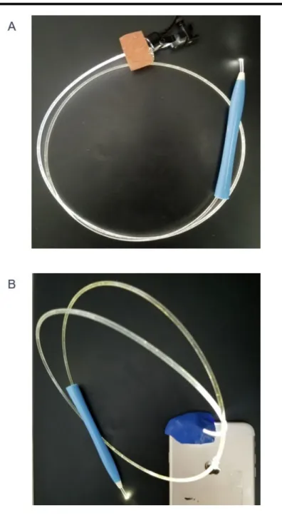
Figure 1: A. Prototype device with attachment for loupes light. B.
Prototype attachment for iPhone ... ..8
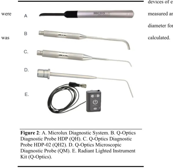
Figure 2: A. Microlux Diagnostic System. B. Q-Optics Diagnostic Probe HDP (QH). C. Q-Optics Diagnostic Probe HDP-02 (QH2). D. Q-Optics Microscopic Diagnostic Probe (QM). E. Radiant
Lighted Instrument Kit (Q-Optics) ... 10
Manuscript 2: Assessment of LED Transilluminator Properties for Evaluation of Cracked Teeth
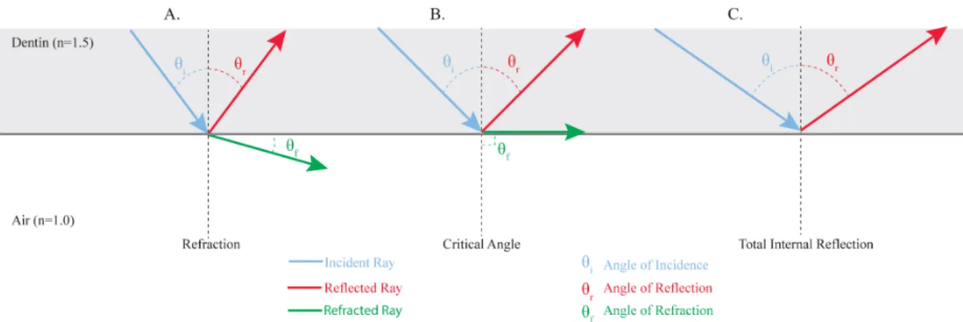
Figure 1: A. Light at fracture line with incident angle less than the critical angle resulting in reflected ray returning through dentin and refracted ray continuing through crack. B. Light at fracture line at critical angle resulting in reflected ray through dentin and refracted ray parallel to the crack line. C. Light at fracture line with incident
angle greater than critical angle resulting in total internal reflection ... 15
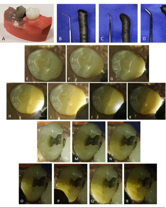
Figure 2: A. Sample tooth mounted for evaluation. B. Q-Optics Microscopic Diagnostic Probe (QM). C. Q-Optics Diagnostic Probe HDP-02 (QH2). D. Q-Optics Diagnostic Probe HDP (QH). Coronal Assessment: E. Magnification with microscope. F. Small probe at low intensity. G. Small probe at high intensity. H.
Medium probe at low intensity. I. Medium probe at high intensity. J. Large probe at low intensity. K. Large probe at high intensity. Endodontic Assessment: L. Magnification with microscope. M. Small probe at low intensity. N. Small probe at high intensity. O. Medium probe at low intensity. P. Medium probe at high intensity.
Q. Large probe at low intensity. R. Large probe at high intensity ... 19
Figure 3: Sensitivity and 95% Confidence Intervals (A) Coronal
LIST OF ABBREVIATIONS
DOM Dental Operating Microscope LED Light Emitting Diode
lm Lumens
MT Microlux Transilluminator
NI iPhone Attachment
NL NoMag
LED Attachment
No Magnification group
QH Q-Optics Diagnostic Probe HDP QH2 Q-Optics Diagnostic Probe HDP-02 QM Q-Optics Microscopic Diagnostic Probe
THESIS INTRODUCTION
Dental practitioners are often challenged with diagnosis and treatment planning of cases
involving fractured teeth (1). Many different methods are proposed for these diagnostic efforts
including a complete dental history, visual examination, vitality and bite testing, periodontal
probing, radiographic examination staining, transillumination, and surgical assessment (2);
however, there is no “catch-all” for this problem. Recently, the American Association of
Endodontists has claimed that transillumination is “the method that provides the most
information, and easily and graphically represents whether a crack is present” (3). The literature
has recommended the use of transillumination in the detection of cracks and fractures of teeth as
early as the 1970s (4,5).
Transillumination is the “passage of a beam of light through a tooth or other tissue for
diagnostic purposes especially in defining fractures” (6), and it is based on the principles of
physics. These physical properties include refraction, reflection, absorption, scatter, and others.
The basic laws dictate that light will continue to travel in a straight path until it encounters
something to change that path; the change that is compelled on light is determined by what type
of impediment to its path is encountered. The most relevant laws to transillumination of teeth are
those of refraction and reflection. Refraction is the change in direction of light as it passes from
one substance density to another; similarly, reflection results in a change of direction of the light
at the interface of two mediums, but, in this case, the result is a return of the light through the
between refraction and reflection is affected by the refractive index, which is given for
any substance as “the ratio of the velocity of light in a vacuum (or air) to its velocity in the
medium” (7). As the light, or incident ray, encounters a change in medium, the relative
differences of the refractive indices of two substances at an interface will determine the direction
of the refracted ray and reflected ray in relation to the “normal”, which is a line perpendicular to
the interface. There is a “critical angle” for any two substances at, and beyond, where total
internal reflection occurs and none of the beam’s energy is transmitted to the second medium;
this phenomenon is dependent upon the difference of their refractive indices and is what makes a
fracture line obvious for the clinician performing the diagnostic test. A continuous movement of
the light around all the tooth surface will maximize angle exposures and will thus enhance the
chances of encountering the critical angle to visualize the fracture.
Transillumination of teeth follows these laws to aid in the detection of cracked teeth. Due
to the differences of refractive indices of dentin and air (1.5 and 1.0, respectively) (8), rays of
light that encounter a crack in a tooth will have their course altered based on the angle of
incidence. If the light encounters the crack at the critical angle or greater, no light will continue
through the crack, resulting in one part of the tooth being illuminated and the other part
remaining dark.
There are many devices currently available commercially that are marketed for the
purpose of transillumination in dentistry, and there are many more tools being used off-label for
the same intention. Many of these modern devices are utilizing light-emitting diodes (LEDs) as
their source of illumination. LEDs have been used for over 100 years, but recent advancements
in the technology have resulted in products that are more efficient, cost effective, smaller, and
current is passed through the device; by manipulating the materials chosen for the
semiconductor, the energy released can be controlled. The LEDs chosen for transilluminators
release energy in the visible light spectrum (~400-700 nm).
The amount of incident light that is transmitted from a transilluminator to a tooth can be
measured in a number of ways. Photometry is the measurement of light in relation to a specific
detector, the human eye. The photometric principles of luminous flux, luminous intensity,
illuminance, and luminance are all part of the function of transillumination. Luminous flux
measures the total output of the light source. Luminous intensity measures the luminous flux in a
given direction. Illuminance is the intensity of light that is incident on a given surface.
Luminance is the amount of light reflected or transmitted by a sample (9).
Many instruments have been used for transillumination in the oral cavity, including
commercially available dental transilluminators, the light from the optical fiber of the high speed
hand piece, curing lights, and rifle bore lights. However, there is no data regarding the physical
and photometric properties of these devices to understand what specifications are optimal for
aiding in accurate detection of cracked teeth. This gap in knowledge may result in clinicians
misdiagnosing cracked teeth, whether that be missing cracks that are present or visualizing
cracks that are not really there at all.
It is important to diagnose cracks accurately, and early, to improve the treatment options
and prognosis of the tooth. In a paper published by Krell and Rivera in 2007 (10), it was
suggested that cracks identified before causing significant pulpal damage can be treated with a
crown and avoid endodontic therapy in almost 80% of cases. Likewise, it is understood that
cracks will continue to propagate without proper treatment, which can eventually result in pulpal
length of cracks along occlusal surfaces and correlated those findings to the extent of the cracks
along the proximal surfaces concluded that these measurements may offer valuable prognostic
value for the tooth in question; specifically, longer cracks on occlusal surfaces resulted in longer
cracks along the proximal surfaces (12). This again shows that early identification of cracks can
result in improved prognosis for the tooth in question.
Despite many recommendations for the use of transillumination in the detection of
cracked teeth and many options for transilluminators being available, there is no research
regarding the tip diameter or luminous flux that is optimal for accurate diagnosis. The purpose of
this thesis was to analyze common transilluminators’ tip diameters and luminous flux, and to
REFERENCES
1. Lubisich EB, Hilton TJ, Ferracane J. Cracked teeth: a review of the literature. J Esthet Restor Dent. 2010 Jun;22(3):158–67.
2. Cracking the Cracked Tooth Code. Endodontics: Colleagues for Excellence (Summer). 2008;1–7.
3. American Association of Endodontists. Transillumination the "Light Detector". AAE 2008;1:1–2.
4. Reynolds RL, Aduddell AC. A clinical evaluation of fiber optics in diagnosis. J South Calif Dent Assoc. 1971 Nov;39(11):896–900.
5. Cooley RL, Barkmeier WW. Diagnosis of the incomplete tooth fracture. Gen Dent. 1979 Apr;27(2):58–60.
6. American Association of Endodontics. AAE Glossary of Terms 9th Edition 2016.pdf [Internet]. 2016. Available from:
https://www.aae.org/specialty/clinical-resources/glossary-endodontic-terms/
7. Sakaguchi RL, Powers JM. Craig’s restorative dental materials [Internet]. St. Louis, Mo.: Elsevier/Mosby; 2012 [cited 2019 Jan 29]. Available from:
http://search.ebscohost.com/login.aspx?direct=true&scope=site&db=nlebk&db=nlabk&A N=1151609
8. Hariri I, Sadr A, Nakashima S, Shimada Y, Tagami J, Sumi Y. Estimation of the enamel and dentin mineral content from the refractive index. Caries Res. 2013;47(1):18–26. 9. Bass M, Optical Society of America, editors. Handbook of optics. 2nd ed. New York:
McGraw-Hill; 1995.
10. Krell KV, Rivera EM. A six year evaluation of cracked teeth diagnosed with reversible pulpitis: treatment and prognosis. J Endod. 2007 Dec;33(12):1405–7.
11. Rivera EM, Walton RE. Longitudinal tooth cracks and fractures: an update and review. Endod Topics. 2015 Nov 1;33(1):14–42.
12. Chen M, Fu K, Qiao F, Zhang X, Fan Y, Wang L, et al. Predicting extension of cracks to the root from the dimensions in the crown. JADA. 2017 Oct;148(10):737–42.
MANUSCRIPT 1: Luminous Flux of Fiberoptic Transilluminators Introduction
The detection and diagnosis of cracks and fractures in teeth is a constant challenge in
dental practice. Accurate identification of cracks is often made difficult by the ambiguity of
reported symptoms, the lack of radiographic evidence, existing restorations, and other obstacles
(1,2). When early detection and intervention occurs, there is a reduced risk of pulpal
involvement, which would subsequently require root canal therapy (3,4) or result in tooth loss.
Among various detection techniques for cracks in teeth, transillumination is reported to be the
most effective diagnostic test (5). Yet, there is a notable lack of research into the physical
characteristics of light sources, i.e., transilluminators, that best enable practitioners to detect
cracks and fractures.
Defects that may compromise the integrity and are identified in the coronal tooth
structure may be classified as craze line, fractured cusp, cracked tooth, or split tooth (4). Craze
lines are nearly ubiquitous in adult teeth and generally non-serious. In these defects, the damage
is present only in enamel and is unlikely to spread to dentin, but they can lead to misdiagnosis as
more significant longitudinal fractures, which may result in unnecessary treatment. Fractured
cusps are most often found with a mesiodistal and faciolingual component, originating at the
occlusal surface and extending subgingivally near the cervical margin of the root; these fractures
are associated with symptoms from biting forces and cold sensitivity and have a good prognosis
direction with varying extension towards, or onto, the root surface. Symptoms and
treatment for cracked teeth are widely variable based on the extent of the crack; the prognosis
ranges from questionable to poor. With split tooth being a situation where extraction is often the
only treatment option, it is key to diagnose the defect in early stages of development for best
prognosis.
Transillumination relies on principles of physics, including reflection and refraction, that
dictates the nature with which light will travel through a given medium (6). The path of the light
remains constant until it encounters a change in the medium. Thus, when light reaches a crack in
a tooth, it interacts with the interface in such a way to alter its path. The change in path often
results in a clear definition with one part of the tooth illuminated and the other part dark.
Many transillumination devices are available currently, and they vary greatly one to
another in design, cost, and performance. A novel prototype, designed to be very low cost and
easy to use, was constructed for the purposes of this study. The aim of this study was to assess
the illuminance and instrument tip diameter of common dental fiberoptic LED transilluminators
and novel prototypes.
Materials and Methods
A novel prototype transillumination device; the Microlux Diagnostic System (AdDent,
Inc., Danbury, CT); and three devices part of the Radiant Lighted Instrument Kit (Q-Optics,
Duncanville, TX) were utilized in this study. An exempt status for the study was approved by the
University of North Carolina Institutional Review Board of Human Research Ethics, IRB
#17-1884.
The novel prototype was constructed to include a fiberoptic cable and an attachment
iPhone 8 (Apple Inc., Cupertino, CA). The attachment segments were connected using polyvinyl
siloxane impression material applied directly to each source. Before setting of the impression
material, a fiber optic cable was inserted to the depth of the LED light source; complete patency
of the PVS was verified following the final set by removing the LED light source and visualizing
that the cable was unobstructed by any remaining material. A plastic tube to facilitate handling
was added to the apparatus near the diagnostic end of the tool (Figure 1). The MicroLux and
Three available tip
sizes of Q-Optics device (QH, QH2, QM) and the 3mm glass Light Guide of the AdDent device
were evaluated. The illuminance of each transilluminator was tested using a digital lux meter
(PM6612L Digital Luxmeter, Peakmeter, China) under standardized conditions. All devices were Figure 1: A. Prototype device with attachment for loupes light.
tested in a dark room to block out any ambient lighting. Each unit was placed onto the lens of the
digital lux meter, slight changes in angulation of the device were made for five seconds, and the
maximum illuminance was recorded. This process was completed ten times for each device, and
the means and standard error of the output were calculated. Five devices of each brand were
tested to determine inter-device reliability, and each source was tested ten times to account for
intra-device error.
The tip of each device, defined as the area that comes into contact with the tooth surface,
was measured using an analog caliper, accurate to within 0.001 inch. The size of tips of five
devices of each brand
were measured and the average
diameter for each device
was calculated.
Results
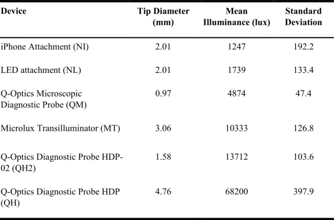
The tip diameter of the test devices was 0.97 mm (QM), 1.58 mm (QH2), 2.01 mm (NI
and NL), 3.06 mm (MT), and 4.76 mm (QH). The mean illuminance (lux) of the test devices was
1247 (NI, 95% CI, 1009-1485); 1739 (NL, 95% CI, 1573-1904); 4874 (QM, 95% CI,
4106-5642); 10333 (MT, 95% CI, 6996-13670); 13712 (QH2, 95% CI, 12028-15395); 68200 (QH,
95% CI, 58550-77849). See Table 1 for additional results.
Table 1: Physical Characteristics of Transilluminator Devices
Device Tip Diameter
(mm) Illuminance (lux) Mean Deviation Standard
iPhone Attachment (NI) 2.01 1247 192.2
LED attachment (NL) 2.01 1739 133.4
Q-Optics Microscopic
Diagnostic Probe (QM) 0.97 4874 47.4
Microlux Transilluminator (MT) 3.06 10333 126.8
Q-Optics Diagnostic Probe
HDP-02 (QH2) 1.58 13712 103.6
Q-Optics Diagnostic Probe HDP
Discussion
The purpose of this study was to assess the physical characteristics of commonly used
transillumination devices and to introduce a novel prototype attachment. Our findings showed
that there is a significant variation in illuminance and tip diameter of instruments.
The prototype device was designed to improve availability and costs associated with
transillumination as these may be obstacles for clinicians in the process of crack detection. By
providing an attachment apparatus to nearly ubiquitous light sources such as dental loupes lights
and cell phone LEDs, nearly all clinicians are already equipped to allow for instant
implementation of such a system into their routine practice. The fiberoptic fiber associated with
the prototype allows for greater adaptation of the tooth, including reaching further into
interproximal area due to its flexibility and relatively small diameter. The ability to sterilize the
device through an autoclave and the low cost associated with the prototype offer additional
benefits with this design. However, the illuminance of this device when attached to either the
dental loupes light or the iPhone LED was low relative to the other devices inspected. Whether
or not the intensity is enough to identify cracked teeth requires further evaluation.
The devices analyzed in this study varied in tip diameter between 0.97 mm to 4.76 mm
and produced illuminance levels ranging from 4874 to 68200 lux. These discrepancies between
devices may have a significant effect on the ability to accurately diagnose cracked teeth. This
data shows that further research is required to assess if this wide variation in transillumination
Conclusions
There is considerable variability in illuminance and tip diameter of commonly used LED
transilluminators available on the market. Further research is required to determine the clinical
REFERENCES
1. Lubisich EB, Hilton TJ, Ferracane J. Cracked teeth: a review of the literature. J Esthet Restor Dent. 2010 Jun;22(3):158–67.
2. Abbott P. Assessing restored teeth with pulp and periapical diseases for the presence of cracks, caries and marginal breakdown. Aust Dent J. 2004 Mar;49(1):33–9.
3. Krell KV, Rivera EM. A six year evaluation of cracked teeth diagnosed with reversible pulpitis: treatment and prognosis. J Endod. 2007 Dec;33(12):1405–7.
4. Rivera EM, Walton RE. Longitudinal tooth cracks and fractures: an update and review. Endod Topics. 2015 Nov 1;33(1):14–42.
5. Cracking the Cracked Tooth Code. Endodontics: Colleagues for Excellence (Summer). 2008;1–7.
MANUSCRIPT 2: Assessment of LED Transilluminators Properties for Evaluation of Cracked Teeth
Introduction
Cracked teeth are encountered by all dental providers on a regular basis (1), and the
diagnosis of these cracks can very often present a significant challenge. The extent of the crack
can have wide ranging effect on the treatment recommendation and prognosis of the tooth (2).
Many authors have attempted to define or classify cracks in effort to standardize this diagnostic
difficulty (3-8). Others have focused on the effectiveness of available diagnostic tests and
methods to accurately detect cracked teeth (9-11), including transillumination, bite and vitality
tests, staining, periodontal probing, removal of existing restorations, radiographic examination,
and surgical assessment. (12-14). Transillumination has been mentioned as an effective method
of crack detection from as early as the 1970s (15), and it continues to be recommended as the
method of detection offering the most information, and best graphic representation, of cracks
well into the 21st century (12).
Transillumination is the “passage of a beam of light through a tooth or other tissue for
diagnostic purposes especially in defining fractures” (16). This method relies on the principles of
physics, including reflection, refraction, absorption, scatter, and others, which determine light’s
path through any substance; light will continue to travel through any medium until it interacts
with particles contained in that medium or reaches an interface where it passes through to
through a tooth and encounters a fracture. Changes in the refractive indices between the
dentin and fracture line result in refraction and reflection of the light at that interface; at angles
greater than the “critical angle”, calculated by the difference in refraction indices, total internal
reflection will occur, which will result in no light passing beyond the crack. This phenomenon is
clinically detectable when teeth with cracks are inspected with a transilluminator and the result is
observed as a clear definition between a part of the tooth that is fully illuminated and another
part that is relatively dark. The accuracy of transillumination, however, has been challenged in
cases with existing restorations and without the aid of magnification (7,9,10) as the introduction
of other materials and interfaces will interfere with the transmission of light regardless of the
presence or absence of a crack.
Figure 1: A. Light at fracture line with incident angle less than the critical angle resulting in reflected ray returning through dentin and refracted ray continuing through crack. B. Light at fracture line at critical angle resulting in reflected ray through dentin and refracted ray parallel to the crack line. C. Light at fracture line with incident angle greater than critical angle
Commercially there are many devices that are marketed as transilluminators, and,
additionally, there are many other devices that are being used as such off-label. These devices
differ significantly in design and cost; however, no research has been performed to evaluate the
physical characteristics required for accurate diagnosis of cracks. The wide variability of sizes
and intensities of transilluminators may result in clinicians mistakenly relying on their device for
diagnosis of cracked teeth even if its properties do not produce optimal sensitivity. The aim of
this study was to evaluate the ability of three LED-transilluminator devices with different
diameter heads (Small - 0.97 mm, Medium - 1.58 mm, Large - 4.56 mm) at varying intensities (L
- Low, H - High) to detect cracks in teeth through an in vitro model, in both coronal and
endodontic assessments . The effectiveness of magnification on crack detection was also
analyzed.
Materials and Methods Sample Selection
Forty-four posterior teeth (22 molars and 22 premolars) extracted for reasons unrelated to
this study were selected for analysis. Twenty-nine of the teeth had existing restorations and/or
caries, and 15 of the samples were unrestored and non-carious. Three diagnostic probes with
distinct tip sizes (QM – 0.97 mm, QH2 – 1.58 mm, QH – 4.76 mm) from the Radiant Lighted
Instrument Kit (Q-Optics, Duncanville, TX) were utilized in this experiment. An exempt status
for the study was approved by the Institutional Review Board of Human Research Ethics, IRB
#17-1884. Five positive controls were selected that contained cracks that were obvious without
any adjunctive diagnostic techniques, and five negative controls were selected where no crack
was detected during selection. The “test samples” selected were found to contain cracks only
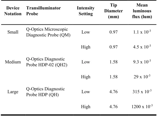
Table 1: Physical Characteristics of Transilluminator Devices
Device
Notation Transilluminator Probe Intensity Setting
Tip Diameter (mm) Mean luminous flux (lum)
Small Q-Optics Microscopic Diagnostic Probe (QM) Low 0.97 1.1 x 10⁻³
High 0.97 4.5 x 10⁻³
Medium Q-Optics Diagnostic Probe HDP-02 (QH2) Low 1.58 9.3 x 10⁻³
High 1.58 29 x 10-3
Large Q-Optics Diagnostic Probe HDP (QH) Low 4.76 315 x 10-3
High 4.76 1200 x 10-3
“Silver” Standard for Coronal Assessment
There is not an established gold standard for confirmation of the presence of cracks in
teeth; therefore, this experiment utilized a “silver” standard, which was an out of socket
examination using high magnification (19.4x) (7) with the aid of Large-High transilluminator to
confirm the presence of a crack.
Sample Preparation and Device Masking
Each sample was mounted into a typodont segment (Acadental, Inc, Overland Park, KS)
using mounting wax and Poly-Vinyl Siloxane (Aquasil Ultra, Dentsply Sirona, York, PA) to the
level of the cemento-enamel junction to simulate a clinical experience during evaluation (see
Figure 1). Samples were stored in purified saline (B. Braun Medical Inc., Irvine, CA) to prevent
The transilluminators were masked for evaluation by placing the diagnostic probe
through silicon straws and injecting PVS impression material to fill the voids. The tip of each
device was trimmed to a similar angle and wrapped with insulating tape (see Figure 2).
Coronal Assessment
Each sample was analyzed by two endodontists with 10+ years of experience under
standardized conditions. The teeth were assessed using the following settings: a standard
overhead dental light (A-dec, Inc, Newberg, OR) with direct vision, the dental operating
microscope (Labo America, Inc, Fremont, CA) set to 7.5x with the light intensity set at
maximum (80,000 lux), and the dental operating microscope set to 7.5x with the light off and
transillumination performed with each of the three diagnostic probes (QM, QH2, QH). Each
device was measure at two distinct intensities (L – Low, H – High), which were adjusted by
changing the settings on the light source to either the maximum or minimum intensity. A
randomized order was generated (www.randomizer.org) for sample assignments. Evaluators
were given 20 s to assess each sample under each of the aforementioned conditions, and their
Endodontic Assessment
Samples remained mounted in the experimental model, and all restorations and/or caries
were excavated (10) and endodontics accesses were prepared using a high speed handpiece under
water irrigation. The two endodontists again inspected each specimen to evaluate for cracks
extending to the internal surfaces of the tooth using the overhead light with no magnification,
dental operating microscope (7.5x), and dental operating microscope (7.5x) with each of the
diagnostic probes following a randomized examination order.
The teeth were removed atraumatically from the typodont segments and stained with
methylene blue dye for final evaluation. Direct inspection of all surfaces of each tooth was
completed out of socket using 19.4x and transillumination to confirm the true presence/absence
of cracks.
Statistical Analysis
Sensitivity and specificity of each device were calculated for both examiners. Sensitivity
of each transilluminator by each examiner were then compared using McNemar’s analysis. Level
of significance was set at 0.05.
Results
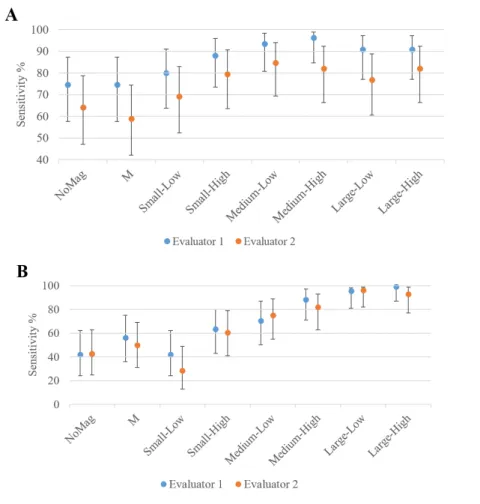
Of the 44 included teeth 39 (89%) were cracked, and 5 (11%) were undamaged (negative
control). Sensitivity for each evaluator following each phase of the study are shown in Figure 2.
In both parts of the study there is an overall trend for increased sensitivity with increased
diameter and luminous flux observed.
In the coronal assessment (see Table 2), the medium-sized light (QH2) exhibited the
highest sensitivity by both examiners (82-97%), but these sensitivities were not statistically
comparison the sensitivities of Small-High, Medium-Low, Medium-High, and Large-High were
found to be significantly higher than the no magnification (64-77%) or dental operating
microscope only (59-77%) groups (see Figure 2).
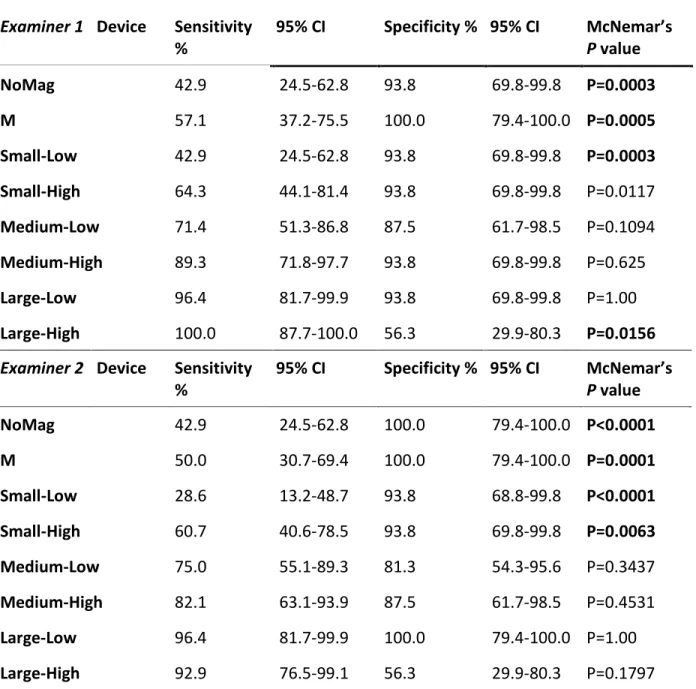
Evaluation of the samples in the endodontic assessment (see Table 3) demonstrated that
the device with the largest diameter (QH) had the highest sensitivity (93-100%). Pairwise
comparison showed that Large-High (93-100%) and Large-Low (96%) were statistically superior
to Medium-High (82-89%), and Medium-High was statistically more sensitive Medium-Low
(71-75%), Small-Low (29-43%), M (50-57%), and NoMag (43%) groups.
Specificity was overall very high for all devices in both the coronal and endodontic
assessments. All groups demonstrated a specificity >81%, except for the Large-High group in the
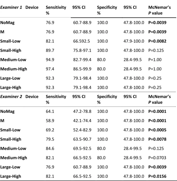
Table 2. Sensitivity and Specificity of Transilluminators in Coronal Assessment
Examiner 1 Device Sensitivity
% 95% CI Specificity % 95% CI McNemar’s P value
NoMag 76.9 60.7-88.9 100.0 47.8-100.0 P=0.0039
M 76.9 60.7-88.9 100.0 47.8-100.0 P=0.0039
Small-Low 82.1 66.592.5 100.0 47.9-100.0 P=0.0082
Small-High 89.7 75.8-97.1 100.0 47.8-100.0 P=0.125
Medium-Low 94.9 82.7-99.4 80.0 28.4-99.5 P=1.00
Medium-High 97.4 86.5-99.9 80.0 28.4-99.5 P=1.00
Large-Low 92.3 79.1-98.4 100.0 47.8-100.0 P=0.25
Large-High 92.3 79.1-98.4 100.0 47.8-100.0 P=0.25
Examiner 2 Device Sensitivity
% 95% CI Specificity % 95% CI McNemar’s P value
NoMag 64.1 47.2-78.8 100.0 47.8-100.0 P=0.0001
M 58.9 42.1-74.4 100.0 47.8-100.0 P<0.0001
Small-Low 69.2 52.4-82.9 100.0 47.8-100.0 P<0.0005
Small-High 79.5 63.5-90.7 100.0 47.8-100.0 P=0.0078
Medium-Low 84.6 69.5-92.5 80.0 28.4-99.5 P=0.125
Medium-High 82.1 66.5-92.5 80.0 28.4-99.5 P=0.0703
Large-Low 76.9 60.7-88.9 100.0 47.8-100.0 P=0.0039
Large-High 82.1 66.5-92.5 100.0 47.8-100.0 P=0.0156
Discussion
The aim of this study was to evaluate the ability of three LED-transilluminator devices
with different diameter heads at varying intensities to detect cracks in teeth and to analyze the Table 3. Sensitivity of Transilluminators in Endodontic Assessment
Examiner 1 Device Sensitivity
% 95% CI Specificity % 95% CI McNemar’s P value
NoMag 42.9 24.5-62.8 93.8 69.8-99.8 P=0.0003
M 57.1 37.2-75.5 100.0 79.4-100.0 P=0.0005
Small-Low 42.9 24.5-62.8 93.8 69.8-99.8 P=0.0003
Small-High 64.3 44.1-81.4 93.8 69.8-99.8 P=0.0117
Medium-Low 71.4 51.3-86.8 87.5 61.7-98.5 P=0.1094
Medium-High 89.3 71.8-97.7 93.8 69.8-99.8 P=0.625
Large-Low 96.4 81.7-99.9 93.8 69.8-99.8 P=1.00
Large-High 100.0 87.7-100.0 56.3 29.9-80.3 P=0.0156
Examiner 2 Device Sensitivity
% 95% CI Specificity % 95% CI McNemar’s P value
NoMag 42.9 24.5-62.8 100.0 79.4-100.0 P<0.0001
M 50.0 30.7-69.4 100.0 79.4-100.0 P=0.0001
Small-Low 28.6 13.2-48.7 93.8 68.8-99.8 P<0.0001
Small-High 60.7 40.6-78.5 93.8 69.8-99.8 P=0.0063
Medium-Low 75.0 55.1-89.3 81.3 54.3-95.6 P=0.3437
Medium-High 82.1 63.1-93.9 87.5 61.7-98.5 P=0.4531
Large-Low 96.4 81.7-99.9 100.0 79.4-100.0 P=1.00
Large-High 92.9 76.5-99.1 56.3 29.9-80.3 P=0.1797
amount of light output by a transilluminator and the use of increased magnification during
examination affect the ability to detect cracks.
This is the first study that correlates the amount of light output of transilluminators with
their sensitivity in tooth crack detection. When using devices with greater light intensity,
evaluators exhibited a higher overall sensitivity. It is interesting to note that the smaller diameter
and lower output lights (Small-Low, Small-High, Medium-Low, Medium-High) performed
significantly poorer in examining for cracks on internal surfaces of the tooth; this may be
explained by inadequate intensity to penetrate into the deeper areas of tooth structure.
Increased magnification was shown to increase sensitivity after removal of restorations
and/or caries and endodontic access (7). However, this study demonstrates that magnification
alone was significantly less sensitive compared to the medium and large transilluminator groups
(Medium-Low, Medium-High, Large-Low, Large-High), suggesting that adequate
transillumination is an essential adjunct to magnification for accurate evaluation of cracked teeth.
Despite a reported 80,000 lux output from the microscope’s manufacturer (Labo America, Inc,
Fremont, CA), which should be adequate illumination to detect cracks, the microscope itself did
not supply light in a uniform direction as is done by a transilluminator. Transillumination
requires that the incident ray be primarily in one direction to allow for the physical properties
previously discussed to highlight crack lines within teeth. The multi-directional orientation of
light coming from the microscope is one possible explanation for increased sensitivity with use
of a transillumination device over the microscope alone.
One limitation of this study is that there is not a gold standard for the detection of cracked
teeth. Alternative imaging techniques have been proposed, including scanning electron
One inadequacy with both of these methods is that they require the tooth to be extracted for
testing, which is not clinically relevant. In addition, the preparation of the samples for scanning
electron microscopy is very damaging to the teeth due to the dehydration process required prior
to imaging (19), and this process may induce cracks into the teeth and confound the results.
Micro-CT, though non-destructive to the examined teeth, may not offer adequate sensitivity to
detect microcracks present in the samples (20). Therefore, the “silver” standard was utilized in
coronal assessment portion of this study to confirm the presence of cracks in the samples using
out of socket evaluation with high magnification and transillumination. Endodontic utilized
methylene blue dye as an adjunct to aid transillumination (21, 22) in confirmation of the
presence or absence of fracture lines within the samples; unfortunately, this technique could not
be utilized in coronal assessment because the dye would have been difficult, or impossible, to
remove prior to evaluation of the teeth in Part II (23).
The specificity for all devices, in both coronal and endodontic assessment, were very
high (80%-100%), except for Large-High group (56% in endodontic assessment for both
examiners independently). Low specificity indicates an increased chance for false negatives,
which means that cracks that are present go undetected by transillumination. One possible
explanation for this may be that the Large-High device had luminous flux that was too great,
enabling the light to mask the presence of a true crack by illuminating beyond the fracture line.
This may be an indication to the ideal luminous flux is less than 1.2 lumens; the specificity for
the Medium-Low (80-94%) and Large-Low (94-100%) groups were very high while still
demonstrating high sensitivity (Medium-High 82-97%, Large-Low 77-96%). Future research
should be conducted to evaluate light sources with luminous flux between those of
Conclusion
Within the limitations of this study, LED transillumination aided significantly in
detection of cracked teeth, and sensitivity increased with larger diameter and increased luminous
flux; magnification was also found to significantly improve a clinician’s ability to diagnose
REFERENCES
1. Lubisich EB, Hilton TJ, Ferracane J. Cracked teeth: a review of the literature. J Esthet Restor Dent. 2010 Jun;22(3):158–67.
2. Krell KV, Caplan DJ. 12-month success of cracked teeth treated with orthograde root canal treatment. J Endod. 2018 Apr;44(4):543–8.
3. Pruden WH. Treatment of the cracked tooth. J N J Dent Assoc. 1971 Apr;42(4):22–23. 4. Talim ST, Gohil KS. Management of coronal fractures of permanent posterior teeth. J
Prosthet Dent. 1974 Feb;31(2):172–8.
5. Luebke RG. Vertical crown-root fractures in posterior teeth. Dent Clin North Am. 1984 Oct;28(4):883–94.
6. Williams J. Incomplete vertical tooth fracture. J Mass Dent Soc. 1988;37(1):13-20. 7. Clark DJ, Sheets CG, Paquette JM. Definitive diagnosis of early enamel and dentin cracks based on microscopic evaluation. J Esthet Restor Dent. 2003;15(7):391–401. 8. Kahler W. The cracked tooth conundrum: terminology, classification, diagnosis, and
management. Am J Dent. 2008 Oct;21(5):275–82.
9. Abou-Rass M. Crack lines: the precursors of tooth fractures - their diagnosis and treatment. Quintessence Int Dent Dig. 1983 Apr;14(4):437–47.
10. Abbott P. Assessing restored teeth with pulp and periapical diseases for the presence of cracks, caries and marginal breakdown. Aust Dent J. 2004 Mar;49(1):33–9.
11. Ailor JE. Managing Incomplete tooth fractures. JADA. 2000 Aug;131(8):1168–74. 12. Cracking the Cracked Tooth Code. Endodontics: Colleagues for Excellence (Summer).
2008;1–7.
13. Hilton TJ, Funkhouser E, Ferracane JL, Gilbert GH, Baltuck C, Benjamin P, et al. Correlation between symptoms and external characteristics of cracked teeth: Findings from The National Dental Practice-Based Research Network. JADA. 2017
Apr;148(4):246-56.
14. Coelho MS, Card SJ, Tawil PZ. Visualization Eenhancement of dentinal defects by using light-emitting diode transillumination. J Endod. 2016 Jul;42(7):1110–3.
15. Cooley RL, Barkmeier WW. Diagnosis of the incomplete tooth fracture. Gen Dent. 1979 Apr;27(2):58–60.
16. American Association of Endodontics. AAE Glossary of Terms 9th Edition 2016.pdf [Internet]. 2016. Available from:
17. Sehy C, Drummond JL. Micro-cracking of tooth structure. Am J Dent. 2004 Oct;17(5):378–80.
18. PradeepKumar AR, Shemesh H, Chang JW-W, Bhowmik A, Sibi S, Gopikrishna V, et al. Preexisting dentinal microcracks in nonendodontically treated teeth: an ex vivo micro-computed tomographic analysis. J Endod. 2017 Jun;43(6):896–900.
19. Shemesh H, Lindtner T, Portoles CA, Zaslansky P. Dehydration induces cracking in root dentin irrespective of instrumentation: a two-dimensional and three-dimensional study. J Endod. 2018 Jan;44(1):120–5.
20. Tawil PZ, Arnarsdottir EK, Coelho MS. Root-originating dentinal defects:
methodological aspects and clinical relevance. Evidence-Based Endodontics. 2017 Dec;2(1):8.
21. Ghorbanzadeh A, Aminifar S, Shadan L, Ghanati H. Evaluation of three methods in the diagnosis of dentin cracks caused by apical resection. J Dent (Tehran). 2013
Mar;10(2):175–85.
22. Wright HM, Loushine RJ, Weller RN, Kimbrough WF, Waller J, Pashley DH. Identification of resected root-end dentinal cracks: a comparative study of transillumination and dyes. J Endod. 2004 Oct;30(10):712–5.
THESIS SUMMARY
Diagnosis of cracked teeth remains a challenging endeavor for dental providers despite
frequent encounters with patients suffering from this condition (1). Many diagnostic tests have
been recommended for aiding in the detection of cracks, including transillumination, bite tests,
removal of restorations, staining, radiographs, wedging, vitality testing, and surgical exploration.
Transillumination and complete restoration removal have been reported as being the most useful
techniques in visualization of the cracks (2,3,4), and the market is saturated with devices sold
specifically as transilluminators for this purpose. However, there is a complete lack of evidence
regarding the physical characteristics of these devices, including tip diameter and light output,
and their ability to aid in the diagnosis of cracked teeth. In this series of experiments, we sought
to evaluate the ability of three LED-transilluminator devices with different diameter heads at
varying intensities to detect cracks in teeth and to analyze the effective of magnification on crack
diagnosis.
After testing crack detection using the overhead dental light with no magnification, the
DOM, and three transilluminator devices at two distinct intensities, we determined that LED
transillumination increased the sensitivity of examiners in detection of cracks. There was an
obvious trend of increased sensitivity with greater light diameter and luminous flux both before
and after removing caries and/or restorations and endodontic access. The largest and brightest
lights tested were statistically significantly more sensitive when attempting to diagnose cracks on
highest intensity resulted in a relatively low specificity, which may result in
underdiagnosing cracked teeth.
These results offer the first attempt to correlate physical properties of LED
transilluminators to the ability to detect cracks in teeth. It is demonstrated that increasing
luminous flux and diameter of transilluminators can increase their sensitivity. Observation with
an overhead light or with a DOM alone is not sufficient to detect cracked teeth, and we
recommend the use of transillumination in this pursuit. Future studies should look for a limit to
the luminous flux which will result in optimal sensitivity and specificity as this experiment
described that too much flux can decrease the specificity significantly. All of these findings can
help to increase the understanding of physical properties of transilluminators and their effect on
accurate diagnosis, which can assist practitioners in hopefully detecting more cracks early in the
REFERENCES
1. Lubisich EB, Hilton TJ, Ferracane J. Cracked teeth: a review of the literature. J Esthet Restor Dent. 2010 Jun;22(3):158–67.
2. Abou-Rass M. Crack lines: the precursors of tooth fractures - their diagnosis and treatment. Quintessence Int Dent Dig. 1983 Apr;14(4):437–47.
3. Cracking the Cracked Tooth Code. Endodontics: Colleagues for Excellence (Summer). 2008;1–7.