Live-cell imaging approaches for the investigation of
xenobiotic-induced oxidant stress
☆,,☆☆Phillip A. Wagesa,1, Wan-Yun Chengb,c,1, Eugene Gibbs-Flournoyb,d,1, and James M.
Sametd,*
aCurriculum in Toxicology, University of North Carolina at Chapel Hill, NC, USA bOak Ridge Institute for Science and Education, Oak Ridge, TN, USA
cIntegrated Systems Toxicology Division, National Health and Environmental Effects Research
Laboratory, Research Triangle Park, NC, USA
dEnvironmental Public Health Division, National Health and Environmental Effects Research
Laboratory, Research Triangle Park, NC, USA
Abstract
Background—Oxidant stress is arguably a universal feature in toxicology. Research studies on
the role of oxidant stress induced by xenobiotic exposures have typically relied on the
identification of damaged biomolecules using a variety of conventional biochemical and molecular techniques. However, there is increasing evidence that low-level exposure to a variety of toxicants dysregulates cellular physiology by interfering with redox-dependent processes.
Scope of review—The study of events involved in redox toxicology requires methodology
capable of detecting transient modifications at relatively low signal strength. This article reviews the advantages of live-cell imaging for redox toxicology studies.
Major conclusions—Toxicological studies with xenobiotics of supra-physiological reactivity
require careful consideration when using fluorogenic sensors in order to avoid potential artifacts and false negatives. Fortunately, experiments conducted for the purpose of validating the use of these sensors in toxicological applications often yield unexpected insights into the mechanisms through which xenobiotic exposure induces oxidant stress.
General significance—Live-cell imaging using a new generation of small molecule and
genetically encoded fluorophores with excellent sensitivity and specificity affords unprecedented
☆Disclaimer: The research described in this article has been reviewed by the National Health and Environmental Effects Research
Laboratory, U.S. Environmental Protection Agency, and approved for publication. The contents of this article should not be construed to represent agency policy, nor does mention of trade names or commercial products constitute endorsement or recommendation for use.
☆☆This article is part of a Special Issue entitled Air Pollution, edited by Wenjun Ding, Andrew J. Ghio and Weidong Wu.
*Corresponding author at: 104 Mason Farm Rd., EPA Human Studies Facility, Chapel Hill, NC 27514, USA. [email protected]
(J.M. Samet).
1These authors contributed equally to this work.
HHS Public Access
Author manuscript
Biochim Biophys Acta
. Author manuscript; available in PMC 2017 March 22.Published in final edited form as:
Biochim Biophys Acta. 2016 December ; 1860(12): 2802–2815. doi:10.1016/j.bbagen.2016.05.017.
A
uthor Man
uscr
ipt
A
uthor Man
uscr
ipt
A
uthor Man
uscr
ipt
A
uthor Man
uscr
spatiotemporal resolution that is optimal for redox toxicology studies. This article is part of a Special Issue entitled Air Pollution, edited by Wenjun Ding, Andrew J. Ghio and Weidong Wu.
Keywords
Live-cell imaging; Oxidant stress; Toxicology; Xenobiotic; Reactive oxygen species; Glutathione; Redox potential; Fluorescence
1. Introduction
Oxidant stress is one of the most commonly cited mechanistic features of the adverse action of xenobiotics. Methodological approaches of varying efficiency have shown a wide variety of oxidant events associated with the toxicological effects of environmental contaminants [1, 2], pharmaceutical agents [3,4] and natural toxins [5,6]. In addition, the field of redox biology continues to elucidate physiological redox processes that represent previously unappreciated targets for toxicological disruption.
As with any analytical endpoint, the detectability of xenobiotic oxidant stress is a function of the magnitude, duration and specificity of the signal that the effect of interest produces, as well as the sensitivity, processing speed and resolution of the method used to detect it. Experimentally, it naturally follows that more sensitive methods allow the detection of oxidant effects induced by lower levels of exposure that may more closely approximate real-world exposure scenarios. This review will focus on imaging methods available for
toxicological research that employ a new generation of fluorogenic small molecule and genetically encoded sensors that, when combined with light microscopy, enable monitoring of reactive species and markers of intracellular oxidant stress in living cells with high specificity and sensitivity, offering unparalleled spatiotemporal resolution that is optimal for the study of xenobiotic-induced oxidant stress.
2. Redox toxicology
The term “oxidant stress” is often used non-specifically in the toxicological literature to refer to a wide variety of chemical reactions involving electrophilic attack on biomolecules, as well as disparate biological outcomes. “Oxidant stress” is used to refer to the generation of supra-physiological levels of reactive oxygen or nitrogen species (ROS, RNS), free radical-mediated damage to macromolecules, oxidation of glutathione and other intracellular “antioxidants”, and the activation of signaling cascades, most notably the KEAP1/Nrf2 pathway.
In practical terms and for the purposes of this review, oxidant stress is defined as an increase in the concentration of oxidants or oxidized biomolecules in the cell relative to a
homeostatic baseline condition. Fundamentally, this stress is understood to be the result of an accumulation of oxidants or oxidant damage at a rate that exceeds the homeostatic capacity of the cell to dissipate or repair it. In toxicology, this damage may be caused directly by the xenobiotic compound itself [7–11], and/or secondarily by oxidant species produced by cellular processes [12–16], or by endogenous oxidant species that are of cellular origin [17–20]. Although the concept is evolving [21], a diminished capacity of
A
uthor Man
uscr
ipt
A
uthor Man
uscr
ipt
A
uthor Man
uscr
ipt
A
uthor Man
uscr
defense and repair processes in the cell, as occurs in aging [22,23], may also be considered a form of oxidant stress.
Exposure to a broad range of structurally disparate environmental and therapeutic agents has been reported to induce oxidative modification of cellular biomolecules [3,16,24–36]. A significant fraction of these xenobiotics or their metabolites have been reported to generate reactive oxygen species through redox cycling [37–40], by perturbing energy metabolism [41–45], or by altering redox-dependent processes in the cell [46–48], including the
induction of “reductive stress”, a situation in which limited availability of electron acceptors leads to the reduction of oxygen to form ROS [49]. Collectively, the study of the adverse outcomes induced by the reductive or oxidative effects of xenobiotic exposure can thus be referred to as redox toxicology.
3. Methodological approaches to redox toxicology
Given the widespread relevance of oxidant stress endpoints to the toxicology of xenobiotics, there is a constant need for improved methodologies for the detection of oxidant species and stress markers. Analytically, the study of oxidative stress in vitro has most commonly been based on the quantification of concentrations of reactive species [50], such as superoxide [51], hydrogen peroxide [52], hypochlorite [53], free radicals [54], and peroxynitrite [55].
Alternatively, in vitro evidence of oxidant stress has also been based on the measurement of levels of oxidized biomolecules, principally oxidized and adducted proteins [56–65], lipids [66–68], and nucleic acids [69–72]. Another commonly employed approach relies on the measurement of the concentrations and form of intracellular antioxidants [73] such as glutathione (GSH) [74–76], ascorbate [77,78], tocopherols [79,80], carotenoids [79,81,82], and the energy intermediates NADPH and NADH [83,84].
More recently, functional measurements based on the effects of oxidant stress on cells have been developed. For instance, oxidant stress has been shown to activate intracellular signaling through multiple mechanisms, including the activation of kinases and the loss of phosphatase activity [85–88]. The state of activation of signaling intermediates in oxidant-sensitive pathways (e.g., KEAP1/NrF2) is increasingly used as a marker of oxidant stress [89–91]. Similarly, induction of transcriptional expression of hemoxygenase 1 (HO-1) [92– 95] and NADPH quinone oxidoreductase 1 (NQO1) [96,97], both of which are genes regulated by the transcription factor NrF2, is gaining in acceptance as a relatively specific marker of the cellular response to oxidant stress. A genomic approach that builds on this concept monitors profiles of gene expression known to be involved in the sensing, signaling and response to oxidant stress [98–100].
A major limitation inherent in all of the aforementioned biochemical and analytical assays used for the study of oxidant stress is that they consume the sample, requiring the use of extraction procedures to isolate the oxidized biomolecules, proteins or mRNA of interest, which precludes repeated collection of data points from the same cells undergoing a response over time. Moreover, disrupting compartments within the cell inevitably removes physical and kinetic constraints that normally modulate the equilibration of redox pairs and
A
uthor Man
uscr
ipt
A
uthor Man
uscr
ipt
A
uthor Man
uscr
ipt
A
uthor Man
uscr
regulate interaction between enzymes and their substrates, thus potentially leading to loss of information and, more pernicious, the introduction of artifacts [101].
4. Advantages of live-cell imaging for redox toxicology studies
The generation of reactive oxygen species and the cellular responses induced by oxidant stress are often short-lived events that can be difficult to capture. This may be especially true in cases where the duration of the oxidant event is insufficient to result in the death of the cell but instead initiates adverse cellular responses, a scenario that is often of concern in toxicology. The ability to monitor events by collecting readouts at close time intervals in the same cells is therefore essential to capturing transient oxidative events. Examples include a
sharp increase in the concentration of an ROS of interest such as H2O2, or a change in the
relative concentration of glutathione (GSH) and its oxidized derivative (GSSG). The transient and localized nature of biochemical redox reactions places unprecedented demands on the spatial and temporal resolution required to study them under physiological and toxicological conditions.
Live-cell imaging is a light microscopy technique for monitoring physiological parameters in living cells in real time [102,103]. Environmental control hardware is used to maintain physiological temperature, pH and humidity levels to support cell viability throughout the period of data collection [103]. By preserving the integrity of the cell throughout the experiment, live-cell imaging avoids many of the limitations of extractive techniques, including loss of spatial information and the introduction of methodological artifacts. Since cellular compartments are not disturbed, live-cell imaging shows the effects of oxidant stress as they unfold in time and space relative to a physiological, baseline or resting state
established during a suitable period just prior to the introduction of the exposure agent. This approach obviates the need to consider artifacts or aberrations introduced by sample extraction and analysis as potential alternative explanations for the effects observed.
In addition to capturing fleeting events, the high temporal resolution of live-cell microscopy systems can provide valuable insights on kinetic indices of interest, which can produce evidence of cause and effect by establishing the existence of a time lag or temporal
difference between events that may appear to occur simultaneously when observed by other techniques. Properly equipped live-cell imaging systems are capable of temporal resolution that is impractical to replicate using non-imaging analytical approaches, making this approach in many cases the gold standard for the study of oxidative events associated with xenobiotic exposure. Similarly, given the intrinsic spatial resolution of microscopy techniques for examining subcellular sites and structures, live-cell imaging is an optimal technique for identifying cellular organelles that are targeted by oxidant stress, or compartments that are involved in the production of reactive species. Spatiotemporal information afforded by live-cell microscopy can contribute comprehensively towards a mechanistic understanding of the role of oxidant events in cellular responses to a physiological or xenobiotic stimulus.
A
uthor Man
uscr
ipt
A
uthor Man
uscr
ipt
A
uthor Man
uscr
ipt
A
uthor Man
uscr
5. Fluorogenic sensors for live-cell imaging of redox toxicology
While the sensitivity and dynamic range of a live-cell assay are characteristics determined by the performance specifications of the components of the imaging system (e.g., light source, quality of the optics, detector sensitivity), specificity, selectivity and dynamicity are properties imparted entirely by the sensor molecule that reports the event of interest. Thus, the potential of live-cell microscopy is largely dependent on the availability and performance of sensor molecules that can report the endpoint of interest with suitable efficiency.
Most of the sensors used in live-cell microscopy are fluorogenic. These sensors are designed to respond to a specific physicochemical characteristic in their environment with a
proportional change in the intensity of the fluorescence light that they emit when excited with an appropriate electromagnetic energy source (e.g., laser light). Some sensors have two excitation maxima, meaning that their fluorescence emission can be induced by excitation light of two different wavelengths, permitting their fluorescence intensity to be expressed as a ratio. As detailed below, such “ratiometric” sensors are highly desirable as they offer important advantages. Specifically, ratiometry allows for correction of a number of problematic artifacts and aberrations in fluorescence microscopy. For instance, expressing fluorescence intensity data ratiometrically corrects for photodamage (also known as “photobleaching”) to the fluorophore that can be caused by repeated cycles of excitation with high energy (short) wavelengths. Similarly, ratiometric data are resistant to variations in fluorescence intensity caused by differences in the concentration of the probe within the cells secondary to alterations in cell morphology, rearrangement of cytoskeletal proteins, or a compartmentalization of probe molecules, the premise being that the intensity of
fluorescence excited by each wavelength is affected equally by these factors.
Experimentally, in a live-cell imaging study the ratiometric sensor is excited sequentially with two wavelengths corresponding to its excitation maxima (or close to them). This allows the ratio of the intensity of the fluorescence emitted upon excitation with each wavelength to be monitored. When normalizing to the maximal signals obtained by positive controls (e.g.,
full oxidation with H2O2 and full reduction with DTT) applied to the cells at the end of the
experiment, the following equation may be used to obtain a measure of the sensor response
where I is the fluorescence intensity obtained with excitation at each wavelength (λ).
Depending on the purpose of the experiment, it may be more informative to express changes induced by exposure relative to “at rest” or baseline readings. Alternatively, it may be preferable to show the magnitude of the change induced by an exposure relative to the maximal signal output possible. Thus, both ways are useful and consideration must be given to each in selecting the most appropriated way of expressing the data to suit the objective of the study.
A
uthor Man
uscr
ipt
A
uthor Man
uscr
ipt
A
uthor Man
uscr
ipt
A
uthor Man
uscr
6. Small molecule sensors used in oxidant stress research
At the time of this publication, a general search of the term “oxidative stress” on the Life Technologies (Molecular Probes, ThermoFisher Scientific) on-line catalog returns over 90 small molecule sensors that are marketed for the assessment of intracellular “ROS” or “oxidant stress”. Since the development of the earliest fluorescent oxidant sensors, the use of some of these probes has become commonplace in the study of xenobiotic-induced oxidant stress. The general design involves the use of a parent compound that acts as a membrane permeable precursor that is typically non-fluorescent until it becomes oxidized, ostensibly by a specific ROS, to yield a fluorescent product [104]. The fluorescence intensity is thus proportional to the concentration of the ROS. Among sensors or this type are variants of dichlorodihydrofluorescein (DCF), N-Acetyl-3.7-dihydroxyphenoxazine (Amplex Red), and CellROX. Each of these fluorophores was independently developed for the assessment of “oxidative stress” endpoints, claiming specificity (at least initially) for a single ROS species.
One of the most commonly used small molecule fluorophores for imaging of xenobiotic-induced oxidant stress is the fluorescein variant dichlorodihydrofluorescein diacetate
(DCFH2-DA). DCFH2-DA was originally developed for acellular detection and
measurement of hydrogen peroxide (H2O2), but has been utilized in numerous physiological
and toxicological studies for the non-specific detection of “ROS” in cells for nearly 50 years [105,106]. Unfortunately, despite their popularity, DCF-family sensors have been
demonstrated repeatedly to have characteristics that impact their utility significantly. First, DCF and its congeners have been shown to react with multiple ROS species either directly
or indirectly, including H2O2, superoxide anion ( ), hydroxyl radical (HO•),
peroxynitrite (ONOO−) and/or RNS [104,105,107]. Moreover, older “ROS sensors” in the
DCF family show relatively high susceptibility to photobleaching and photo-oxidation, redox cycling, and a short dynamic range. These deficiencies not only present challenges in experimental settings but, more troubling, can also confound the interpretation of results. In recognition to their demonstrated lack of specificity, the DCF-family probes are sometimes used as “general oxidative stress” sensors which, while adequate for some purposes, are far less informative than the specificity offered by modern sensors. For an in-depth discussion of the benefits and shortcomings of these probes, the reader is referred elsewhere
[50,104,105,107–109]. Despite the widely acknowledged limitations of DCF-based sensors, they offer ready commercial availability and relative ease of use. It is not surprising, therefore, that these fluorophores continue to find utility as a tool for the investigation of oxidant stress, including in toxicological studies. However, as explained below, when it comes to small molecule sensors for the detection of ROS species, better choices exist.
7. Advanced fluorogenic sensors for oxidant stress measurements
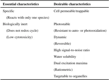
The fact that DCF-family probes continue to be used widely is also evidence of the persistent demand for fluorogenic small molecule sensors to detect intracellular oxidative events. The properties of an ideal small molecule fluorophore include high sensitivity and relative specificity for one reactive species (oxidant or redox pair of interest), membrane permeability and trapping, efficient localization to a defined cellular compartment, wide dynamic range, low toxicity, photostability with repeated cycles of excitation, dynamic
A
uthor Man
uscr
ipt
A
uthor Man
uscr
ipt
A
uthor Man
uscr
ipt
A
uthor Man
uscr
response to changing concentrations of the analyte, water solubility, and insensitivity to pH, ionic strength and temperature (Table 1). While no fluorogenic sensor exists that possesses all these qualities, recent developments in redox chemistry have led to a new generation of advanced fluorogenic sensors specifically designed for oxidant stress measurements with improved performance characteristics. This section highlights a few of these small-molecule fluorophores, grouped by the reactive species to which they respond. (See Table 2.)
7.1. Hydrogen peroxide sensors
While it is typically thought of as an injurious ROS in toxicology, H2O2 is also increasingly
recognized as a signaling molecule and effector of protein modification (i.e., cysteinyl
sulfenylation) with pivotal roles in normal physiology [110,111]. Thus, monitoring H2O2 is
important in elucidating mechanisms of toxicity involving damage to macromolecules as well as the perturbation of normal signaling processes.
7.1.1. Peroxy Green 1 (PG-1)—Developed by the laboratory of Christopher Chang at
UC Berkeley, Peroxy Green 1
(9-(4-methoxy-2-methyl-phenyl)-6-(4,4,5,5-tetramethyl-1,3,2-dioxaborolan-2-yl)xanthen-3-one, PG-1) was the first fluorogenic H2O2 indicator to offer
sufficient sensitivity to detect H2O2 production in non-phagocytic cells responding to
physiological signals [112,113]. PG-1 consists of fluorescein conjugated to a
“chemoselective boronate switch” that responds to H2O2 with high specificity [112,113].
Although PG-1 is not targeted to specific intracellular compartments, it can be used to
identify the intracellular source of H2O2 production when used in conjunction with
well-characterized metabolic inhibitors [114]. It has to be noted that PG-1 is relatively susceptible to photobleaching, has been reported to respond to two-electron transfer by peroxynitrite
[50], and has a slower reaction time compared to more recently developed H2O2-specific
protein-based sensors such as HyPer (discussed below). Despite these shortcomings, PG-1 is
a worthy small molecule fluorophore that is sensitive and relatively specific for H2O2.
Additionally, the Chang laboratory has modified the PG-1 molecule to emit at different
colors with the H2O2-specific sensors PY-1 (Peroxy Yellow 1) and PO-1 (Peroxy Orange 1)
in order to allow multiplex detection with green-emitting fluorophores [115].
7.1.2. PF6-AM—Building upon the boronate-based chemistry of PG-1,
peroxyfluor-6-acetoxymethyl ester (PF6-AM) was developed to be a cytosolically-trappable H2O2 sensor
[116]. Like PG-1, PF6-AM consists of a carboxyfluorescein core that is conjugated to a single phenolic boronate group that masks the fluorescent properties of the fluorophore. As an improvement over PG-1, PF6-AM employs the strategy used in the design of other sensors (e.g., DCF-AM) by introducing two acetoxymethyl ester groups to cap phenol and carboxylic acid functionalities that were engineered into the structure of this sensor in order to enhance its intracellular retention [116]. While attached, the lipophilic AM esters make this fluorophore membrane-permeable. Entry into the cell exposes the AM groups to cleavage by nonspecific intracellular esterases yielding a negatively-charged boronated fluorophore, PF6, that remains confined within the cytoplasm.
Just as with PG-1, exposure to H2O2 removes the sole boron group that serves to quench the
fluorescence of the reporter in its inactive state, thus permitting the fluorescein core to emit
A
uthor Man
uscr
ipt
A
uthor Man
uscr
ipt
A
uthor Man
uscr
ipt
A
uthor Man
uscr
upon excitation with 482 nm light [116,117]. The primary advantage to using this
fluorophore over other boronated H2O2 sensors is the added ability to sensitively detect
H2O2 localized within cells over sustained periods of time. In addition, Dickinson and
colleagues report a nearly two-fold increase in the florescence of PF6 in comparison to PG-1 using the same methodological parameters [116]. A relatively new fluorophore, PF6-AM has
been used to examine H2O2 responses under physiological conditions in several studies that
have followed the original report by Dickinson and colleagues [116,118–122].
7.1.3. MitoPY1—Mitochondrial PY-1 (MitoPY1) is a H2O2-specific organelle-targeting
fluorogenic sensor. This fluorophore uses the same boronate chemistry as described
previously to detect H2O2, but it has an additional positively-charged phosphonium moiety,
tetraphenylphosphonium (TPP+), that targets the probe to the mitochondria of treated cells
[109,123–126]. As the site of oxidative phosphorylation, mitochondria also represent a potential source of partially-reduced oxygen species that can contribute to xenobiotic-induced oxidant stress. Since its introduction, studies have used MitoPY1 in the
investigation of mitochondrial H2O2 production across a variety of physiological and
toxicological contexts [118,127–131]
7.2. Superoxide sensors
The detection of superoxide presents a significant challenge due to the fact that the sensor must compete with the very high enzymatic activity of superoxide dismutases that exists in the cell [132]. Additionally, superoxide anion can act as a reductant as well as an oxidant, which can make the development of a specific superoxide sensor more complex.
7.2.1. Hydroethidine—Hydroethidine
(2.7-diamino-10-ethyl-9-phenyl-9.10-dihydrophenanthridine, HE; also known as dihydroethidine, DHE) is a reduced form of the red-fluorescent compound ethidium (ET) that has become a widely-used standard for measuring superoxide generation within cells [50,104,133–136]. The oxidation of HE by
yields the red-fluorescing hydroxylated form of ethidium (HO-ET). Unfortunately, HE can also be oxidized enzymatically within the cell to form the ET, which is spectrally very similar to HO-ET. One solution to this problem is the use of HPLC to isolate HO-ET from ET [136]. However, this approach is cumbersome and incompatible with live-cell
microscopy. More recently, Robinson and colleagues reported that although both species fluoresce when excited at 514 nm, a second wavelength, 405 nm, can be used to excite HO-ET with considerably greater efficiency relative to HO-ET [133]. Although the emission spectra for HO-ET and ET overlap significantly, their peaks are more than 5 nm apart. In principle, this should make it possible to resolve HO-ET from ET fluorescence in living cells using spectral unmixing [137]. An additional application of HE-based dyes for the detection of
involves the use of the cell impermeable sensor hydropropiridine (HPr+). Since the undesirable oxidation of HE occurs largely as a result of intracellular enzymatic reactions, it
does not affect the detection of extracellular levels of , which can be monitored using
this cell-impermeable HE analog, [50, 138].
A
uthor Man
uscr
ipt
A
uthor Man
uscr
ipt
A
uthor Man
uscr
ipt
A
uthor Man
uscr
7.3. NO sensors
Interest in nitric oxide (NO) has expanded from its adverse effects as an air pollutant to include its role as an important intra- and inter-cellular signaling mediator. Cells maintain a baseline level of NO for normal physiological functions including signal transduction, neurotransmission, and immune responses [139]. High levels of NO induce
pathophysiological effects involved in diseases including cancer, diabetes, and stroke [140]. Non-invasive, intracellular NO measurement is therefore critical for understanding the mechanisms of NO-induced adverse effects. The characteristics desirable in an NO sensor are the same as those for an ROS sensor (Table 1).
The backbone of NO sensors utilizes 1.2-diaminobenzene as a functional group to react with
NO oxidation products (typically N2O3) for the conversion of diamine to triazole [141], with
an emission maximum of 510 nm that can be detected using fluorescein compatible instruments (e.g., flow cytometers, microscopes, microplate readers). Probes based on 2.3-diaminonapthalene (DAN) for NO detection were developed in the 1990s by the Nagano group [142,143]. This probe is converted to the fluorescent compound,
2.3-naphththotriazole, when exposed to NO in an aerobic environment. Under a series of
optimization experiments, DAF-FM DA (4-Amino-5-methylamino-2′,7′-difluorofluorescein
diacetate) was demonstrated to have high specificity for NO and possess desirable sensor characteristics (cell membrane permeability, water solubility, photostability, and bio-compatibility, with excitation and emission wavelengths in the visible range [142,143]). However, the DAF sensors have a limited functional pH range, between 5.8 and above. The same group reported another series of NO probes based on rhodamine that are usable over a broader pH range (pH 4 and above) [144]. In particular, DAR-4MAM has a low detection limit (7 nM), high specificity, high membrane permeability, and is trappable inside the cell through cleavage of the acetoxymethyl ester by endogenous esterases [144]. Regarding optical properties, DAR-4MAM is excited with longer wavelengths (diamine 543 nm, triazole 554 nm) than DAF-FM DA (diamine 487 nm, triazole 495 nm). Although DAR-4MAM is not as bright as DAF-FM DA, the emission maxima of 574 nm is distant from the emission range of cellular auto-fluorescence and hence exhibits a greater signal-to-noise ratio than DAF-FM DA.
Another small molecule probe for NO detection was designed based on the popular
fluorogenic center boron dipyrromethene (BODIPY) [145,146]. Among these, the DAMBO (8-(3.4-diaminophenyl)-2.6-bis(2-carboxyethyl)-4.4-difluoro-1,3,5,7-tetramethyl-4-bora-3a, 4a-diaza-s-indacene) sensors exhibits desirable features including high water solubility, high signal to noise ratio, and compatibility with popular instrumentation (excitation for diamine 495 nm, triazole 497 nm) and emission (triazole 510 nm) with a well-established
microscopy filter system (fluorescein-4-isothiocyanate (FITC)). DAMBO congeners are also stable over a broad pH range (pH 4–12).
Many of the NO fluorescent probes employ the conversion of 1.2-diaminobenzene to triazole (with an emission maximum of 515 nm) for NO detection. In the presence of NO inhibitors, no triazole production was observed for the three sensors (DAF-FM DA, DAR-4MAM, DAMBOO-T) which demonstrates the specificity of these sensors. However,
A
uthor Man
uscr
ipt
A
uthor Man
uscr
ipt
A
uthor Man
uscr
ipt
A
uthor Man
uscr
it is notable that co-localization with other oxidants such as , ONOO−, and NO− can modulate the detection of NO with these sensors [147–149]. In addition, the probes do not
interact with NO directly, but rather the oxidation products, such as and N2O3 [150].
As a result, this limits NO detection based on the concentration of oxygen in the medium. Therefore, it is important to verify findings obtained using this sensor with additional experiments using NO inhibitors in order to modulate functional effects.
7.4. Specialized oxidant sensors
With continued progress in sensor design, the development of new sensors that can be used to investigate a broader range of oxidant species and relevant endpoints can be anticipated. A survey of the current literature reveals several examples of unconventional oxidant sensors that have been applied in physiologically- and toxicologically-relevant studies. For instance, in 2009 the Koide group described a novel fluorophore capable of detecting the ubiquitous
tropospheric air pollutant ozone (O3) in biological and atmospheric samples [151]. Of the
various iterations of O3 sensor reported, a fluorophore designated as “Compound 8” (C8)
was demonstrated to have specificity for this potent oxidizing air pollutant in cultured lung epithelial cells. However, a major disadvantage limiting its use is that this fluorophore is not confined to the intracellular space, which likely contributed to the observed loss of C8
fluorescence intensity immediately following a short O3 exposure period [151]. Despite this
drawback, this is a useful sensor that demonstrates the feasibility of using fluorogenic probes to target even the most highly reactive oxidant species.
Several groups have recently described specific sensors to detect peroxynitrite (ONOO−)
[152–155]. This is an important development because ONOO− is not only a reactive species
produced in response to a broad range of stimuli, but it is also often cited as a confounding
reactive species in the detection of H2O2, O2•− and NO by several small-molecule sensors.
Thus, fluorescent probes designed to specifically detect ONOO− offer additional utility in
that they can be used for validating measurements made by other fluorogenic sensors.
Lastly, another emerging fluorescent probe technology is the packaging of small-molecule sensors into “inert” nanosized particles. A primary advantage in the development of these “nanoprobes” is the ability to combine multiple probes onto a single delivery platform in efforts to monitor various, potentially unrelated, endpoints simultaneously. Similarly, the encapsulation of fluorogenic sensors into nanomaterials may overcome the inherent
cytotoxicity issues of certain sensors, while protecting the loaded sensor(s) from nonspecific interactions with the intracellular environment [109]. Although the loading of nanoparticle matrices such as silicate sol-gel is not a particularly new technology, recent applications have generated a line of sol-gel nanoprobes called Photonic Explorer for Bioanalysis with Biologically Localized Embedding (PEBBLEs) for the specific detection of ROS
[109,156,157]. The transparent nature of the silicate matrix used to generate PEEBLEs has been demonstrated to have minimal cellular effects, while actually enhancing the
photoprotection of the loaded fluorogenic reporter so as to minimize photobleaching [109,157]. Pushing this technology even further, Yang and colleagues report the generation
of a new fluorogenic nanosensor capable of reporting fluctuations in mitochondrial H2O2
and pH [158]. Ultimately, the development of more sophisticated scientific questions
A
uthor Man
uscr
ipt
A
uthor Man
uscr
ipt
A
uthor Man
uscr
ipt
A
uthor Man
uscr
regarding oxidant stress will continue to drive the evolution of next-generation sensors needed for both physiological and toxicological studies of oxidant processes.
8. Genetically-encoded sensors for redox toxicology
The development, characterization, and application of the growing family of genetically encoded redox sensors based on Aequora victoria-derived green fluorescent protein (GFP) congeners has been extensively reviewed [101,159–162]. Since their introduction, these sensors have spurred a rapid expansion of our understanding of the role of intracellular redox events in physiology [163,164], and have afforded significant insight into the role of oxidant stress in toxicology [137,165–169]. Using these sensors involves introduction of an expression vector (wherein the coding sequence is typically placed downstream of a viral promoter) into the cell using either transfection or by transduction with a viral promoter (e.g., adenovirus or lentivirus). An added advantage of live-cell microscopy is that this approach is tolerant of the low expression efficiencies that, since the cells that express the sensor are readily identified visually and can be selected for study. A variety of xenobiotics of interest have been tested in a range of relevant cell culture models refining our
understanding of the localization and effect of xenobiotic-induced oxidant stress in toxicologically relevant contexts. These studies are cited in the following section, which reviews genetically-encoded sensors available to monitor oxidative events.
8.1. Glutathione redox potential (EGSH) sensors
In terms of absolute concentration, the GSSG/GSH couple is the dominant redox pair in the cell, since it is present in millimolar concentrations in most cells [101,170]. The relative concentration of members of a redox pair can be calculated with the Nernst equation
using with the known standard redox potential (E°), and the measured redox potential (E) for the pair, in millivolts, and the Faraday and universal gas constants. While the standard redox
potential for glutathione (E°GSH) is −280 mV [170], EGSH values in the cytosol of resting
cells are considerably more negative, reading −320 mV [101,162], implying that cytosolic glutathione exists largely in the reduced form. Indeed, biochemical studies confirm that healthy cells maintain a large ratio of GSH/GSSG that is supported primarily through the activity of glutathione reductase at the expense of NADPH derived from the pentose phosphate shunt. A practical advantage of the very high ratio of GSH/GSSG in cells is that a
small increase in the concentration of GSSG can be sensed as a large change in the EGSH.
EGSH sensors were originally developed to respond to thiol-disulfide equilibria. The original
families of genetically-encoded sensors that are used to monitor EGSH were derived
independently from YFP and GFP, and are named rxYFP [171] and roGFP [172],
respectively. rxYFP and roGFP were subsequently demonstrated to respond to EGSH by
inserting into a redox relay through which they ultimately equilibrate with the concentration of GSSG (Fig. 1). The physical readout for these sensors is based on a relative change in the
A
uthor Man
uscr
ipt
A
uthor Man
uscr
ipt
A
uthor Man
uscr
ipt
A
uthor Man
uscr
510 nm emission intensity of the fluorogenic center evoked by excitation with 488 nm and, in the case of roGFP, 405 nm light. Beyond their innovative utility, the introduction of these sensors has revolutionized the approach to study oxidant processes by enabling the
quantitative monitoring of EGSH, as a pivotal and well-defined redox event with high
spatiotemporal resolution [173–175].
8.1.1. The yellow fluorescent protein-based redox sensor rxYFP—rxYFP was
constructed by introducing substitutions at amino acids 149 and 202 in YFP to cysteines
[171]. rxYFP was demonstrated to monitor EGSH by equilibrating with the GSSG/GSH
redox pair through its interaction of glutaredoxin-1 (Grx1)-dependent interaction,
information that was subsequently utilized to construct a chimeric protein linking rxYFP to Grx1. SinceGrx1-rxYFP is not dependent on interaction with endogenous Grx1, the
resulting Grx1-rxYFP fusion protein is a sensor with improved EGSH-sensing kinetics
[176]). The utility of rxYFP has been expanded by modified versions that target its
expression to a variety of subcellular compartments [177,178]. However, all types of rxYFP are sensitive to pH changes that can be expected to occur within a physiological range, which imposes limitations and validation requirements on their use [101]. The rxYFP sensors also suffer from an additional disadvantage, namely the fact that they are not ratiometric, which renders them susceptible to artifacts caused by photobleaching, variations in intracellular distribution of the sensors and differences in cell morphology (e.g., cell thickness) [101]. On the other hand, oxidized and reduced forms of rxYFP, can be resolved successfully by specialized immunoblot-ting, which is useful to confirm the results obtained from live-cell imaging experiments [179]. The expansion of the color palette available to analyze oxidant events with genetically-encoded sensors is yet another contribution to the field, as in the case of rxRFP, wherein a cpRFP scaffold was utilized to develop a red fluorescent redox sensor. rxRFP is useful for monitoring the general redox health of the cell,
since it has been used to detect GSSG, , and ONOO− in live-cell studies [180]. Sugiura
et al. recently reported the development of a group of proteins similar to rxYFP, known as oxidation balance sensed quenching (Oba-Q) proteins [181]. Although these sensors are only excitable at one wavelength (and therefore not ratiometric), they are all based on variant chromophores of GFP including Sirius (Oba-Qs), CFP’ (Oba-Qc), and BFP (Oba-Qb), allowing for co-expression of multiple sensors in the same cell without overlap of fluorescence emission spectra.
8.1.2. The green fluorescent protein-based sensor roGFP—The roGFPs are
arguably the most important family of sensors to be developed for redox research and, as such, also hold the greatest potential for redox toxicology. roGFP1 was engineered by
substituting cysteines at glutamine204 and serine147 to allow the formation of a disulfide
bond between the beta strands near the fluorogenic center of GFP under oxidizing conditions [182]. It was subsequently discovered that roGFP interacts only slowly with oxidant species
such as H2O2, and that this sensor preferentially equilibrates with the GSH/GSSG redox pair
through the intervention of Grx1 [183]. The family of roGFPs has been expanded to generate sensors with midpoint potentials that are useful in the range of glutathione redox potentials that exist in various cellular compartments, including the endoplasmic reticulum ([184, 185] Fig. 2). Organelle targeting versions of roGFP have also been developed to direct
A
uthor Man
uscr
ipt
A
uthor Man
uscr
ipt
A
uthor Man
uscr
ipt
A
uthor Man
uscr
the sensor to the cytosol, mitochondrial matrix, mitochondrial intermembrane space, nucleus, peroxisome, endosomes and lysosomes [114,162,164,186–189]. In addition, expression of roGFPs in transgenic animals has successfully resulted in the capability to
specifically assess the EGSH of tissues and even specific cells in organ systems [173–175].
(See Fig. 3.)
roGFP1 and roGFP2 (S147C, Q204C, S65T) are currently the most widely used members of the family of roGFPs. While roGFP1 has the advantage of being less sensitive to pH relative to roGFP2, roGFP2 has a wider dynamic range [182]. Additionally, one group has utilized six of the seven mutations of the super-folder GFP with an additional two directed sites on roGFP2 to produce roTurbo-GFP, a sensor that is notably brighter than other roGFPs, which may increase its usefulness in certain applications [190]. Unlike the rxYFPs, all roGFP congeners show two excitation maxima, excitable with the widely accessible 405 and 488 nm lasers found in many imaging systems. As mentioned earlier, probes with fluorescence emission that can be excited with multiple wavelengths are said to be ratiometric, because the intensity of the fluorescence induced by each excitation wavelength can be expressed as a ratio. As also mentioned above, ratiometry offers several important advantages.
Although roGFP has proved to be a revolutionary sensor, its response time to changes in
EGSH is rather slow because it is limited by the rate of its interaction with endogenous Grx1.
In order to improve the response kinetics of roGFP, the group led by Tobias Dick in Heidelberg developed chimeric proteins linking roGFP2 to Grx1 (roGFP2-Grx1 and Grx1-roGFP), the idea being to increase the effective concentration of Grx1 experienced by the sensor molecule [183]. Both iterations of the fusion of roGFP2 to Grx1 succeeded in increasing the response time kinetics. However, only Grx1-roGFP2 targets well to
subcellular compartments such as the mitochondrial matrix in mammalian cells, Drosophila and Arabidopsis [191,192]. The construction of another chimeric protein that linked roGFP2 to mycoredoxin-1 (Mrx1), the analog of Grx1 in mycobacterium, is another interesting application further expanding the uses of roGFP [193]. This chimeric protein
(roGFP2-Mrx1) monitors the mycothiol redox potential (EMSH) of mycobacterium, useful as a
readout of the pathogen/host interactions and the efficacy of antibiotics. The introduction of this sensor expanded the use of roGFP to monitor redox metabolism in prokaryotic models.
8.2. Genetically-encoded sensors of hydrogen peroxide
8.2.1. HyPer—Belousov and colleagues developed a genetically-encoded fluorogenic
sensor, which they named HyPer, to specifically detect intracellular levels of H2O2.
Structurally, HyPer was formed through circular permutation of YFP (cpYFP) inserted into
the bacterial H2O2-sensing protein OxyR1 [194]. The experimental utility of this sensor has
been established in a series of studies in which HyPer was targeted to a variety of subcellular organelles, including the mitochondria [195]. HyPer has also been shown to be successfully
expressed in zebrafish, revealing the role of H2O2 in developmental growth and wound
healing [196, 197]. HyPer has been chimerically linked to the growth factor receptors EGFR
and PDGFR in order to create proteins positioned to sense localized increases in H2O2
production [198]. Studies using HyPer have demonstrated that xenobiotic induction of H2O2
is specific to subcellular compartments [114,195,199]. The group that developed HyPer
A
uthor Man
uscr
ipt
A
uthor Man
uscr
ipt
A
uthor Man
uscr
ipt
A
uthor Man
uscr
recently reported an improved version, called HyPer-3, that contains point mutations that increase the sensor dynamic range [197].
HyPer is highly sensitive to pH within a physiological range, with a change in pH as small as 0.2 units sufficient to effect a deflection in the excitation ratio reflecting full reduction or excitation of the sensor [194]. Thus it is imperative to monitor pH when using HyPer to
monitor H2O2. This can be done using pH specific sensors such as such as pHluorin or
pHluorin2 [200], albeit these sensors also emit in the green channel, requiring separate experiments or spectral unmixing [137]. pHRed is an alternative pH sensor that emits in the red channel and can therefore be co-expressed and monitored concurrently with HyPer in the same cells [199,201]. However, the ideal pH sensor to use as a control for HyPer is SypHer, which is a version of HyPer that contains a single point mutation (C199S) that renders it
unable to sense H2O2 [202]. Recently a SypHer-2 sensor has been developed which is
reported to be a significant improvement upon the original SypHer sensor [203]. It has been
argued [204] that cpYFP molecules, including HyPer, may also detect which, if
verified, would limit its use as an H2O2 sensor. However, not only is there sufficient
evidence to suggest that HyPer is significantly more sensitive to H2O2 than [161], but a
recent study demonstrated that cpYFP itself could not be utilized as a sensor [205,206],
which should alleviate this concern.
8.2.1.1. Orp-roGFP: For situations where there is a persistent concern regarding the
influence of pH or , an alternative to HyPer exists in the form of Orp1-roGFP2.
However, Orp1-roGFP2 has not been fully characterized yet (i.e., the reductive pathway has not been identified), and it has yet to be broadly used in mechanistic studies [101,191].
Beyond H2O2, there are other protein ROS sensors under development, including those that
sense H2S and ONOO−, two reactive species implicated in signaling as well as oxidant stress
[207,208]. Once these sensors are fully characterized, they will be instrumental in
developing an integrated understanding of the role of oxidant stress and oxidant signaling in toxicology.
9. Limitations and caveats of using protein sensors in redox toxicology
At least one group has demonstrated that laser excitation can produce ROS through
physiological cellular processes and chemical reactions with cell media, potentially creating artifacts and modulating sensor responses [209]. Among the ways to optimize laser-induced fluorescence, such as lowering power and dwell time, the most effective may be to limit the frequency of successive laser excitations to 60 s or longer, a frequency that is well suited for monitoring changes associated with intracellular redox events.
Another aspect that needs to be considered when utilizing genetically encoded sensors is the potential for cellular stress induced by the expression of the sensor. The vector encoding the sensor is introduced into cell cultures through transfection or viral vector transduction and the expression is typically driven by a cytomegalovirus (CMV)-promoter placed upstream of the open reading frame. The use of CMV, SV40 and similar viral promoters is an efficient means of effecting a high level of transcriptional expression in mammalian cells, which can
A
uthor Man
uscr
ipt
A
uthor Man
uscr
ipt
A
uthor Man
uscr
ipt
A
uthor Man
uscr
in turn result in the synthesis of a high concentration of the encoded protein, in some cases reaching micromolar levels (Cheng, W-Y et al. unpublished data). However, co-opting cells to express high levels of recombinant proteins, including GFP has been shown to induce cytotoxic effects [210]. Therefore, it is possible that cells that express high levels of the reporter protein have differential sensitivity to xenobiotic exposures that affect their survival and responses to xenobiotic challenge. This survival disadvantage may explain the
observation of a decreasing fraction of cells that express GFP in a stably transduced cell line over time [211]. Reducing the heterogeneity in the level of sensor expression in the cells (e.g., through cell sorting selection) or titrating sensor expression may decrease the variability in the response to the xenobiotic challenge.
As identified in this review, some of the sensors available for oxidant stress studies rely on fluorescence that is sensitive to pH, and the possibility that the readout reported by the sensor is an artifact of a change in pH should be investigated with appropriate controls. Although minimally invasive ways to measure intracellular pH exist in the form of dyes or other genetically encoded sensors, the aim should be to maintain constant pH during the experiment by using appropriate environmental conditions (i.e., proper buffers in the exposure media). It should also be noted that some xenobiotic exposures lead to cellular acidification by disrupting the mitochondrial respiratory chain, which could in turn result in driving a redox sensor to its reduced form. This could lead to an impairment or failure to detect an oxidative event by the inactivated sensor.
Another potential problem with expressing redox sensors is that the sensor molecules represents an additional electron sink that could alter the redox relay of interest. In practice, whether sensor expression reaches sufficiently high levels to interfere measurably depends on its concentration relative to cellular relay components. For example, the concentration of roGFP achievable in the cell is expected to be too low relative to those of GSH and other cellular thiols to have much of an impact. On the other hand, it would be difficult to rule out a scenario where the expression of HyPer consumes a significant fraction of the local
concentration of H2O2. Experimentally, the approach is to monitor the influence of probe
expression on general cellular physiology such as a change in cell growth rate, as well as specific endpoints relevant to the responses being studied (e.g., a change in basal GSH levels, HO-1 gene expression).
10. Specific challenges using oxidant stress sensors in redox toxicology
studies
The use of redox sensors in toxicological applications creates a number of uncertainties that require special consideration and experimental validation. With few exceptions (e.g., the ozone-specific sensors) fluorophores appropriate for oxidant stress measurements are developed for applications in cell biology. These sensors are, therefore, designed to report on
specific conditions (e.g., EGSH) or concentrations of reactive species (e.g., H2O2) within a
range expected for cells at rest or responding to a physiological stimulus. However, the magnitude of redox effects that result from xenobiotic exposures can exceed well beyond those observed in pathophysiology. Similarly, xenobiotics may generate a sensor readout
A
uthor Man
uscr
ipt
A
uthor Man
uscr
ipt
A
uthor Man
uscr
ipt
A
uthor Man
uscr
through mechanisms that depart significantly from the established pathway through which the sensor was designed to report. This section will consider issues of concern and illustrate experimental approaches that can be used to validate live-cell imaging data in toxicology studies.
10.1. Dynamic range
As mentioned, many xenobiotics of interest in toxicology have chemical reactivities (e.g., electrophilicity) that may exceed grossly the redox potential of a physiologically relevant
oxidant species such as H2O2 that a sensor such as HyPer or PG-1 was designed to detect.
Similarly, highly reactive environmental compounds may induce EGSH changes that are
outside of the dynamic range within which roGFP is designed to report. In practical terms,
this means that exposure to a strong electrophile could result in a shift in the cytosolic EGSH
that is too far from the midpoint potential of roGFP to be measured accurately (typically a 40 mV window centered on the sensor midpoint potential [101]. Similarly, localized
accumulation of H2O2 generated by a redox-active agent could in theory reach levels that
produce a rate of probe oxidization in which essentially every available HyPer molecule is oxidized, thus exhausting the dynamic range of the probe and the signal that can be
generated, a point beyond which further increases in the concentration of H2O2 would
become unresolvable.
10.2. Artifacts
Beyond dynamic range limitations, the use of oxidant stress sensors in toxicological studies also incurs the potential for generating multiple types of artifacts. For instance, it is theoretically plausible for a xenobiotic to react directly with cellular molecules targeted by the sensor (e.g., cysteinyl thiols in GSH, or Grx) by oxidizing or coordinating them, making them unavailable to participate in redox relays with which the fluorophore equilibrates. Additionally, a toxicant may also attack the sensor molecule itself, damaging the fluorogenic
center, decreasing its sensitivity, or rendering it susceptible to other species (e.g., H+, Ca2+).
Alternatively, the sensor could possibly be oxidatively altered in a manner that mimics the modification that induces the change in fluorescence under normal conditions, thereby short-circuiting the pathway and creating a potential false positive signal.
In interpreting results from toxicological studies using fluorogenic sensors, it is important to distinguish between frank artifacts and off-target reactivity of a xenobiotic compound that is properly reported by the sensor. As an example, we may consider the case of a hypothetical dithiocarbamate derivative that inhibits Grx, the proximal enzyme in the relay through which
roGFP senses the oxidant effect of H2O2 on EGSH (Fig. 1, [101,212]). Since the effect of
Grx inhibition would be to sever the ability of roGFP to sense EGSH, any oxidant effect that
this dithiocarbamate derivative may have on EGSH, either through the production of H2O2 or
by direct reactivity with GSH, could not be reported by the sensor, resulting in a false negative result. In contrast, however, if dithiocarbamate exposure interferes with the pentose phosphate pathway, the effect would be to decrease the production of NADPH. Reduced availability of NADPH, in turn, effectively lowers the reductant tone in the redox relay by decreasing the rate of GSSG reduction, which would be reported by roGFP as an increase in
EGSH. In this last scenario, the roGFP sensor responds correctly, notwithstanding the fact
A
uthor Man
uscr
ipt
A
uthor Man
uscr
ipt
A
uthor Man
uscr
ipt
A
uthor Man
uscr
that the oxidant stress that induces the change in EGSH is secondary to a metabolic
impairment rather than, for example, increased production of H2O2 working through the
relay.
Thus, interpretation of effects observed in cells exposed to xenobiotics requires detailed consideration of alternative mechanisms that can explain the sensor readout, including alternative reactions that would not be considered plausible in a physiological context. On the bright side, experiments conducted to validate the readout of a sensor responding to a xenobiotic exposure often yield data that inform the mechanism of action of the agent of interest, sometimes beyond what the sensor data alone could provide.
10.3. Validating sensor readouts
Experimentally, the genetically encoded sensor readouts can be validated by verifying the role of each enzyme in the redox relay leading to the terminal response sensed by the fluorophore. In the case of stimuli that induce changes in roGFP fluorescence, this means testing the involvement of Gpx and Grx (Fig. 1). Our laboratory used this approach in a
recent study [212] that investigated the basis for roGFP2-reported increases in EGSH induced
in human airway epithelial cells undergoing real time exposure to O3, a highly reactive
environmental electrophile. The requirement for Grx in the O3-induced EGSH change was
tested two different ways. First, cells were pretreated with 2-AAPA, an inhibitor of Grx
activity, which resulted in a complete suppression of the O3-induced increase in EGSH
reported by roGFP. This was followed by comparing cells expressing roGFP2 and
Grx1-roGFP2, which confirmed that the rate of O3-induced EGSH sensed by roGFP is limited by
Grx availability. The requirement for Gpx in the roGFP redox relay can be conveniently tested in most cultured cell types by exploiting the inducibility of Gpx with selenium supplementation of the culture medium. Overnight preincubation with 100 μM sodium selenate increased intracellular levels of Gpx1 and significantly accelerated the rate of the
EGSH increase induced by exposure to O3, demonstrating that Gpx1 has a role in the redox
relay that leads to changes in roGFP fluorescence in cells exposed to O3.
On the reductive side of the relay, glutathione reductase (GR) activity to reduce GSSG is dependent on the concentration of NADPH, which can be restricted metabolically by the withdrawal of glucose from the culture medium for 2–4 h prior to exposure. Our
experiments showed that glucose deprivation effectively sensitized the cells to increases in
EGSH induced by a subsequent exposure to O3 [212], without a detectable alteration in basal
EGSH. Depletion of intracellular levels of GSH using an inhibitor of glutathione synthesis,
such as buthionine sulfoximine (BSO), is also an effective tool that can be brought to bear in studies aimed at scrutinizing the integrity of the roGFP redox relay in cells exposed to xenobiotics [199].
As an adjunct to the use of species-specific sensors, the involvement of H2O2 in a
xenobiotic-induced response may be demonstrated by overexpressing catalase in cells. This approach has been used to support data obtained using HyPer in human respiratory cells exposed to the ambient air electrophile 1.2-naphthoquinone [114,213]. A subsequent study
investigated the role of H2O2 in Zn2+-induced oxidative responses using cytosolic as well as
ectopic mitochondrial expression of catalase in airway epithelial cells co-expressing HyPer
A
uthor Man
uscr
ipt
A
uthor Man
uscr
ipt
A
uthor Man
uscr
ipt
A
uthor Man
uscr
in either compartment, thus providing independent spatial information to confirm the imaging data [199]. This approach could also be used to validate responses reported by small molecule sensors.
11. Future directions in live-cell imaging of toxicological oxidant stress
The growing popularity of the new generation of small molecule and genetically-encoded redox and ROS sensors discussed in this review has generated interest in the development of versions that can be used in specific applications. An example of this is the introduction of roGFP sensors that have midpoint potentials that make them usable in compartments such as the ER that are more oxidizing than the cytosol [101] (Fig. 2). Similarly, the ability to target protein or small molecule sensors to membranes or subcellular compartments is a useful property for a sensor as it leverages the spatial resolution of microscopy, and it can therefore be anticipated that new targetable variants of existing sensors will become available in the future.
Various aspects of the dynamic relationship between the concentration of ROS species and any specific redox status is of considerable mechanistic interest in toxicology research. The ideal approach to investigating these issues is to monitor the behavior of multiple sensors introduced in the same cell. However, for historical and practical reasons, most of the redox sensors available currently are variants of either green fluorescent protein or fluorescein, two green fluorophores with overlapping spectra that are not well resolved using the typical bandpass filters with which microscopy systems are typically equipped. Faced with this challenge, our laboratory developed dynamic spectral unmixing microscopy (DynSUM),
specifically to resolve fluorescence signals related to changes in EGSH and H2O2
concentrations reported by roGFP and HyPer, respectively, in human lung cells
co-expressing both sensors [137]. Spectral unmixing requires a specialized detector capable of capturing a wide segment of the combined fluorescence spectra emitted by the sample, which is then “unmixed” into its constituent spectra using a statistical algorithm. Future introduction of new sensors based on fluorogenic centers with a broader range of emission spectra will facilitate simultaneous monitoring of oxidative endpoints of interest. New sensors will likely be restricted to red-shifted fluorophores in order to make use of longer excitation wavelengths that are less likely to cause photobleaching. Undoubtedly, redox toxicology research will benefit as the repertoire of sensors to monitor oxidative endpoints in living cells is expanded to include targets of ROS such as lipids, nucleic acids and proteins. A small molecule fluorogenic sensor for the detection of cysteinyl sulfenylation, a pivotal protein modification in redox biology, was described recently [214].
The inherent non-invasiveness of live-cell microscopy permits the behavior of reporters to be monitored with spatiotemporal resolution unrivaled by other experimental techniques. An important advantage of the high temporal resolution afforded by reporter-based live-cell imaging is that it allows access to the mechanistic information encoded in the kinetic relationships between exposure and response. Metrics such as the lag time in the response to an exposure, the rate of the response, the magnitude of the signal achieved, the duration of the peak response, the rate of resolution, etc., all carry quantitative information that can be modeled mathematically to generate an integrated picture of the pathways through which
A
uthor Man
uscr
ipt
A
uthor Man
uscr
ipt
A
uthor Man
uscr
ipt
A
uthor Man
uscr
xenobiotic exposure causes adverse effects. Such computational uses of laboratory data represent a growing sector of toxicology research and live-cell imaging data are particularly amenable to this application.
Given the pervasiveness of oxidative effects as toxicological mechanisms and the emergence of cellular redox processes as toxicological targets, the demands placed on methodologies to assess redox toxicology can be expected to grow. The sensor-based live-cell imaging approach described in this review should make it a powerful tool with broad application in the investigation of the multifaceted aspects of xenobiotic-induced oxidant stress.
References
1. Cross CE, Valacchi G, Schock B, Wilson M, Weber S, Eiserich J, van der Vliet A. Environmental oxidant pollutant effects on biologic systems: a focus on micronutrient antioxidant-oxidant interactions. Am J Respir Crit Care Med. 2002; 166:S44–S50. [PubMed: 12471088]
2. Stringer B, Kobzik L. Environmental particulate-mediated cytokine production in lung epithelial cells (A549): role of preexisting inflammation and oxidant stress. J Toxicol Environ Health A. 1998; 55:31–44. [PubMed: 9747602]
3. Jaeschke H, McGill MR, Ramachandran A. Oxidant stress, mitochondria, and cell death
mechanisms in drug-induced liver injury: lessons learned from acetaminophen hepatotoxicity. Drug Metab Rev. 2012; 44:88–106. [PubMed: 22229890]
4. Wolf MB, Baynes JW. The anti-cancer drug, doxorubicin, causes oxidant stress-induced endothelial dysfunction. Biochim Biophys Acta. 2006; 1760:267–271. [PubMed: 16337743]
5. Chakraborti S, Das S, Chakraborti T. Oxidant-mediated activation of cytosolic phospholipase a(2) in pulmonary endothelium: role of protein kinase C alpha and a pertussis toxin-sensitive protein. Endothelium. 2005; 12:121–131. [PubMed: 16291515]
6. Zengin S, Al B, Yarbil P, Taysi S, Bilinc H, Yildirim C, Aksoy N. Oxidant/antioxidant status in cases of snake bite. J Emerg Med. 2013; 45:39–45. [PubMed: 23623287]
7. Pham HT, Maccarone AT, Campbell JL, Mitchell TW, Blanksby SJ. Ozone-induced dissociation of conjugated lipids reveals significant reaction rate enhancements and characteristic odd-electron product ions. J Am Soc Mass Spectrom. 2013; 24:286–296. [PubMed: 23292977]
8. Wisthaler A, Weschler CJ. Reactions of ozone with human skin lipids: sources of carbonyls, dicarbonyls, and hydroxycarbonyls in indoor air. Proc Natl Acad Sci U S A. 2010; 107:6568–6575. [PubMed: 19706436]
9. Kumagai Y, Shinkai Y, Miura T, Cho AK. The chemical biology of naphthoquinones and its environmental implications. Annu Rev Pharmacol Toxicol. 2012; 52:221–247. [PubMed: 21942631] 10. Iwamoto N, Sumi D, Ishii T, Uchida K, Cho AK, Froines JR, Kumagai Y. Chemical knockdown of
protein-tyrosine phosphatase 1B by 1.2-naphthoquinone through covalent modification causes persistent transactivation of epidermal growth factor receptor. J Biol Chem. 2007; 282:33396– 33404. [PubMed: 17878162]
11. Abdel-Malek ZA, Kadekaro AL, Swope VB. Stepping up melanocytes to the challenge of UV exposure. Pigment Cell Melanoma Res. 2010; 23:171–186. [PubMed: 20128873]
12. Skillrud DM, Martin WJ 2nd. Paraquat-induced injury of type II alveolar cells. An in vitro model of oxidant injury. Am Rev Respir Dis. 1984; 129:995–999. [PubMed: 6732057]
13. Berisha H, Pakbaz H, Absood A, Foda HD, Said SI. Nitric oxide mediates oxidant tissue injury caused by paraquat and xanthine oxidase. Ann N Y Acad Sci. 1994; 723:422–425. [PubMed: 8030904]
14. Kang SA, Jang YJ, Park H. In vivo dual effects of vitamin C on paraquat-induced lung damage: dependence on released metals from the damaged tissue. Free Radic Res. 1998; 28:93–107. [PubMed: 9554837]
15. Aroun A, Zhong JL, Tyrrell RM, Pourzand C. Iron, oxidative stress and the example of solar ultraviolet A radiation. Photochem Photobiol Sci. 2012; 11:118–134. [PubMed: 21986918]