CHARACTERIZING THE BIOCHEMICAL DETERMINANTS GOVERNING MERS-CORONAVIRUS HOST RANGE
Kayla M. Peck
A dissertation submitted to the faculty at the University of North Carolina at Chapel Hill in partial fulfillment of the requirements for the degree of Doctor of Philosophy in the
Department of Biology.
Chapel Hill 2016
ABSTRACT
Kayla M. Peck: Characterizing the Biochemical Determinants Governing MERS-Coronavirus Host Range
(Under the direction of Christina Burch and Mark Heise)
Coronaviruses are a diverse family of viruses that infect a wide range of hosts, including both mammalian and avian species. Within recent history, coronaviruses have expanded their host range into humans, with four emergence events resulting in infections that cause only mild disease. However, two additional emergence events resulted in outbreaks of severe disease, causing heightened concern for public health. The 2003 severe acute respiratory syndrome coronavirus (SARS-CoV) emerged in Southeast Asia and rapidly spread around the world with a 9 percent mortality rate before being controlled by public health intervention strategies. In 2012, Middle East respiratory syndrome coronavirus (MERS-CoV) emerged from its zoonotic
reservoir. To date, it has infected over 1800 people with a 36 percent mortality rate and is still circulating in the population. Due to the emergence of coronaviruses with pandemic potential, it is important to understand how these lineages have been able to expand their host range to infect new species. One key determinant of viral host range is the interaction between the virus spike protein and the host cell receptor. For MERS-CoV specifically, the virus can infect bats, camels (the putative intermediate host species), and humans, but is unable to infect mice or other
MERS-CoV receptor binding domain and the mouse cell receptor; 2) investigate biochemical determinants that govern infection for other species; 3) attempt to generate a mouse-adapted MERS-CoV; and 4) present an approach to investigate potential evolutionary mechanisms of coronavirus host range expansion. This work has contributed to the development of a small animal model, allowing us to begin pathogenesis studies. Additionally, understanding the
ACKNOWLEDGMENTS
I have been extremely fortunate to be a part of three amazing labs. Christina Burch has been the best mentor a student could hope for, providing me with an environment of kindness, independence, and overwhelming faith in me. Mark Heise graciously accepted my eclectic skillset into his group and gave me the best confidence-boosting pep talks. Ralph Baric forged my dissertation project and gave me access to his space and resources that advanced my academic career. While most people had no idea where I belonged or who I was actually affiliated with, thank you to all of the labs who accepted me into their group, the hybrid that I was. This also includes Mark Tanaka who generously hosted me for two summers and provided me with invaluable computational biology experience.
Thanks to Adam Cockrell for training me in molecular virology, to Boyd Yount who taught me to be overly liberal with ethanol in the BSL3, and to Trevor Scobey who was always willing to help me with experiments, training, and finding things in the ever-rearranging Baric lab. Thanks to Jenny McGraw for being my desk partner and soup day companion, as well as my inspiration for working harder.
I could not have gotten through graduate school without the support of my amazing friends. Thank you to everyone who suffered through my Eevee, potato, raccoon, Louis Pasteur, zombie apocalypse, and Shrek obsessions. All the love in the world to my tortuga, Hailey.
TABLE OF CONTENTS
LIST OF TABLES ... viii
LIST OF FIGURES ... ix
LIST OF ABBREVIATIONS ... xi
INTRODUCTION ... 1
0.1 Virus Evolution ... 1
0.2 Host Range Expansion ... 4
0.3 Coronaviruses ... 6
0.4 Overview of Chapters... 12
CHAPTER 1: BIOCHEMICAL DETERMINANTS OF MOUSE DPP4 PERMISSIVITY TO MERS-CORONAVIRUS... 15
1.1 Introduction ... 15
1.2 Identify key mutations in mouse DPP4 that allow it to support MERS-CoV infection (modified from Cockrell et al. 2014) ... 16
1.3 Identify key biochemical determinants that prevent mouse DPP4 from acting as a valid receptor (modified from Peck et al. 2015b) ... 24
1.4 Removing glycosylation from mDPP4 ... 31
2.1 Introduction ... 36
2.2 Determine the role of glycosylation in additional DPP4 orthologs ... 37
2.3 Identify key mutations and determinants of DPP4 ortholog permissivity to MERS-CoV infection ... 46
2.4 Adaptation to alternate receptor molecules ... 52
CHAPTER 3: MOUSE-ADAPTATION OF MERS-CORONAVIRUS ... 58
3.1 Introduction ... 58
3.2 Adaptation of MERS-CoV to glycosylated DPP4 receptors ... 60
4. FUTURE DIRECTIONS ... 72
4.1 Development of a MERS-CoV mutator ... 72
4.2 Potential evolutionary mechanisms for coronavirus host range expansion (modified from Peck et al. 2015c). ... 85
CONCLUSION ... 90
APPENDIX A: PHYLOGENETIC TREE CORONAVIRUS STRAINS ... 94
APPENDIX B: PHYLOGENETIC TREE DPP4 ORTHOLOGS ... 96
LIST OF TABLES
Table 1: Amino acid identities at five DPP4 ortholog residues
LIST OF FIGURES
Figure 0.1: Virus evolution rate against mutation rate ... 3
Figure 0.2: Phylogenetic tree of whole-genome length coronavirus sequences ... 8
Figure 0.3: Positive selection on coronavirus host cell receptors ... 10
Figure 1.1: MERS-CoV infection utilizing hDPP4 and mDPP4 ... 17
Figure 1.2: mDPP4 as a backbone can support MERS-CoV infection... 19
Figure 1.3: MERS-CoV infection is dependent upon specific amino acids in DPP4 ... 21
Figure 1.4: Human and chimeric DPP4 molecules can support MERS-CoV infection in hamster and mouse cells ... 23
Figure 1.5: Is charge or glycosylation important for mediating mouse DPP4 permissivity? ... 26
Figure 1.6: Glycosylation can act to dramatically reduce infection by MERS-CoV ... 28
Figure 1.7: Glycosylation is more important than charge in mediating MERS-CoV infection... 29
Figure 1.8: DPP4 construct expression in HEK 293T cells ... 30
Figure 1.9: Tunicamycin treatment of DPP4 molecules ... 32
Figure 1.10: PNGase F treatment of DPP4 molecules ... 33
Figure 2.1: Permissivity of DPP4 orthologs to MERS-CoV ... 38
Figure 2.2: Sequence and structural comparison of nonpermissive DPP4 orthologs ... 39
Figure 2.3: DPP4 ortholog glycosylation knockout mutants ... 41
Figure 2.4: Glycosylation knockout panel in haDPP4 ... 42
Figure 2.5 Guinea pig and bat DPP4 glycosylation knockout mutants ... 43
Figure 2.7: Many amino acid changes are required to make fDPP4
and haDPP4 permissive to MERS-CoV infection ... 48
Figure 2.8: Fine-tune mapping of fDPP4 determinants for MERS-CoV permissivity ... 49
Figure 2.9: Fine-tune mapping of haDPP4 permissivity to MERS-CoV... 51
Figure 2.10: DPP4 family members... 54
Figure 2.11: FAP as a backbone for supporting MERS-CoV infection ... 55
Figure 2.12: Comparison of BtCoV-HKU4 and MERS-CoV RBD ... 57
Figure 3.1: Host invasion adaptation strategy using hDPP4 as a backbone ... 62
Figure 3.2: Host invasion adaptation strategy using mDPP4 as a backbone ... 63
Figure 3.3: DPP4 panel tested on human, mouse, and hamster cell lines... 64
Figure 3.4: DPP4 construct expression in stably expressing NIH 3T3 cells ... 65
Figure 3.5: Growth curve of permissive NIH 3T3 stably-expressing cell lines ... 66
Figure 3.6: Host invasion adaptation of MERS-CoV to hDPP4 + gly ... 67
Figure 3.7: Host invasion adaptation of MERS-CoV to mDPP4 288... 67
Figure 3.8: Virus titer and percent permissive host cells for host invasion adaptation experiments ... 68
Figure 3.9: Infection panel of passage 25 virus from two adaptation experiment treatments ... 69
Figure 4.1: Sequence and structural alignment of SARS-CoV and MERS-CoV nsp14 ... 77
Figure 4.2: Pooled and plaque isolated rMERS-CoV-RFP D2 mutator virus ... 80
LIST OF ABBREVIATIONS ACE2: Angiotensin-converting enzyme 2
APN: Aminopeptidase N
bDPP4: Bat dipeptidyl peptidase IV BHK: Baby hamster kidney cells BSL3: Biosafety level 3
cDPP4: Camel DPP4
DBT: Delayed brain tumor cells DNA: Deoxyribonucleic acid DPP4: Dipeptidyl peptidase IV DPP8: Dipeptidyl peptidase VIII DPP9: Dipeptidyl peptidase IX E: Envelope protein ExoN: Exoribonuclease
FAP: Fibroblast activation protein fDPP4: Ferret DPP4
FMDV: Foot and mouth disease virus GII.4: Genogroup II genotype 4 gly: Glycosylation site
gpDPP4: Guinea pig DPP4 haDPP4: Hamster DPP4 HCoV: Human coronavirus
hDPP4: Human dipeptidyl peptidase IV HEK 293T: Human embryonic kidney 293T cells HIV: Human immunodeficiency virus hpi: Hours post-infection
Huh7: Human hemochromatotic cells IFA: Immunofluorescence assay
kb: Kilobases
kDa: Kiladaltons M: Matrix protein
mDPP4: Mouse dipeptidyl peptidase IV
MERS-CoV: Middle East respiratory syndrome coronavirus MHV: Murine hepatitis virus
mL: Milliliter
MOI: Multiplicity of infection N: Nucleocapsid protein NoV: Norovirus
nsp10: Nonstructural protein 10 nsp14: Nonstructural protein 14 ORF: Open reading frame PCR: Polymerase chain reaction PFU: Plaque-forming units PNGase F: Peptide-N-glycosidase F poly-A: Poly-adenosine
RBD: Receptor binding domain
RdRp: RNA-dependent RNA polymerase RFP: Red fluorescent protein
RIPA: Radioimmunoprecipitation assay rMERS: Recombinant MERS-CoV RMS: Root mean square
RNA: Ribonucleic acid
S: Spike protein
s/n/c: Substitutions per nucleotide site per cell infection s/n/y: Substitutions per nucleotide site per year
TBST: Tris-buffered saline plus Tween
Tu: Tunicamycin
µL: Microliter
INTRODUCTION 0.1 Virus Evolution
Viruses are small infectious agents that replicate by infecting the cells of other organisms. Their general structure includes genomic material, RNA or DNA, a protein coat (capsid) that protects the genome, and sometimes an envelope that surrounds the capsid. Despite the small number of components, viruses accomplish a surprising amount of diversity, with breadth in what organisms they infect, their life-cycle strategies, and the impact they have on their hosts. In fact, estimates suggest that there are over 320,000 different viral species that infect mammals (Anthony et al. 2013a). If this number is extrapolated out to include all vertebrate species, the estimated number of viral species rises to over 3.6 million, representing an extraordinary amount of diversity. In addition to this remarkable diversity, viruses are also the most prevalent
severe consequences for public health. However, despite these negative aspects of rapid viral evolution, it also allows us to use viruses as model systems for investigating and understanding evolutionary processes.
One of the fundamental facets of evolution is the rate at which populations evolve. Unfortunately, the dynamics that govern the rate of molecular evolution for viruses are not quite straightforward. For other organisms, the rate of molecular evolution roughly follows neutral theory, which proposes that mutation rate, specifically the rate of neutral mutations, is the sole predictor of evolution rate (Kimura 1984). The success of this theory in estimating rates of evolution (Li et al. 1987, Kimura 1991, Bromham and Penny 2003) does not universally hold true for viruses, with molecular clock dynamics both supported (Gojobori et al. 1990, Leitner and Albert 1999) and refuted (Jenkins et al. 2002). The diversity and complexity of viral life cycles complicates our ability to understand the precise relationship between viral evolution rate and neutral theory.
the error or extinction threshold (Eigen 1993, Bull et al. 2005). At this threshold, the population is overwhelmed with abundant transient deleterious mutations which arise more rapidly than can be removed by natural selection (Holmes 2003, Bull et al. 2007), causing the population to die out. Including the proportion of deleterious mutations into a model of evolution rate greatly improves the ability to explain empirical evolution rates seen for rapidly mutating viruses (Sanjuan 2012). Additionally, because of the diversity of virus life cycles, including within-host parameters, specifically the within-host reproductive ratio, also improves explanatory power (Peck et al. 2015a). With the increasing amount of available data on empirically estimated parameter values, we can improve our ability to understand what components influence the current evolution rates of viruses.
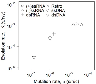
Figure 0.1: Virus evolution rate against mutation rate
One important component of virus evolution is host range expansion, or the ability to infect a new species. The infection of a new species, whether by adaptation or by exposure from geographic proximity, represents a new niche that a virus can explore, promoting adaptive radiation (Nichol et al. 1993, Rainey and Travisano 1998, MacLean and Bell 2002, Chow et al. 2004). Many viruses that infect the human population originated from a zoonotic source (Daszak et al. 2000, Taylor et al. 2001). The prevalence of zoonotic viruses that have gained the ability to
infect humans is largely due to their rapid evolution rate; approximately one new emerging virus disease occurs each year (Howard and Fletcher 2012). RNA viruses, in particular, represent the majority of viral species that have emerged into humans (Howard and Fletcher 2012). This is often attributed to their high mutation rates, which result in a faster evolution rate (Figure 0.1). These higher rates of evolution enhance the ability with which viruses can expand into
previously uninfected species. Unfortunately, the specific selective pressures that precede a host range expansion event are unknown; additionally, many of the evolutionary mechanisms that facilitate host range expansion for viruses still remain to be elucidated. Here, I examine virus evolution specifically in the context of viral host range expansion in order to better understand the dynamics that link mutation rate, evolution rate, emergence, and public health.
0.2 Host Range Expansion
Host range expansion is a complex process with many components determining which viruses can expand their host range and which species those hosts may comprise. Simply, there are three main steps for a pathogen to successfully expand its host range. The first step is exposure. In many cases, host jumps are caused by ecological factors that either change the
The second step is for the pathogen and host to be compatible on a number of levels (e.g. by route of transmission, cell receptor, and/or infectious dose). Arguably, the most crucial barrier in this step for viruses is utilization of a new host species cell receptor by the viral spike protein, which is responsible for attachment and entry into the host cell. However, even compatibility between these two components may not be enough; for example, some viruses require specific host cell factors like proteases to facilitate viral entry (Gierer et al. 2013, Shirato et al. 2013, Millet and Whittaker 2015). Physiology can also play an important role; differences in the temperatures of human and avian airways are a crucial barrier in preventing avian influenza A viruses from replicating efficiently in humans (Scull et al. 2009).
The third step is for the pathogen to be efficiently transmitted between individuals of the new host species (Woolhouse et al. 2005). Most zoonotic pathogens are not effectively
transmitted between humans (Woolhouse and Gowtage-Sequeria 2005), causing humans to act as a dead end host in these scenarios. However, if a zoonotic virus is compatible with human cells and is able to transmit efficiently between humans, it has the potential to result in a severe
outbreak. For example, although the avian influenza A virus H5N1 has been transmitted multiple times to humans from avian species, its inability to transmit reliably between humans is a
efficiently infecting a wide range of mammalian species (Rupprecht et al. 2002). Note that while rabies virus is not commonly transmitted between humans, this is likely due to behavioral
dynamics and not due to the biological inability of the virus to be transmitted. Previous studies have investigated what potential drivers can lead to the selection of a specialist or generalist virus population (Bono et al. 2012) with advantages and disadvantages of both infection strategies.
0.3 Coronaviruses
Here I focus on coronaviruses, a diverse family of viruses that has expanded its host range many times over the course of its evolutionary history. Coronaviruses are enveloped, single-stranded positive-sense RNA viruses with particularly large genomes (28-32 kb) that can infect a wide range of avian and mammalian hosts. Genomes contain a 5’ cap and 3’ poly-A tail, with the genome organization divided into nonstructural protein genes and structural and
accessory genes. The core structural proteins include S (spike), E (envelope), M (matrix), and N (nucleocapsid) proteins. While accessory genes vary among coronaviruses, and may include some strain-specific structural glycoproteins, the order of the structural proteins is highly conserved as S, E, M, and N. The ~180 kDa spike glycoprotein (S) mediates entry into the host cell and surrounds the virus particle, yielding a crown-like appearance. Coronaviruses utilize a variety of cellular proteins as receptors (Perlman and Netland 2009, Graham et al. 2013), with cleavage of the spike protein crucial for mediating virus-host membrane fusion and subsequent entry into the cell.
Bats, rodents, and birds act as the natural reservoir species for many coronaviruses (Li et al. 2005a, Woo et al. 2009, Wang et al. 2015), but host range expansion has been prevalent, with
coronaviruses (HCoV) have been identified, with each of these hypothesized to have originated as a zoonotic strain that underwent host range expansion. Of these six HCoVs, four are
associated with mild respiratory disease: 229E, HKU1, NL63, and HCoV-OC43. Two of these (HCoV-229E and HCoV-NL63) are thought to have originated from bats. While the standing hypothesis for HCoV-OC43 was that it likely emerged from a bovine
reservoir species (Vijgen et al. 2005), new analyses suggest that the original host of this lineage, and perhaps HCoV-HKU1 as well, may have been a murine species (Lau et al. 2015). In addition to HCoVs that cause mild symptoms, two strains have emerged to cause severe disease,
including severe acute respiratory syndrome coronavirus (SARS-CoV) and Middle East respiratory syndrome coronavirus (MERS-CoV). These two viruses combined have resulted in over 1,100 deaths, with MERS-CoV still circulating in the human population, causing
heightened concern due to the lack of vaccines or therapeutics. The threat to public health caused by the emergence of these highly pathogenic strains into humans draws attention to the
importance of understanding both the biochemical and evolutionary mechanisms of coronavirus host range expansion.
The selective pressures that drive coronavirus host range expansion are not yet
understood. Additionally, a pattern of coronavirus host range characteristics or biases is not yet evident. Some coronaviruses appear to be generalists, capable of infecting many different orders of mammals. For example, Betacoronavirus 1 has been detected in dogs, humans, and numerous ungulate species (Erles et al. 2003, Hasoksuz et al. 2007, Alekseev et al. 2008). Other
Figure 0.2: Phylogenetic tree of whole-genome length coronavirus sequences
immunology, physiology, and ability to traverse wide geographic regions during seasonal migrations (Dobson 2005, Calisher et al. 2006). Due to this, metagenomics analyses have focused on examining the diversity and prevalence of coronaviruses in bats. Studies have found varying levels of coronavirus diversity in bat populations in North America (Donaldson et al. 2010) and China (Ge et al. 2012, Wu et al. 2012b), as well as detecting individual strains in bat populations worldwide (reviewed in Drexler et al. 2014). Furthermore, novel coronaviruses continue to be discovered globally, with recent findings coming from Mexico (Anthony et al. 2013b), Brazil (Goes et al. 2013), and South Africa (Ithete et al. 2013). Continued efforts to sample bat populations worldwide will help us better estimate the diversity and prevalence of coronaviruses, as well as more thoroughly map their phylogenetic relationships.
The biggest threat to the public health comes from coronaviruses that emerge into the human population to cause severe disease. The first of these, SARS-CoV, emerged from bats into the human population in 2003 and infected over 8,000 people with a 9.6% mortality rate before it was controlled by public health measures (Cherry 2004). Despite the awareness raised for coronaviruses during this outbreak, reconstructing the evolutionary path that led to the host range expansion of SARS-CoV has been surprisingly difficult. SARS-CoV is closely related to bat coronaviruses, with up to 92% nucleotide sequence identity to its closest detected relative (Li et al. 2005a). However, no virus identical to SARS-CoV has ever been isolated from bats, raising
origin hypothesis include the fact that, initially, no bat coronaviruses were found to utilize ACE2 or any ACE2 ortholog (Ren et al. 2008). In fact, it took over a decade before researchers were able to identify a SARS-like coronavirus capable of utilizing human, civet, and Chinese horseshoe bat ACE2 for cell entry (Ge et al. 2012). The discovery of this virus (bat SL-CoV- WIV1) provides strong evidence that SARS-CoV originated from a bat reservoir (see Figure 0.2). Currently, the best supported hypothesis is that an intermediate host species, specifically the civet, played an important role in facilitating the host range expansion event, either by imposing a specific selective pressure or by putting the virus into close proximity with humans (reviewed in Wang and Eaton 2007, Plowright et al. 2015). However, the identification of bat SL-CoV-WIV1 suggests that an intermediate host may not have been required for emergence of SARS-CoV into humans. Further research is needed to determine the precise mutational and cross-species path of SARS-CoV in order to help reveal how new human pathogens emerge.
Figure 0.3: Positive selection on coronavirus host cell receptors
In 2012, MERS-CoV emerged into the human population in Saudi Arabia. As of November 2016, there were 1,813 confirmed cases with a 36% mortality rate (WHO 2016). MERS-CoV is grouped phylogenetically into the C betacoronavirus clade along with the bat coronaviruses BtCoV-HKU4 and BtCoV-HKU5 (Figure 0.2) (van Boheemen et al. 2012, Drexler et al. 2014, Peck et al. 2015c). Unlike SARS-CoV, MERS-CoV utilizes dipeptidyl peptidase 4 (DPP4) as an entry receptor (Raj et al. 2013). To date, only MERS-CoV and BtCoV-HKU4 have been found to utilize DPP4 (Wang et al. 2014, Yang et al. 2014); the presence of a closely related bat coronavirus that uses the same host cell receptor as MERS-CoV provides strong support for the emergence of MERS-CoV from a bat lineage. Adaptation of MERS-CoV to humans specifically is supported by the fact that MERS-CoV utilizes human DPP4 (hDPP4) more efficiently than bat DPP4 (bDPP4), yet BtCoV-HKU4 utilizes both with equivalent
efficiency (Yang et al. 2014). Thus far, the host cell receptors of BtCoV-HKU5 and other closely related group 2c coronaviruses have not been identified.
The similarity between MERS-CoV and group 2c bat coronaviruses (Figure 0.2) strongly suggests that MERS-CoV originated from bats. Additionally, similar to the SARS-CoV story, bDPP4 has been found to be under strong positive selection (Figure 0.3), which may be due to the circulation of DPP4-utilizing coronaviruses in bats (Cui et al. 2013). It is not clear whether this signal is from the progenitor of MERS-CoV or from other coronaviruses that utilize DPP4, such as BtCoV-HKU4. As with SARS-CoV, the ancestral virus has been elusive, with no
been detected in geographic regions very distant from its original emergence location in Saudi Arabia. RdRp gene sequences with 96.5% and 99.6% amino acid identity to MERS-CoV were detected from bat populations in Mexico (Anthony et al. 2013b) and South Africa (Ithete et al. 2013), respectively. These discoveries emphasize the importance of further metagenomics analyses of bat viromes in widespread geographic locations.
Looking at the phylogenetic tree of coronaviruses (Figure 0.2), the lineages that have emerged into humans occupy several clades. This suggests that there is not a single lineage of zoonotic coronaviruses that is capable of expanding its host range into humans. Instead, many lineages have been able to emerge into the human population. Additionally, these lineages utilize different host cell receptors for entry into the cell. SARS-CoV and HCoV-NL63 utilize ACE2 (Li et al. 2003, Hofmann et al. 2005), MERS-CoV utilizes DPP4 (Raj et al. 2013), and HCoV-229E utilizes a protein known as aminopeptidase N (APN) (Yeager et al. 1992). The receptor molecules of HCoV-HKU1 and HCoV-OC43 have not yet been identified, however the presence of O-acetylated sialic acid has been shown to serve as a receptor determinant (Krempl et al.1995, Huang et al. 2015). The diversity of receptors utilized, combined with the large number of species that coronaviruses have evolved to infect, poses the question of whether coronaviruses have an increased capacity for host range expansion relative to other RNA viruses. Perhaps there are unique characteristics of coronaviruses that increase their ability to emerge into new species, providing an interesting case study of the evolution of host range.
0.4 Overview of Chapters
interactions between the MERS-CoV receptor binding domain (RBD) and the human and mouse DPP4 orthologs are analyzed both computationally and experimentally to provide data on what particular residues and residue properties are important for mediating MERS-CoV permissivity. I find that two residues are capable of mediating this interaction; two amino acid changes within the mouse DPP4 (mDPP4) backbone allow it to successfully support infection. The biochemical explanation behind these two changes is 1) the strengthening of a hydrophobic core in the MERS-CoV RBD and 2) the removal of a non-conserved glycosylation site. Data gathered from this chapter helped generate a transgenic mouse model that will be utilized to study MERS-CoV pathogenesis.
Chapter 2 investigates whether there is a broader signature of permissivity in additional
DPP4 orthologs. Other traditional small animal model species, namely ferrets, guinea pigs, and hamsters, are also nonpermissive to MERS-CoV infection. Using the insight gained in my previous experiments, I determine whether the same biochemical mechanisms that conferred permissivity of mDPP4 to MERS-CoV are applicable to these other species. Many
nonpermissive species have glycosylation sites near the location that aligns with the mDPP4 motif, suggesting that this may potentially be a broadly acting mechanism for blocking MERS-CoV infection. I find that although successful infection is never achieved without removal of glycosylation in these orthologs, the interaction dynamics are more complex, suggesting that there are other important key determinants that will require more work to reveal.
Chapter 3 turns to the field of evolutionary biology in order to determine how
mutations, including remodeling of the RBD to bind around the mDPP4 glycan, there was no adaptation of MERS-CoV to wildtype mDPP4. One potential hypothesis is that there was not enough genetic variation present in the population to achieve the multiple mutations required for successful binding (contingent on it being biologically possible). In Chapter 4, I present future directions for generating a MERS-CoV mutator that can be utilized in adaptation experiments to enhance the sequence space accessible by the viral population. Mutations in the nsp14 gene of coronaviruses have been found to generate elevated mutation rates, allowing mutators to be a possible characteristic that permits coronaviruses to expand their host range more frequently, and with more success, than other viruses.
The increasing prevalence of emerging pathogens heightens the need to understand the biochemical and evolutionary dynamics of host range expansion events. Coronaviruses, particularly, draw attention due to the recent emergence of highly pathogenic strains that lack effective vaccines or therapeutics. For many viruses, including SARS-CoV and MERS-CoV, the specific factors contributing to the host range expansion into humans are still unclear.
Elucidating the selective pressures imposed on the virus populations will help reveal the specific path of emergence of SARS-CoV and MERS-CoV. Understanding the adaptation pathway of coronaviruses to new species will help us better catalogue the selective pressures driving host range expansion and will provide a framework for which we can set up ecological and/or geographical preventative measures. Although the limited number of outbreak events in
CHAPTER 1: BIOCHEMICAL DETERMINANTS OF MOUSE DPP4 PERMISSIVITY TO MERS-CORONAVIRUS
1.1 Introduction
Wit et al. 2013a, van Doremalen et al. 2014). This species restriction prevents the adequate study of MERS-CoV pathogenesis and limits the development of vaccine strategies or alternate
therapeutics. Understanding the determinants that mediate MERS-CoV permissivity in these species will provide novel insights into interactions between the MERS-CoV RBD and DPP4, as well as help in the development of new small animal models.
1.2 Identify key mutations in mouse DPP4 that allow it to support MERS-CoV infection (modified from Cockrell et al. 2014)
In order to investigate the relationship between MERS-CoV and DPP4, we designed an ectopic expression system utilizing the 945ΔRRE expression vector, a lentiviral vector derived from pTK945, to constitutively express the human and mouse DPP4 genes in human embryonic kidney 293T (HEK 293T) cells. These cells lack detectable endogenous expression of hDPP4 (Zhao et al. 2013). Both orthologs were expressed either as full-length proteins or as fusions to the Venus protein at the carboxy terminus as a marker of green fluorescence. HEK 293T cells were transfected with 3 µg of the appropriate DPP4 expression plasmid. At ~24 hours post-transfection, cells were infected at a multiplicity of infection (MOI) of 5 with a recombinant MERS-CoV strain designed to express tomato red fluorescent protein (rMERS-CoV-RFP) (Figures 1.1A and B). This rMERS-CoV strain is derived from the original EMC2012 isolate (van Boheemen et al. 2012) and was previously demonstrated to infect and replicate in a manner similar to wildtype MERS-CoV (Scobey et al. 2013). Cells were imaged ~24 hours
post-infection (hpi) by rMERS-CoV-RFP. High transfection efficiency was seen by visualizing the DPP4-Venus fusion constructs (Figures 1.1A and B).
overexpression of mDPP4 showed levels of infection that were equivalent to control HEK 293T cells (Figure 1.1B). To confirm that mDPP4 was being expressed, and a negative infection
Figure 1.1: MERS-CoV infection utilizing hDPP4 and mDPP4
readout was not based on absence of the protein, we performed Western Blot analyses to detect the presence of mDPP4 after transfection into HEK 293T cells (Figure 1.1C). Bands present at ~110 kDa for both hDPP4 and mDPP4 indicate successful expression of both constructs. Additionally, Western blot analysis for MERS-CoV spike (S) and nucleocapsid (N) proteins show detection of both viral proteins in cells expressing hDPP4 but not mDPP4 (Figure 1.1D). These results support hDPP4, but not mDPP4, as a functional receptor for MERS-CoV (Cockrell et al. 2014).
infection. Thus, there must be important differences between blades IV and V of hDPP4 and mDPP4 that determine permissivity.
Figure 1.2: mDPP4 as a backbone can support MERS-CoV infection
In order to further explore the differences between hDPP4 and mDPP4 in this region, we threaded the mDPP4 molecule using I-TASSER (Zhang 2008) to produce a predicted protein structure. Overlaying the resulting molecule with hDPP4 and employing three-dimensional (3D) visualization using PyMOL (Molecular Graphics System, Version 1.6.0.0 Schrodinger, LLC) shows that overall, these two proteins are highly similar in structure (Figure 1.2C), keeping in mind that we are using a predicted structure for mDPP4. This supports our experimental findings that the mDPP4 molecule can act as a backbone to support infection and that there are important differences at the interface of the receptor and the virus RBD.
Due to a lack of obvious structural differences, we computationally interrogated the interface of mDPP4 and the MERS-CoV RBD to determine what amino acid differences
between hDPP4 and mDPP4 might explain the differences in permissivity. Based on analysis in Rosetta (Rohl et al. 2004), we identified five functionally variant surface amino acids in this region of mDPP4 that may affect MERS-CoV binding. Using overlap extension PCR, we
mutations includes two located on blade IV (T330R and V340I) and three on blade V (P282T, A288L, and R289I). These two sets of mutations were introduced independently into mDPP4 in
Figure 1.3: MERS-CoV infection is dependent upon specific amino acids in DPP4
(A) Vector NTI protein sequence alignment of hDPP4 (top strand) with chDPP4 (middle strand) and mDPP4 (bottom strand) indicating positions of introduced human mutations with red arrows. (B) HEK 293T cells were transfected with the indicated DPP4 molecule. At ~20 hours post-transfection, cells were infected with rMERS-CoV-red virus at MOI of 1, and infection was assessed ~18 hpi by fluorescence microscopy. (C) In an independent experiment, cells
order to test their impact on permissivity to rMERS-CoV-RFP. Neither set recapitulated the levels of infection seen when all five mutations were included (Figure 1.3B). Further investigation led us to introduce each mutation singly and determine the impact on receptor permissivity. This experiment revealed that A288L and T330R were partly responsible for the observed increase in infection from each group (on blades V and IV, respectively) (Figure 1.3B). This conclusion is supported by Western blot analysis, which shows the detection of N protein in infected cells expressing the A288L or T330R construct, but not the other three individual mutants (Figure 1.3D). Combining these two mutations into a single construct (chDPP4 288, 330) results in high levels of infection by rMERS-CoV-RFP that are comparable to when all five mutations are included (Figure 1.3B). Quantifying the number of MERS-CoV-infected red cells reveals that chDPP4 288, 330 exhibits nearly a 1.5-log increase in infection compared to mDPP4 (Figure 1.3C). Western blot analysis also supports this conclusion (Figure 1.3D). Although no mDPP4 mutants achieved a level of infection quantitatively similar to hDPP4, the dramatic increase in infection offers strong support for the identified residues playing an important role in mediating permissivity.
cells (Figure 1.4A and B, respectively), suggesting that additional rodent-specific factors are not responsible for preventing MERS-CoV infection, at least in an in vitro context.
Overall, our results indicate that successful infection by MERS-CoV requires a combination of at least two mutations in mDPP4 (A288L and T330R) (Cockrell et al. 2014), lying within blades V and IV, respectively. These results are in agreement with previous crystal
Figure 1.4: Human and chimeric DPP4 molecules can support MERS-CoV infection in hamster and mouse cells
(A) BHK cells were electroporated with the indicated DPP4 molecules. At ~20 hours
structure data from hDPP4 and the MERS-CoV RBD, which suggest the hDPP4 equivalents (L294 and R336) are critical residues for binding and successful infection of MERS-CoV (Lu et al. 2013, Wang et al. 2013).
Using this information, we generated a transgenic mouse using the CRISPR/Cas genome editing technique that engineers these two mutations into mDPP4. Whereas other mouse models have been developed, each comes with its own caveat that reduces the effectiveness of
pathogenesis research. The first transgenic mouse was generated by transient adenovirus-mediated hDPP4 expression and resulted in susceptibility to MERS-CoV (Zhao et al. 2014). However, the transient nature of this model, as well as immune responses that can be triggered by the adenovirus delivery system make this model less than ideal. The second mouse model produced showed global expression of hDPP4 and is also successfully infected by MERS-CoV (Agrawal et al. 2015). However, this model results in high viral titers in most organs including the brain, suggesting that additional improvements are needed to more faithfully phenocopy the human disease model. In addition, the enzymatic activity of DPP4 can have detrimental effects, particularly when the protein is overexpressed (Takasawa et al. 2010), and the impact of these effects in the transgenic mouse model should be explored. Our mouse model is still in the validation phase, and while it may come with its own set of caveats, the endogenous expression of mDPP4 behind its natural promoter suggests that we may overcome some of the previous complications. Overall, the production of an accurate mouse model will provide a new tool for studying pathogenesis and developing potential therapeutics.
1.3 Identify key biochemical determinants that prevent mouse DPP4 from acting as a valid receptor (modified from Peck et al. 2015b)
small animal models are nonpermissive, including mice (Cockrell et al. 2014, Coleman et al. 2014), ferrets (Raj et al. 2014), and hamsters (de Wit et al. 2013a). The relevance of MERS-CoV as an emerging pathogen and the importance of small animal models for studying pathogenesis and for developing vaccines and therapeutics led us to identify the determinants of interactions between the MERS-CoV RBD and mDPP4. Interactions between DPP4 and the MERS-CoV RBD are primarily restricted to blades IV and V of the DPP4 N-terminal β-propeller domain (Lu et al. 2013, Wang et al. 2013). Recently, we found that two key residues in mDPP4 (A288L and
T330R) could permit infection by MERS-CoV when mutated to the hDPP4 amino acids
(Cockrell et al. 2014). These residues lie within blades IV and V of the β-propeller domain (see Lu et al. 2013, Wang et al. 2013). The importance of A288L can be understood by recognizing that there is a strong hydrophobic region in the MERS-CoV RBD that engages the equivalent hDPP4 residue (L294) (Wang et al. 2013). In fact, all permissive DPP4 orthologs have a leucine residue at this site (i.e. bat, camel, human, marmoset). This interaction, however, is altered in mDPP4, potentially making this hydrophobic region less amenable to interacting with the MERS-CoV RBD.
On blade IV, the T330R substitution in mDPP4 regulates two potentially critical virus-host cell receptor interaction events. First, the 330 arginine provides a highly conserved charge that is present in all known permissive hosts, but missing from all known nonpermissive hosts (Figure 1.5A). In hDPP4, the interaction between this residue (R336 relative to hDPP4
numbering) and the MERS-CoV RBD Y499 has been previously noted as a key interaction (Lu et al. 2013, Wang et al. 2013). The absence of this interaction could be a primary factor behind
analysis is consistent with the loss of glycosylation at this site, as evidenced by a ~2.5 kDa downward shift in the mDPP4 T330R mutant (Figure 1.5B). Considering these two potentially important effects, we hypothesized that either the introduction of the conserved charge or the removal of glycosylation was crucial for regulating mDPP4 permissivity to MERS-CoV infection.
Figure 1.5: Is charge or glycosylation important for mediating mouse DPP4 permissivity?
(A) MEGA6 (Tamura et al. 2013) protein sequence alignment of DPP4 for various permissive (human, camel, bat) and nonpermissive (mouse, ferret, hamster, guinea pig) species, visualized in GeneDoc. Residue numbers are relative to mDPP4. The mutation T330R in mDPP4
introduces a conserved positive charge for permissive hosts, but also knocks out a glycosylation site. NCBI accession numbers: human, NP_001926.2; camel, AIG55259; bat, AGF80256.1; mouse, NP_034204.1; ferret, ABC72084.1; hamster, AIG55262.1; guinea pig, XP_003478612.2. (B) The downward shift in the mDPP4 T330R band is consistent with the removal of
To test the impact of glycosylation versus charge on the ability of mDPP4 to support infection by MERS-CoV, we generated a panel of DPP4 mutants (Figures 1.5C and 1.5D) contained within the 945ΔRRE expression vector, a lentiviral vector derived from pTK945. DPP4 constructs were expressed in HEK 293T cells that lack detectable expression of
endogenous hDPP4 (Zhao et al. 2013). At ~18 hours post-transfection with 3 µg of the DPP4 expression plasmid, cells were infected with rMERS-CoV-RFP which encodes tomato red fluorescent protein in place of ORF5 (Scobey et al. 2013). Cells were imaged ~24 hours post-infection to assess the number of positive cells as a readout for MERS-CoV post-infection.
A set of hDPP4 mutants were generated and assayed for permissivity to MERS-CoV infection in order to first assess the importance of glycosylation versus charge in the human context. We generated two mutants: one that included a glycosylation site and one that removed the charge. First, we swapped the three residues of the NLT mDPP4 putative glycosylation site with residues 334 to 336 of hDPP4 (hDPP4 + gly). This addition shows a severe reduction in infection (Figures 1.6A and 1.6B), with an upward shift in the Western blot band consistent with successful introduction of the glycosylation site (Figure 1.6C). However, this mutation impacts both the glycosylation site and the charged 336 residue (aligning to residue 330 in mDPP4, Figure 1.5A). Therefore, our second mutant introduces the R336T mutation by itself, which removes the positive charge without introducing glycosylation. While we do observe a decrease in infection, it is not comparable in magnitude to the decrease seen when glycosylation is
included (Figures 1.6A and 1.6B), suggesting that the presence of a positively charged residue at position 336 is not essential for hDPP4-mediated MERS-CoV infection. Additionally, the
surface (Figure 1.8). These results show that glycosylation can act to inhibit infection by MERS-CoV and that the positive charge is not a crucial interaction in the context of hDPP4.
In order to directly assess the relative contribution of charge versus glycosylation in the context of mDPP4, we evaluated whether the presence of glycosylation or charge at the 330 site regulates mDPP4 receptor activity. For these studies, mutations were evaluated singly and in the presence of the secondary mutation (A288L), which is essential for high levels of MERS-CoV receptor activity. Importantly, introduction of the charged residue at 330 simultaneously destroys the glycosylation site, preventing us from testing whether the presence of the charged residue at 330 can enhance mDPP4 receptor activity in the presence of a glycosylation site. However, we can remove the glycosylation site without introducing a charged residue with the mutation
Figure 1.6: Glycosylation can act to dramatically reduce infection by MERS-CoV
N328A, which disrupts the N of the NXT motif (Figures 1.5A and 1.5D). When we assessed the N328A mutant in the context of the A288L background we observed high levels of infection (Figure 1.7A) that are not statistically different from mDPP4 A288L, T330R (Figure 1.7B). Both glycosylation knockout mutants have levels that are statistically greater than mDPP4 but
statistically less than hDPP4 (Figure 1.7B). All mutants containing the T330R or N328A mutation show a ~2.5 kDa downward shift in the Western Blot, consistent with the loss of glycosylation (Figure 1.7C). Importantly, surface staining for mDPP4 and hDPP4 signifies that
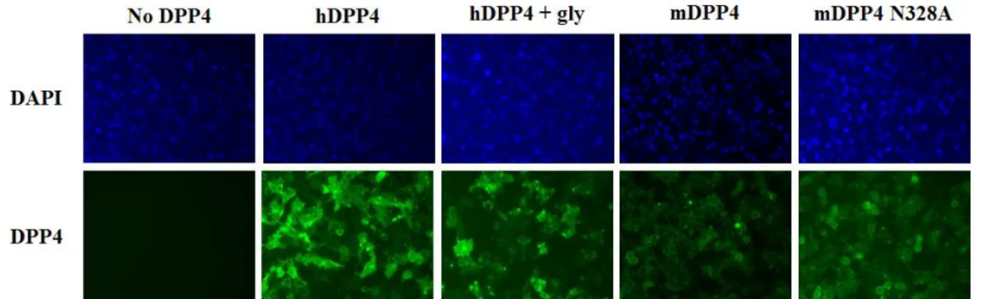
Figure 1.7: Glycosylation is more important than charge in mediating MERS-CoV infection
(A) Cells were transfected and infected following the protocol detailed in Figure 1.6A. Neither mDPP4 N328A, nor mDPP4 T330R can confer permissivity to MERS-CoV, however both result in strong levels of infection when coupled with A288L. (B) Red cell counts were calculated as in Figure 1.6B with the following MOIs: hDPP4, 0.001; mDPP4, mDPP4 288, mDPP4 328,
mDPP4 330, no DPP4, 0.1; mDPP4 288, 328 and mDPP4 288, 330, 0.01. All DPP4 constructs are significantly greater than no DPP4 and mDPP4 (*, p < 0.05, Student’s t-test) and
all derivatives of the DPP4 receptors are expressed at the cell surface and available to interact with the MERS-CoV RBD (Figure 1.8). Together, these results indicate that removal of the glycosylation site, rather than addition of the charged residue at position 330, is responsible for regulating the ability of MERS-CoV to utilize mDPP4 as a functional receptor. The secondary mutation, A288L, also plays an important role in MERS-CoV permissivity because high levels of infection are only seen when the glycosylation mutants are combined with the A288L substitution (Figures 1.7A and 1.7B). Together, this suggests that while glycosylation is an important barrier, its removal is not sufficient to permit infection in the absence of the A to L modification at position 288. Overall, glycosylation can act to block MERS-CoV infection, yet other determinants are also important for mediating permissivity. Further research will determine the relative contributions of these determinants and whether they act as a broader signature of permissivity among DPP4 orthologs.
Figure 1.8: DPP4 construct expression in HEK 293T cells
1.4 Removing glycosylation from mDPP4
The above experiments analyzed the removal of glycosylation from mDPP4 using PCR mutagenesis. Knockout of the glycosylation site was evaluated by Western blot, with a
downward shift of the protein band by ~2.5 kDa indicating successful removal of the glycan. However, additional resources are available for removing (or preventing) the glycosylation of protein molecules. Tunicamycin acts by inhibiting the formation of N-linked glycans (Esko and Bertozzi 2009). PNGase F, on the other hand, acts to cleave the glycan from the amino terminus after formation (Mulloy et al. 2009). I tested both of these methods in order to determine whether the previously discovered phenotype, that removing glycosylation from mDPP4 can confer permissivity to MERS-CoV, could be recapitulated with various methods of glycan removal. One caveat to these experiments is that DPP4 is known to have many putative glycosylation sites. hDPP4, for example, has nine N-linked glycosylation sites throughout the molecule, eight of which are clustered to blades II through V of the β-propeller domain (Rasmussen et al. 2003). The presence of multiple glycosylation sites within DPP4 may cause the use of tunicamycin or PNGase F to be too disruptive for proper folding and function of the molecule.
tunicamycin treatment, suggesting successful inhibition of glycan formation (Figure 1.9A). However, the infection results show that mDPP4 does not gain permissivity to rMERS-CoV-RFP when glycosylation is inhibited and hDPP4 loses its ability to confer infection (Figure 1.9B). The ablation of permissivity in hDPP4 suggests that other glycosylation sites are critical to DPP4 folding and/or function, specifically for MERS-CoV RBD interactions. The reduction in molecule size of both mDPP4 and hDPP4 suggests that they have a similar number of
glycosylation sites and that the additional glycosylation sites in mDPP4 may also be crucial for proper functionality. These results rule out my ability to use tunicamycin in future glycosylation analysis experiments for DPP4.
Figure 1.9: Tunicamycin treatment of DPP4 molecules
(A) Western blot analysis of HEK 293T cells transfected with either mDPP4 or hDPP4 (fused to venus) and treated with 1 ug/mL tunicamycin (Tu) ~2 hours post-transfection. Protein lysates were harvested ~24 hours post-treatment with Tu. Shifted bands indicate the successful
inhibition of glycosylation. Western blot protocol follows Figure 1.1 (Cockrell et al. 2014). (B) HEK 293T cells transfected with mDPP4 or hDPP4 and treated with Tu ~2 hours
To test the efficacy of PNGase F, HEK 293T cells were transfected with 3 µg of hDPP4 or mDPP4 following previously detailed protocols. At ~24 hours post-transfection, cells were washed twice with 1X PBS. PNGase F (NEB) was diluted in 1X PBS to final concentrations of 0, 2500, and 5000 units/mL. Cells were incubated at 37 ºC for 1 hour and then rinsed twice with 1X PBS. Protein lysates were then harvested using the previously describe 1X RIPA buffer protocol and analyzed by Western blot (Figure 1.10). Unfortunately, even at a concentration using 5000 units/mL, no downward shift in protein size is visible (Figure 1.10). This indicates that 1) the concentration of PNGase F may need to be higher, although this puts it outside of the range found to be effective for other proteins, or 2) the surface glycans on DPP4 do not meet the requirements needed for PNGase F to be effective. Namely, the enzyme requires at least one amino acid at both the amino and carboxyl terminus of the asparagine (N) in order for proper cleavage (Mulloy et al 2009). Additionally, the traditional PNGase F protocol treats proteins
Figure 1.10: PNGase F treatment of DPP4 molecules
after they have been harvested from the cells. However, because I was interested in treated monolayers and testing for infection, I treated the cells directly. This protocol adjustment could also explain the failure of PNGase F to remove glycans. Because of the lack of evidence for glycan removal (protein size shifting via Western blot) after treatment (Figure 1.10), PNGase F was not used in subsequent experiments on DPP4 glycosylation and MERS-CoV permissivity.
CHAPTER 2: BIOCHEMICAL DETERMINANTS OF DPP4 ORTHOLOG PERMISSIVITY TO MERS-CORONAVIRUS
2.1 Introduction
Among viruses for which the host receptor has been identified, there is an association between host range and phylogenetic conservation of that receptor (Woolhouse 2002). This result is consistent with previous species-level studies that have shown that the more
phylogenetically related two species are, the more likely it is that a virus will be able to jump between them (deFilippis and Villarreal 2000). These observations confirm that the host receptor is a primary determinant of host range expansion and also that receptor conservation can
potentially act as a screen to identify viruses that are likely to jump into humans.
The link between DPP4 sequence conservation across species and permissivity to MERS-CoV infection, however, is not obvious. While only a small subset of species have been tested in vitro (Muller et al. 2012, de Wit et al. 2013a, de Wit et al. 2013b, Ohnuma et al. 2013, Barlan et
al. 2014, Cockrell et al. 2014, Coleman et al. 2014, Eckerle et al. 2014, Falzarano et al. 2014,
Raj et al. 2014, van Dormalen et al. 2014), there is no clear phylogenetic clustering of
permissive and nonpermissive hosts when analyzing the DPP4 gene tree (Cui et al. 2013, Peck et al. 2015c). The lack of a clear pattern of permissivity among closely-related DPP4 genes
To understand the interactions between the MERS-CoV RBD and DPP4 and to better characterize the host range expansion events of MERS-CoV, both past and potential, I examined the permissivity of various DPP4 orthologs. After establishing the characteristics and
permissivity of these orthologs, I determined whether there was a detectable broad signature of permissivity. Primarily, do all nonpermissive species block MERS-CoV infection using the same mechanisms? Because phylogenetic relatedness did not yield a signal (Cui et al. 2013, Peck et al. 2015c), and analyzing the sequences at previously identified key RBD residues (Cockrell et al. 2014) did not yield any obvious hypotheses (Table 1), I turned to the results found in Chapter 1. Based on the discovery that glycosylation is an important barrier to MERS-CoV infection, I noticed that some nonpermissive species also have glycosylation sites near the site observed in mDPP4 (Figure 1.5A). Thus, I tested whether glycosylation acts to block MERS-CoV infection in other nonpermissive species before investigating alternate determinants.
2.2 Determine the role of glycosylation in additional DPP4 orthologs
First, I validated the panel of DPP4 constructs I had generated by confirming the
permissive or nonpermissive nature of various species orthologs. While hDPP4 and mDPP4 were
282 288 289 330 340
Human T L I R I
Bat T L T K I
Camel V L I R I
Mouse P A R T V
Guinea Pig A I T G T
Ferret T S T S T
Hamster T L T T V
Table 1: Amino acid identities at five DPP4 ortholog residues important for MERS-CoV binding
obtained as a complete plasmid, I generated the remaining DPP4 orthologs using Gibson
assembly (Gibson et al. 2009). This method employs an in vitro recombination method to allow for the rapid and financially tractable assembly of multiple DPP4 constructs.
Following previous methods, I transfected 3 µg of each DPP4 ortholog into HEK 293T cells. At ~24 hours post-transfection, I infected the cells with rMERS-CoV-RFP at an MOI of 1. At ~24 hours post-infection, I visualized the cells to determine whether they were permissive to MERS-CoV. As expected, human, bat, and camel DPP4 molecules are permissive to MERS-CoV infection while mouse, ferret, guinea pig, and hamster are not (Figure 2.1). Although previous work (Cockrell et al. 2014) generated data that was used to develop a mouse model for MERS-CoV, understanding why ferret, hamster, and guinea pig cannot be infected could help create a secondary option for studying MERS-CoV pathogenesis.
Figure 2.1: Permissivity of DPP4 orthologs to MERS-CoV
Aligning the sequences of the permissive and nonpermissive species in a region crucial to MERS-CoV binding reveals the presence of a glycosylation site in hamster DPP4 (haDPP4), ferret DPP4 (fDPP4), and guinea pig DPP4 (gpDPP4) molecules (Figure 2.2A). The
glycosylation site in hamster is identical to that in mouse, suggesting that it may interact with the MERS-CoV RBD in a similar way. The glycosylation site in fDPP4 is slightly upstream, and the glycosylation site in gpDPP4 is slightly downstream (Figure 2.2A). However, the latter is shared by bDPP4 (sequence from species Pipistrellus pipistrelle), suggesting that it might not play as important of a role in blocking infection unless there is a gpDPP4-specific structural effect.
Figure 2.2: Sequence and structural comparison of nonpermissive DPP4 orthologs
The crystal structures for fDPP4, haDPP4, and gpDPP4 have not yet been solved. Utilizing the same threading technique as in Chapter 1, I generated predicted structures for each of these proteins using I-TASSER (Zhang 2008). Overlaying the structures with hDPP4 shows that they are predicted to have highly similar structural backbones (Figure 2.2B). Again, maintaining awareness that the structures are predictions, the RMS scores obtained for fDPP4, haDPP4 and gpDPP4 overlaid with hDPP4 are 0.616, 0.378, and 0.604, respectively. This can be compared to amino acid sequence identity values of 88%, 85%, and 87%, respectively. From these computational results, haDPP4 is predicted to be the most structurally similar to hDPP4 and may be the best option for an alternate transgenic animal model.
Based on my previous discovery that glycosylation plays an important role in blocking MERS-CoV, I knocked out the glycosylation sites in each of these species using PCR
mutagenesis. Each of these knockout mutations changed the N of the glycosylation NXT (or NXS) motif to an alanine and are designated as “-gly” in subsequent figures. Following previous methods, 3 µg of each DPP4 construct was transfected into HEK 293T cells. At ~24 hours transfection, cells were infected with rMERS-CoV-RFP at an MOI of 1. At ~24 hours post-infection, cells were imaged and red fluorescence analyzed as a readout of infection. My results show that removing glycosylation from these three DPP4 orthologs did not result in an increase in infection (Figure 2.3A). The amount of infection that was supported by all three glycosylation knockout molecules was equivalent to their respective wildtype molecules. The glycosylation, and subsequent removal, of each of these sites was confirmed by Western blot analysis (Figure 2.3B), following previous protocols (Peck et al. 2015b).
is directly upstream from the previously studied NLT (starting at residue 332) glycosylation site. As a test to determine whether this site affected permissivity, particularly because the equivalent 288 residue found to be important in mice shares the human amino acid identity in haDPP4, I generated constructs that mutated the N of the NKT motif using PCR mutagenesis, both singly and in combination with the NLT glycosylation knockout. Transfecting the haDPP4
glycosylation variants into HEK 293Ts and infecting with rMERS-CoV-RFP revealed that mutating this additional glycosylation site had no impact on infection (Figure 2.4). Furthermore, mutating both of the glycosylation sites together did not have any impact on infection levels
Figure 2.3: DPP4 ortholog glycosylation knockout mutants
(Figure 2.4). It is possible that the NKT motif is not actually glycosylated; my next step is to validate glycosylation by Western blot before drawing further conclusions.
Whereas fDPP4 and haDPP4 did not share their glycosylation sites with permissive molecules, gpDPP4 shared its downstream glycosylation site with bDPP4. Due to this similarity, I knocked out the glycosylation site in bDPP4 to determine 1) whether it was actually
glycosylated and 2) its impact on the permissivity of bDPP4. Using PCR mutagenesis to mutate the N of the NXS motif to an alanine, I followed previous protocols to express the bDPP4 mutant in HEK 293T cells and visualized the cells ~24 hours post-infection. My results show that
removing glycosylation from bDPP4 has no detectable difference on its ability to support MERS-CoV infection (Figure 2.5). My next step is to perform a Western blot in order to
Figure 2.4: Glycosylation knockout panel in haDPP4
haDPP4 has two glycosylation sites near the site identified as important for mediating MERS-CoV permissivity in mDPP4. Knocking out the NKT (starting at residue 329) or the NLT (starting at residue 332) motif, or both in combination, has no impact on MERS-CoV
determine whether the bDPP4 molecule is actually glycosylated or whether the NXS represents a sequence motif that is not, in actuality, glycosylated in the final molecule.
Overall, glycosylation is not the only determinant that mediates MERS-CoV infection in nonpermissive DPP4 orthologs. This is particularly surprising in the case of haDPP4; this protein not only has a glycosylation site in the same location as mDPP4 (Figure 2.2A), but the secondary residue that was identified to be important for mDPP4 (residue 288) in haDPP4 is the same amino acid identity as hDPP4 (Table 1). Based on this observation, while removing
glycosylation may be a substantial component of permitting infection in these DPP4 orthologs, other determinants clearly play an important role.
The importance of glycosylation in blocking MERS-CoV infection may vary between species. To gain a better intuition about the extent of glycosylation among DPP4 orthologs, I
Figure 2.5 Guinea pig and bat DPP4 glycosylation knockout mutants
constructed a phylogenetic tree of a subset of full-length DPP4 protein sequences (Figure 2.6). Retrieving the sequences from GenBank, I used MAFFT to align the amino acid sequences (Katoh et al. 2002). The phylogenetic tree was generated using maximum likelihood with the PhyML package (Guindon et al. 2010) and visualized using EvolView (Zhang et al. 2012). In the tree, shaded colors indicate the general organism group that each species belongs to - blue: reptiles and amphibians; green: avian species; orange: other mammals; red: Chiroptera (bats); purple: ungulates; gray: rodents; pink: primates (Figure 2.6). The DPP4 protein tree is slightly discordant with the species tree, notably with the horse and African savanna elephant DPP4 sequences not clustering with other ungulate (purple) orthologs.
Plotted adjacent to the phylogenetic tree are glycosylation sites that are either upstream (column 1), at the same site (column 2), or downstream (column 3) of the glycosylation site that is present in mDPP4 (residues 328-330). Based on this tree, only eight other species have
putative glycosylation sites at the same location as in mDPP4; the majority of these are present in the rodent (gray) group, however two are present within the Chiroptera group (black flying fox and large flying fox) (Figure 2.6). Permissivity data is indicated in the far right column, with green squares indicating permissive species and red squares indicating nonpermissive species, based on either in vitro or in vivo data (de Wit et al. 2013a, Barlan et al. 2014, Eckerle et al. 2014, Cockrell et al. 2014, Coleman et al. 2014, Raj et al. 2014, van Doremalen et al. 2014). The small number of data points for species makes it difficult to map potential shifts in permissivity. However, a few interesting observations emerge. First, all non- human primates lack
Figure 2.6: DPP4 protein phylogenetic tree
DPP4 phylogenetic tree based on amino acid sequences. Shaded colors indicate the group each species falls in. Blue: reptiles and amphibians; Green: avian
species; Orange: other mammals; Red: Chiroptera (bats); Purple: ungulates; Gray: rodents; Pink: primates. Colored circles to the right of the species names indicate whether the sequence has a glycosylation site upstream (first column), at the same site (second column), or downstream (third column) of the NXT glycosylation site in mDPP4 (residues 332-334). Numbers inside the circle designate how many amino acids upstream (or downstream) the N of the NXT or NXS putative glycosylation site is. For the second column, a 1 indicates that there is a
nonpermissive. If so, a separate mechanism from glycosylation at our location of interest would be responsible for blocking MERS-CoV infection. Second, whereas other permissive DPP4 orthologs have glyocyslation sites in this region (horse and common pipistrelle DPP4), of note is the lack of a glycosylation site in the nonpermissive domestic pig, sheep, and cattle DPP4
molecules (Figure 2.6). This suggests that glycosylation in this region is not the primary
explanation for why these species do not support MERS-CoV infection. Follow-up studies could explore the mechanism of nonpermissivity in this species specifically, and whether or not it lies at the level of the receptor. Third, glycosylation sites in the designated region are prevalent in rodents, other mammals, and avian species (Figure 2.6). The diversity of glycosylation profiles suggests a lack of strong conservation of the site seen in mDPP4. Further research to determine whether these other orthologs are permissive can help elucidate whether this region plays a broader role in the MERS-CoV infection phenotype. In general, while some trends are seen when looking at the glycosylation profile across many species (e.g. upstream glycosylation sites in the other mammal (orange) group), more data on species permissivity will help determine whether DPP4 phylogenetic relationships can help inform receptor-binding dynamics.
2.3 Identify key mutations and determinants of DPP4 ortholog permissivity to MERS-CoV infection
The mDPP4 data suggest that two mutations are necessary to support MERS-CoV
2.3A). This suggests other determinants are important for conferring permissivity in these other molecules.
Other groups have introduced single point mutations in both haDPP4 and fDPP4 to try to instigate permissivity (Raj et al. 2014, van Doremalen et al. 2016). For haDPP4, other studies found that a minimum of five amino acid mutations allowed the molecule to support MERS-CoV infection (van Doremalen et al. 2016). Focusing on the idea that changes in both blades IV and V of DPP4 are required to confer infection in the context of mDPP4, I constructed chimeric fDPP4 and haDPP4 molecules that made substantial changes on both blades. Using overlap PCR, I constructed a chimeric fDPP4 that swaps out 16 residues on blade V and 11 residues on blade IV to the equivalent human amino acid identities, indicated by their starting residues of 278 and 331, respectively. Transfecting 3 µg of the chimeric fDPP4 constructs into HEK 293T cells and infecting with rMERS-CoV-RFP (MOI 1) at ~24 hours post-transfection yielded results that were imaged ~24 hours post-infection. Results show that while the single glycosylation knockout mutant did not show an increase in infection, the combination of 27 amino acid changes on blades IV and V show a dramatic increase in infection (Figure 2.7A). Western blot analysis shows that each DPP4 variant is being highly expressed; however, because of the large number of amino acid changes present in each mutant, a downward shift in the protein band is visible for each construct (fDPP4 (278), fDPP4 –gly, fDPP4 (278)(331)) (Figure 2.7C), obscuring the usual signal representative of glycan removal.
acids in fDPP4 that could be found in a permissive species were not included in the fine-tuning mutation sets. 278snp includes the mutations E285Q, D290A, A291S, and S293L. 330snp includes the mutations N330D, N331E, and D332S. 334snp includes the mutations N334G, S335R, and R337N. 338snp includes the mutations K338C, P339L, and E340V (Figure 2.8). Each construct was transfected into HEK 293T cells following previous protocols and infected with rMERS-CoV-RDP at an MOI of 1. Results show that all combinations of mutants show
Figure 2.7: Many amino acid changes are required to make fDPP4 and haDPP4 permissive to MERS-CoV infection
lower levels of infection compared to when both blocks (278) and (331) are included together (Figure 2.8). Interestingly, the mutants fDPP4 (278), 334snp and fDPP4 (278), 338snp show increased levels of infection compared to all other combination sets (Figure 2.8). This suggests that 1) the (278) block is an important determinant and cannot be recapitulated with a subset of the four selected amino acids and 2) infection is more robust to amino acid changes on blade IV.
Figure 2.8: Fine-tune mapping of fDPP4 determinants for MERS-CoV permissivity
Sets of mutations were made in fDPP4 for fine-tune mapping of MERS-CoV permissivity determinants. The table indicates which combination of hDPP4 amino acid identities were introduced into fDPP4 on blade V (rows) or blade IV (columns). Blue boxes around the
In fact, the lack of increased infection for fDPP4 (278), 330snp (Figure 2.8) suggests that our hypothesis surrounding glycosylation as an important barrier may not be as applicable to fDPP4. The 330snp subset removes the glycosylation site while the 334snp and 338snp amino acid changes do not. However, we cannot rule out that the 334snp and 338snp amino acid mutations alter the structure of fDPP4 in a way that shifts the glycosylation site and allows proper binding by MERS-CoV. Overall, the determinants that would allow fDPP4 to fully support MERS-CoV infection are complex and require further investigation to fully understand.
In the context of haDPP4, fewer amino acid changes can result in a dramatic increase in the infection levels (Figure 2.7B). Compared to fDPP4, which I found to require 27 mutations (possibly less), haDPP4 can support high levels of infection with just four mutations: E291A, V293M, T295I, and N332A. The first three are present on blade V while the fourth is on IV and removes the glycosylation site (Figure 2.7B). As with fDPP4, these residues were selected based on examining amino acids that were not present in permissive DPP4 orthologs. The four
identified mutations overlap with previous studies that found five amino acid changes in haDPP4 were required for high levels of MERS-CoV infection (van Doremalen et al. 2016). To
determine whether fewer mutations could recapitulate the results seen with all four, I used PCR mutagenesis to make a set of the mutations individually and in combination with the
glycosylation knockout (N332A, designated as ‘-gly’).
work by van Doremalen et al. (2016) which demonstrated that less than five amino acid changes were unable to confer high permissivity to MERS-CoV. Of these, residue 295 overlaps with the results found here, while residue 336 removes a glycosylation site, functionally achieving the same as the 334 mutation presented here (Table 2). Our conclusions show that while
glycosylation is likely an important barrier to MERS-CoV infection, there are additional determinants on blade V that also play a crucial role.
Comparing the data we have so far on the residues that are important for mediating permissivity in hDPP4, mDPP4, haDPP4, it is difficult to discern an obvious pattern (Table 2). The only clear trend is that at least one change is required on both blades IV and V of DPP4. This indicates that there are two key points of interaction between DPP4 and the MERS-CoV RBD that are important for allowing the virus to utilize a new species receptor. Further
understanding of the host range expansion of MERS-CoV will come with further data on which mutations can confer permissivity to DPP4 receptors of currently nonpermissive species.
Figure 2.9: Fine-tune mapping of haDPP4 permissivity to MERS-CoV
Sets of mutations in haDPP4 in order to map determinants of permissivity to MERS-CoV