Downloaded from
http://journals.tums.ac.ir/
on Wednesday, August 15, 2012
Depth Dose Calculation of Holmium-166 for Different Shape
Source by VARSKIN3 Code
Ali Asghar Mowlavi1,2, Azam Afzalifar1, Naser Afzalifar3, Ehsan Kashani4
1
Physics Department, School of Sciences, Sabzevar Tarbiat Moallem University, Sabzevar, Iran.
2
ICTP, TRIL, Trieste, Italy.
3
Physics Department, School of Sciences, Imam Khomeini University, Sabzevar, Iran.
4
Vasei Hospital, Sabzevar Medical University, Sabzevar, Iran.
(Received 25 September 2009, Revised 22 November 2009, Accepted 4 December 2009)
ABSTRACT
Introduction: Using beta emitter radionuclide is a useful therapeutic modality in the treatment of skin cancers in areas which are difficult to cure by other methods. The aim of this research is to evaluate the tissue response to beta rays of 166Ho and determine the feasibility of beta emitting radionuclide for treatment of skin cancers.
Methods: In this work, we have calculated depth dose distribution of 166Ho using Varskin3 code. The code has been run for various input parameters to calculate absorbed depth dose for different shape of source.
Results: Absorbed depth dose variation has been calculated for166Ho beta emitter, in different shape of sources such as slab, spherical, cylindrical and 2-D disk shapes. Comparison of the result for different shape sources has been presented.
Conclusion: The result shows that 2-D disk source induces damage to skin cells more than other shape of sources. These computational and Lee et al. experimental results are shown that 166Ho radionuclide treatment is very useful for skin cancer therapy. One of advantages of using 166Ho radionuclide is that no adverse effect on underlying bone and soft tissue due to the physical characteristics of beta rays, high linear energy transfer or rapid depth dose fall off is observed.
Keywords: Holmium-166, Varskin3 code, Skin cancer, Source shape
Iran J Nucl Med 2010;18(1):32-36
Corresponding author: Ali Asghar Mowlavi, Department of Physics, Sabzevar Tarbiat Moallem University, Sabzevar, Iran.
E-mail: [email protected]
Downloaded from
http://journals.tums.ac.ir/
on Wednesday, August 15, 2012
Ir
an J Nucl Me
d
20
10, Vo
l
18
, No
1 (Ser
ial No
3
3
)
33
INTRODUCTION
Skin cancer is the most common malignancy in human, and more than 95% of basal cell carcinomas, the most common form of skin cancer, occur in patients more than 40 years of age (1). Due to the increasing life span in many countries, the incidence of skin cancer has been increased continuously. In spite of the malignant nature of this cancer, the death rate is not high, because basal cell carcinomas are slowly progressive (2). Radiation therapy of skin cancer using an electron beam (3) X-rays (4) or a neutron beam (5) has been reported. Treating skin cancer with topical application of a radioisotope has also been reported, such as
using 125I-seeds on a gold plaque (6), also a
very effective and convenient brachytherapy
for this application using a 166Ho-patch has
been reported by Chung el al. (7).
However; several factors such as total dose, fractionation regimens, and field size and beam quality affect the treatment outcome. In general, a total dose ranging 35-70Gy with daily fractionation lying in the 2.0- 3.5Gy is accounted for the optimal therapeutic regimen (8-9). The aim of this research is to evaluate the tissue response to
beta rays of 166Ho and determine the
effective shape of source for treatment of skin cancers. In this work, we have run the code to calculate skin absorbed depth dose variation from the skin surface. This code also contained a volume averaged dose model and an offset particle model. The volume averaged dose model that we have used allowed us to calculate the dose averaged over a volume of tissue defined by a cylinder with diameter equal to that of the dose averaging area and bounded at the top and bottom by two selected skin depths. This model can be used to calculate the dose averaged between two depths in tissue, which is useful when characterizing the dose measured by a finite volume dosimeter such as a thermo luminescent dosimeter (10, 11).
METHODS
Varskin3 computer code calculates skin dose due to radioactive skin contamination provided by RSICC in USA. To run the code in windows XP after installation, user can contact the basic information section of the code to select radionuclide. When the desired geometrical parameters and options are selected, the calculation is initiated. The calculation time is greatly affected by the number of radionuclide used in the calculation and the various options that are selected. This code calculates doses using a compiled FORTRAN program while the main program that collects the input data is written in visual basic (10, 11). With running Varskin3 code for calculating dose, the required input parameters contain: activity of source, cover thickness, cover density, air gap thickness, radiation time and different source geometry. We have selected
the default value of 10 cm2 for skin average
area and 60 minutes for exposure time.
166Ho is a beta emitter radioisotope with
Emax=1.84 MeV and half life 26.9 hr. It also
emits the following photons: 0.081MeV with 5.4% and 1.38 MeV with 0.9% intensity per decay.
RESULTS AND DISCUSSION
By running Varskin3 code, we have calculated the absorbed depth dose variation
for 166Ho in different form of sources shape
Downloaded from
http://journals.tums.ac.ir/
on Wednesday, August 15, 2012
Ir
an J Nucl Me
d
20
10, Vo
l
18
, No
1 (Ser
ial No
3
3
)
34
0 1 2 3 4 5 6 7 8
0.0 2.0x10⁻³ 4.0x10⁻³
6.0x10⁻³ 8.0x10⁻³
1.0x10⁻²
D
o
s
e
ra
te
(r
a
d
/h
)
Depth(mm)
Disk Source Cylindrical Source Spherical Source
0 1 2 3 4 5 6 7
0 10 20 30 40 50 60 70 80 90 100 110
No
rm
a
liz
ed
do
se
ra
te
Depth(mm)
2-D Disk Source 166Ho Slab Source 166
Ho
Cylindrical Source 166Ho Spherical Source 166
Ho
accurate dose calculation. Computational result shows the calculated dose is more sensitive to the cylinder. The slab source geometry requires knowledge of three physical dimensions: the x-side length, the y-side length, and the thickness. The slab source geometry requires two to five times more execution time than the cylinder and sphere geometries, and the accuracy of the calculation is significantly lower. The geometry characteristics of different sources with 60 minutes irradiation time, and any cover or air gap have been presented in Table 1.
Figure 1. Schematic representations of the 4 type
source geometry: (a) Disk, (b) Sphere, (c) Cylinder, and (d) Slab.
Table 1. The geometry characteristics of different
sources with any cover or air gap and 60 min irradiation time.
Source
shape Diameter Thickness Other lengths
Cylindrical 1000 μm 1000 μm ---
Spherical 1000 μm --- ---
2-D Disk 1000 μm --- ---
Slab --- 1000 μm X =Y=44 μm
The result of the absorbed depth dose
variation for three different shapes of 166Ho
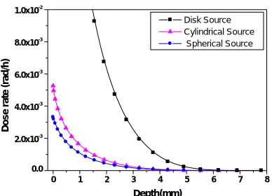
are shown in Figure 2, which the default unit of measure for activity is 1µCi for all sources.
Figure 2. Absorbed depth dose variation for different
shapes of 166Ho with activity 1µCi.
Figure 3. The normalized dose rate variation for all
of the sources.
Downloaded from
http://journals.tums.ac.ir/
on Wednesday, August 15, 2012
Ir
an J Nucl Me
d
20
10, Vo
l
18
, No
1 (Ser
ial No
3
3
)
35
0 1 2 3 4 5 6 7
0 20 40 60 80 100
Norm
ali
z
e
d
dos
e ra
te
Depth(mm)
Cylindrical source of 166Ho Lee et al. result
0 1 2 3 4 5 6 7
0 20 40 60 80 100
N
o
rm
al
iz
ed
do
se
rate
Depth(mm)
Slab source of 166 Ho Lee et al. result
0 1 2 3 4 5 6 7
0 20 40 60 80 100
No
rm
a
lize
d
do
se
r
a
te
Depth(mm)
2-D disk source 166Ho Lee et al. result
0 1 2 3 4 5 6 7
0 20 40 60 80 100
Norm
alized dose rat
e
Depth(mm)
Spherical source of 166 Ho Lee et al. result
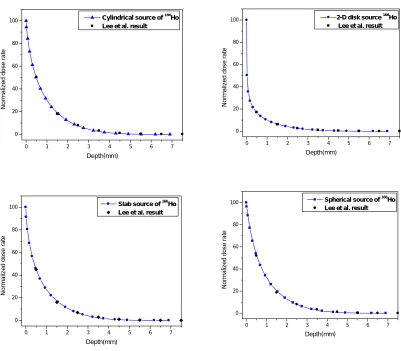
Figure 4. Comparison of the normalized absorbed rate variation against to the depth for 1µCi of 166Ho d with
the result of Lee et al.
Regarding to computational results for the same activity, disk source induces more dose than others to the skin surface and minimum dose is due to the slab source. We have presented the normalized dose rate for all of the sources in Figure 3. We must mention that the absorbed dose rate has been normalized to the dose rate in 0 mm depth for each source in order to show the effect of source geometry on dose rate variation.
Lee et al. (12) used 166Ho source for curing
five patients including 4 women and 1 man in age range 41-95 yr, as well as several
animal models with superficial squamous cell, basal cell carcinoma and Bowen's disease in their experimental research. We have compared our results with Lee et al. experimental data as shown in Figure 4 with a very good agreement.
CONCLUSION
Downloaded from
http://journals.tums.ac.ir/
on Wednesday, August 15, 2012
Ir
an J Nucl Me
d
20
10, Vo
l
18
, No
1 (Ser
ial No
3
3
)
36
good agreement with the experimental data of Lee et al. Our computational study and Lee et al. experimental results revealed that
166Ho radionuclide can be used effectively
for skin cancer therapy. The advantage of
166Ho radionuclide is that no adverse effect
on underlying bone and soft tissue due to the physical characteristics of beta rays, high linear energy transfer or rapid depth dose fall off is seen. Varskin3 code is a very useful tool for skin dosimetry, as well as it is fast, accurate and user friendly. It can be used for dose optimization calculation especially in beta source over the human skin.
AKNOWLEDGEMENT
Authors would like to thank Prof. G. Furlan and D. Treleani in TRIL program at ICTP, Trieste, Italy for their support.
REFERENCES
1. Kopf AW. Computer Computer analysis of
3531 basal-cell carcinomas of the skin. J Dermatol. 1979;6(5):267-281.
2. Fitzpatrick PJ. Skin cancer of the
head--treatment by radiotherapy. J Otolaryngol. 1984;13(4):261-266.
3. Chinn DM, Chow S, Kim YH, Hoppe RT.
Total skin electron beam therapy with or without adjuvant topical nitrogen mustard or nitrogen mustard alone as initial treatment of T2 and T3 mycosis fungoides. Int J Radiat Oncol Biol Phys. 1999;43(5):951-958.
4. Nevrkla E, Newton KA. A survey of the
treatment of 200 cases of basal cell carcinoma (1959-1966 inclusive). Br J Dermatol. 1974;91(4):429-433.
5. Hofmann B, Fischer CO, Lawaczeck R,
Platzek J, Semmler W. Gadolinium neutron capture therapy (GdNCT) of melanoma cells and solid tumors with the magnetic resonance imaging contrast agent Gadobutrol. Invest Radiol. 1999;34(2):126-133.
6. Packer S, Rotman M. Radiotherapy of
choroidal melanoma with iodine-125. Ophthalmology. 1980;87(6):582-590.
7. Chung YL, Lee JD, Bang D, Lee JB, Park KB,
Lee MG. Treatment of Bowen's disease with a specially designed radioactive skin patch. Eur J Nucl Med. 2000 Jul;27(7):842-846.
8. Shimm DS, Wilder RB. Radiation therapy for
squamous cell carcinoma of the skin. Am J Clin Oncol. 1991;14(5):383-386.
9. Petrovich Z, Parker RG, Luxton G, Kuisk H,
Jepson J. Carcinoma of the lip and selected sites of head and neck skin. A clinical study of 896 patients. Radiother Oncol. 1987;8(1):11-17.
10. Durham JS. VARSKIN 3: A computer code
for assessing skin dose from skin dose contamination. US Nuclear Regulatory Commission Office of Nuclear Regulatory Research, Washington DC, NUREG/CR-6918, 2006.
11. Radiation Safety Information Computational
Center, 2009,www-rsicc.ornl.gov
12. Lee JD, Park KK, Lee MG, Kim EH, Rhim