
R E S E A R C H A R T I C L E
Open Access
Gut derived-endotoxin contributes to
inflammation in severe ischemic acute
kidney injury
Jiangtao Li
1, Krishna Rekha Moturi
2, Lirui Wang
1, Kun Zhang
1and Chen Yu
1*Abstract
Background:Recent researches indicate that the intestinal consequences of renal ischemia reperfusion (IR) would predispose to the translocation of gut-derived endotoxin. Here, we designed experiments to test the hypothesis that the gut-derived endotoxin has a potential role in mediating local inflammatory processes in the acutely injured kidney. Methods:Rats were performed sham or renal IR surgery (60 min of bilateral renal ischemia, then 24 h of reperfusion) (n= 5). The intestinal structural and mucosa permeability were evaluated. Serum endotoxin and bacterial load in liver and
mesenteric lymph nodes (MLN) were measured. Separate groups were pretreated with oral norfloxacin 20 mg/kg/day or saline for 4 weeks and divided into sham plus saline, sham plus norfloxacin, renal IR plus saline and renal IR plus norfloxacin group. Serum biochemistry and endotoxin were determined. Kidney pathological changes were scored. Protein or mRNA expression of toll-like receptor 4 (TLR4) and proinflammatory mediators were measured in kidney homogenate.
Results:Renal IR led to marked intestinal integrity disruption and increase in intestinal permeability. These are accompanied by low grade of endotoxemia as well as increased bacterial load in liver and MLN. The group pretreated with norfloxacin showed significant attenuation of the increase in serum urea, ALAT, ASAT and endotoxin. The increased renal protein or mRNA of TLR4 and proinflammatory mediators (IL-6 and MCP-1) in the unpretreated animals was significantly attenuated in the norfloxacin-pretreated animals. However, norfloxacin pretreatment did not produce any protective effects on renal tubular integrity.
Conclusions:Our results show for the first time that gut-derived endotoxin, resulting from an increased intestinal
permeability after severe renal IR, subsequently amplifies intrarenal inflammatory response by activation renal TLR4 signaling. Our study results do not establish that antibiotic administration was effective in improving the overall renal outcome. However, our findings may be the first step to understanding how to tailor therapies to mitigate intrarenal inflammation in select groups of patients.
Keywords:Acute kidney injury, Renal ischemia reperfusion, Inflammation, Intestinal permeability, Endotoxin
Background
Acute kidney injury (AKI) is a serious complication with no special therapies currently. Renal ischemia-reperfusion (IR) is one of the major causes of AKI [1]. The pathophysi-ology of IR-induced renal injury includes oxidative stress, inflammation and damage to the vascular endothelium and tubular epithelium [2]. Among them, inflammation is
possibly the most important pathophysiological factor in-volved in the propagation of renal IR.
The gut bears critical immunologic and barrier func-tions that prevent the passage of substances, such as pathogenic microorganisms and their products, antigens and proinflammatory mediators from the intestinal lumen into the internal milieu. The disruption of gut barrier is proposed to permit the translocation of intestine endo-toxin/bacteria into the portal vein and peripheral circula-tion where host defenses are already compromised, leading to aggravation of the inflammatory status and propa-gation of inflammation [3]. A rather new research using
* Correspondence:[email protected]
1Department of Nephrology, Tongji Hospital, Tongji University School of
Medicine, 389 XinCun Road, Shanghai 200065, China
Full list of author information is available at the end of the article
renal IR mice demonstrated a series of intestinal conse-quence of ischemic AKI, manifested by the increased in-testinal permeability, enhanced inflammation, profound apoptosis and necrosis [4]. However, whether the conse-quences of renal IR would predispose to the translocation of endotoxin and/or bacterial components is not clear.
Toll-like receptors (TLRs) are key members of the in-nate immune system, serving as pattern recognition re-ceptors that identify a wide spectrum of microbial products. TLR4 is one of the key TLRs which critically involved in inflammation [5, 6]. TLR4 is expressed by cells of the immune system, such as dendritic cells, mac-rophages, neutrophils, B cells and NK cells. TLR4 can also be expressed in tissue by cell types, including kidney mesangial cells and tubular epithelial cells in response to injury [7]. Activation of TLR4 by LPS generates an exces-sive production of inflammatory mediator [7, 8]. During the process of renal IR, whether the gut-derived endotoxin would further contribute to renal inflammation through the TLR4 mediated pathway remains to be clarified.
In present study, we used a rat model of severe renal IR and examined (1) whether the translocation of endo-toxin and/or bacterial components would occur as the intestinal consequences of renal IR and (2) whether the gut-derived endotoxin would further contribute to renal inflammation through TLR4 activation.
Materials and methods
Ethical approval and animals
The animal use and care protocol and experimental proce-dures were reviewed and approved by the Experiment Ani-mal Center Committee in Tongji University and the procedures were carried out in accordance to the National Institute of Health Guidelines for the Care and Use of La-boratory Animals (8th edition; available online: https:// www.ncbi.nlm.nih.gov/books/NBK54050/) and to the Euro-pean Community Council Directive for the Care and Use of Laboratory Animals (Directive 2010/63/EU; http:// ec.europa.eu/environment/chemicals/lab_animals/legislatio-n_en.htm). Sprague-Dawley male rats (Shanghai SLAC La-boratory Animal Co., Ltd., Shanghai, PR China), weighing 200-250 g, were bred at a stable temperature of 22–23 °C with 12-h light-dark cycles. They were permitted to get ac-cess to diet and water freely. All procedures were per-formed using sterile techniques under adequate anesthesia.
Experimental protocol
The first set of experiments was performed to test the hypothesis that the intestinal consequences of renal IR would predispose to the translocation of endotoxin and/ or bacterial components. To do so, a well-characterized rat model of AKI induced by severe renal IR was used. Prior to surgeries, all rats were randomly allocated into sham operation group and renal IR group (n= 5 for each
group). Intestinal structural alterations and permeability were assessed. Translocation of gut-derived endotoxin and bacteria was detected.
The second group of experiments was conducted to study whether the gut-derived endotoxin would further contribute to renal inflammation during renal IR. By doing so, norfloxacin was used to reduce the transloca-tion of gut-derived endotoxin through inhibiting intes-tinal flora overgrowth. Briefly, the rats were randomly pretreated with oral norfloxacin (Sigma-Aldrich, USA) 20 mg/kg/day or saline for 4 weeks and then divided into 4 groups (n= 5 for each group), including sham plus sa-line, sham plus norfloxacin, renal IR plus saline and renal IR plus norfloxacin. On any given day, the person administering norfloxacin or saline was blinded to treat-ment. The effects of norfloxacin on endotoxinemia, renal and hepatic dysfunction, pathological changes and in-flammatory response in kidney were investigated.
Since our study is a pilot study and prior information is not available, sample size was not determined by power analysis. However, five animals per group for continuous endpoints is reasonable [9]. Animals randomization in our study was conducted by Excel software (Excel 2003, Microsoft Corporation, One Microsoft Way, Redmond, WA, USA).
Renal IR
Thirty min after intraperitoneal injection of 2% pentobar-bital sodium (50 mg/kg), the rats were fully anesthetized and then fixed on a heating plate to keep a rectal temperature around 37 °C. The renal pedicles were iso-lated after a midline laparotomy. For renal IR induction, vascular clamps were placed around both renal pedicles for 60 min, followed by 24 h of reperfusion. After clamps removal, kidneys were inspected for their original color re-covery. To avoid excessive fluids loss, rats were adminis-trated by 1 mL warm sterile saline intraperitoneally prior to abdomen closing. For sham animals, identical surgical procedures were conducted except the induction of IR.
Collection and preparation of samples
With overdose of 2% pentobarbital sodium, rats were eu-thanized 24 h after surgery. Blood was collected by intra-cardic puncture, centrifuged (4000 g for 10 min at 4 °C) and the serum was stored at−70 °C for further measure-ments. Samples from kidney, liver and mesenteric lymph nodes (MLN) were snap-frozen in liquid nitrogen and stored at −70 °C for further use. Tissues of kidney and ileum were fixed in formalin (10% phosphate-buffered, pH = 7.4) for histological evaluations.
Biochemistry
were determined by standard methods on an Olympus AU 2700 Analyzer (Olympus Optical Co., Ltd., Tokyo, Japan). All determinations were performed blindly with respect to group allocation or treatment.
Serum D-lactate concentration
A serum D-lactate quantitative colorimetric detection kit (Genmed, Boston, United States) was used to deter-mine serum D-lactate levels. Results were expressed as mmol/L. All determinations were performed blindly with respect to group allocation.
Protein levels of cytokines in kidney tissue
The sample of kidney was homogenized and the superna-tants were subjected to protein concentration determina-tions using bicinchoninic acid (BCA) method (Nan jing jian cheng bioengineering institute, Nanjing, China). Quantitative assessment of tumor necrosis factor (TNF-α), interleukin-6 (IL-6) and monocyte chemotactic protein-1 (MCP-1) protein levels were measured by commercial available ELISA kits (MultiSciences, Hangzhou, China). Results were expressed as pg/mg protein. All determina-tions were performed blindly with respect to treatment.
Measurement of endotoxemia
Endotoxin quantification was determined by a Kinetic Turbidimetric LAL Kit (Xiamen Limulus Experimental Reagents Factory, Xiamen, China). Results were expressed as EU/mL. All determinations were performed blindly with respect to group allocation or treatment.
In vitro intestinal permeability
After 24 h surgery, length of 5 cm terminal ileum seg-ment was excised. Ileum lumen was gently flushed, and one end of ileum was fastened by ligation. Next, 200ul of 40 mg/mL FITC-Dextran (MW, 4400 Da, FD-4) (Sig-ma-Aldrich, USA) was poured into ileum lumen and an-other end was fastened. The ileum sac was vibrated carefully in 20 mL of saline under 37 °C for 60 min. Per-meability of ileum wall was assessed in vitro by measur-ing the amount of FITC-dextran in saline [10]. All measurements were performed blindly with respect to group allocation.
Bacterial load quantification
Bacterial DNA load was measured in liver and MLN. Total DNA was extracted from tissues homogenates by DNEASY Blood & Tissue Kit (Qiagen, Germany). The primer sets applied (Table1) in present study targeted a conserved region of the 16srRNA gene present in uni-versal bacteria. The methods for bacterial DNA amplifi-cation has been described by Balamurugan R [11]. To normalize the variable mass of the collected tissue sam-ples, primers for the β-actin gene were used. Bacterial
16srRNA gene expression was normalized to β-actin in each sample and calculated by the 2−ΔΔCt method. All PCR analysis were performed blindly with respect to group allocation.
mRNA levels of cytokines in kidney
Total RNA was extracted by TRI reagent (Sigma-Al-drich, USA) according to the instruction from the manu-facturer. Complementary DNA (cDNA) was synthesized by using a commercial cDNA synthesis kit (Thermo Sci-entific, USA). mRNA expression of TNF-a, IL-6 and MCP-1 was measured by real-time quantitative RT-PCR conducted on a Light Cycler 480 (Roche) with SYBR green PCR master mix (Taraka, USA). Expression of tar-geted genes was normalized to β-actin expression in each sample and calculated using the 2−ΔΔCt method. Primer sequences are listed in Table2. All RT-PCR ana-lysis were performed blindly with respect to treatment.
Western blot
Total proteins from kidney were separated by SDS-PAGE and transferred to nitrocellulose membranes (Bio-Rad). Blots were probed with primary specific antibodies followed by horseradish peroxidase-conjugated goat anti-rabbit IgG antibody (ab97051; Abcam, 1:10000). The immunoreactive bands densities were quantified using NIH Image J software (version 1.47). Primary antibodies were the following: anti-TLR4 (ab22048; Abcam, 1:500) and anti-GAPDH (ab37168; Abcam, 1:1000). All western blot analysis were conducted blindly with respect to treatment.
Histology
Immunohistochemistry (IHC)
IHC was performed on paraffin-embedded sections of kidney specimens. TLR4 was detected with anti-TLR4 (ab22048; Abcam, 1:250) and then incubated with horse-radish peroxidase-conjugated goat anti-rabbit IgG H&L antibody (ab97051; Abcam, 1:150). The samples were stained with a diaminobenzidine staining kit (DAKO). All IHC experiments were carried out blindly with re-spect to treatment.
Statistical analyses
Graphpad Prism 4.0 (GraphPad Software, Inc., San Diego, USA) was used for statistic analysis. Differences between two groups were compared by two-tailed un-paired t test. Comparison among multiple groups was analyzed with one-way ANOVA followed by Bonferroni multiple comparison method. The data were presented as mean ± SD. Values of P< 0.05 were considered as sta-tistically significant.
Results
Intestinal mucosal injury after renal IR
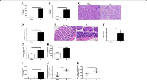
Renal IR led to a pronounced kidney injury, evidenced by a markedly rise in serum urea and creatinine as com-pared to the sham rats (P< 0.001) (Fig. 1a, b). This was associated with evidence of renal morphological damage as reflected by increased necrotic tubules in the renal IR rats compared to the sham rats (Fig.1c, d).
The gut wall forms an anatomical barrier and the breakdown of this barrier potentially leads to the trans-location of microbiota and their products. Therefore, we examined the morphological alterations in ileum. The integrity of ileum mucosa under light microscopy was nearly normal in the sham rats. By contrast, edema and inflammatory cells were observed in the ileal mucosa and submucosa in renal IR rats. The difference in the degree of ileal mucosal injury between groups were sup-ported by the results of Chiu’s scoring, in which renal IR rats presented higher score than the sham rats. (Fig.1e, f ). Taken together, these data clearly show that renal IR
results in a pronounced breakdown of intestinal physical barrier.
Increased intestinal mucosal permeability after renal IR
We next evaluated the impact of renal IR on mucosal permeability by two separate methods. The serum level of the bacterial fermentation product D-lactate has been used as an effective marker to evaluate intestinal muco-sal permeability. Bacterial overgrowth and the disruption of the mucosal barrier, which result in the leakage of bacterial metabolic products into the circulation, are at-tributed to the elevation of serum D-lactate level. In our study, there was a markedly elevation in the serum D-lactate levels detected in the rats subjected to renal IR, when compared with the sham rats (P< 0.01) (Fig. 1g). The permeability of ileum wall was further assessed by using the ex vivo isolated sac method. In the renal IR rats, the amount of FD-4 that passed the wall of ileum was sig-nificantly higher than that of the sham rats (P< 0.001) (Fig.1h). Together these findings show that renal IR leads to an increased intestinal mucosal permeability.
Renal IR leads to endotoxinemia and bacterial translocation
Breakdown of the intestinal barrier potentially leads to the translocation of microbiota and their products. Endotoxin levels in peripheral blood from rats 24 h after renal IR were significantly higher than that of the sham rats (P< 0.001) (Fig.1i). The traditional culture and colony counting method is insufficient to detect all species of microbiota. Therefore, a more sensitive approach using qPCR by targeting bacterial conserved 16srRNA se-quences was employed to quantify bacterial load in liver and MLN. We found that the renal IR rats had signifi-cantly greater bacterial load in liver and MLN, when com-pared with the sham rats (liver:P< 0.001; MLN:P< 0.05) (Fig.1j, k). Overall, these findings show that the disruption of intestinal barrier in renal IR allows the translocation of bacteria and endotoxin.
Table 1List of primers used to quantify bacteria
Gene Forward Primer Reverse Primer
β-actin 5’-TCGTACCACTGGCATTGTGATGGA-3′ 5’-ACCGCTCATTGCCGATAGTGATGA-3′ 16 s rRNA 5’-TCCTACGGGAGGCAGCAGT-3′ 5’-GGACTACCAGGGTATCTAATCCTGTT-3′
Table 2Oligonucleotide primer sets for real-time PCR
Gene Forward Primer Reverse Primer
β-actin 5’-AGATTACTGCCCTGGCTCCTAG-3’ 5’-CATCGTACTCCTGCTTGCTGA-3’
TNF-α 5’-TGCCTCAGCCTCTTCTCATT-3’ 5’-GGGCTTGTCACTCGAGTTTT-3’
IL-6 5’-AGTTGCCTTCTTGGGACTGA-3’ 5’-ACAGTGCATCATCGCTGTTC-3’
Norfloxacin pretreatment shows no protective effects on renal outcome
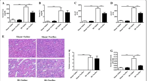
Renal IR led to an increase in serum urea and creatinine (P< 0.001, respectively), along with tubular necrosis in renal tissues (P< 0.001). Norfloxacin pretreatment re-duced the serum urea levels in renal IR rats (renal plus norfloxacin) (P< 0.001) (Fig. 2a). However, no differ-ences were observed with respect to serum creatinine (Fig. 2b) and tubular necrosis scores (Fig.2e, f ) between the renal IR rats (renal IR plus saline) and renal IR rats with norfloxacin pretreatment (renal IR plus norfloxacin). Taken together, these data suggest that norfloxacin pre-treatment has no substantial benefits to renal outcome.
Norfloxacin pretreatment protests against liver dysfunction following renal IR
The ALAT and ASAT values were markedly elevated in the renal IR rats (renal IR plus saline) compared with the sham rats (sham plus saline) (P< 0.001, respectively). Norfloxacin pretreatment reduced the severity of liver dysfunction in renal IR rats (renal IR plus norfloxacin), as shown by a reduction in the serum ALAT and ASAT values (P< 0.001, respectively) (Fig.2c, d). In conclusion, these data indicate that norfloxacin pretreatment pro-tects liver dysfunction.
Norfloxacin pretreatment attenuates renal IR-induced endotoxinemia
As expected, serum endotoxin levels were significantly higher in the renal IR rats (renal IR plus saline) compared with the sham rats (sham plus saline) (P< 0.001). By con-trast, pretreatment of the renal IR rats with norfloxacin (renal IR plus norfloxacin) significantly reduced their serum endotoxin levels (P< 0.001) (Fig. 2g). In summary, these data show that four-week oral norfloxacin pretreatment is effective on attenuating renal IR-induced endotoxinemia.
Norfloxacin pretreatment reduces renal TLR4 protein expression
To further examine whether pretreatment with norflox-acin modulates renal TLR4 expression, we performed IHC and Western blot. IHC revealed that renal IR led to an increase expression of renal TLR4 protein. Pre-treatment of the renal IR rats with norfloxacin (renal IR plus norfloxacin) reduced their renal expression of TLR4 (Fig. 3a). These findings were further confirmed by Western blot (Fig. 3b). Taken together, these results show that renal TLR4 expression is increased in renal IR rats and its expression can be modulated by norflox-acin pretreatment.
Norfloxacin pretreatment decreases renal proinflammatory mediators
Proinflammatory cytokines and chemokines contribute to the pathophysiology of renal IR. To further evaluate the effect of norfloxacin pretreatment on renal inflam-mation, both mRNA and protein levels of TNF-a, IL-6 and MCP-1 in kidney homogenates were detected. The mRNA levels of TNF-a, IL-6 and MCP-1 were markedly increased in the renal IR rats (renal IR plus saline) compared with the sham rats (sham plus saline) (all P< 0.001). Pre-treatment of the renal IR rats with norfloxacin (renal IR plus norfloxacin) significantly reduced mRNA levels of these pro-inflammatory mediators in kidney tissue (IL-6: P< 0.01; MCP-1:P< 0.01) (Fig.3c-e). Furthermore, the protein levels of these proinflammatory mediators assessed by ELISA were consistent with the mRNA results (Fig.3f-h). These findings indicate that norfloxacin pretreatment could abrogate the renal inflammatory signaling cascade.
Discussion
AKI induced-gut injury was first reported in 2011. Over the past years, however, the clinical importance of intes-tinal consequences during AKI went somewhat out of the focus of nephrologists. Here, we showed that gut-derived bacterial endotoxin, resulting from the dis-rupted gut barrier due to renal IR, evokes a TLR4
mediated inflammatory response in the kidney. Our study provides new insights into the mechanism of in-flammation in ischemic AKI.
In a renal IR model of rats, Mehri and colleagues re-ported that a minimum of 45 min ischemia is required to study the impacts of renal injury on distant organs [13]. On the basis of their observations, we selected a renal IR model of 60 min ischemia to address our hy-pothesis. As expected, severe renal injury resulted in striking morphological alterations in ileum. On the other hand, an enhanced permeability of the intestinal mucosa was witnessed by experimental findings in vivo and vitro. Our findings confirmed the previous study reporting that renal IR initiates a complex cascade of events that eventually result in intestine pathological changes and functional alterations [4].
techniques. It seems plausible to assume that serum endotoxin may derive from the intestine other than sur-gical contamination, since the intestine contains the lar-gest number of bacterial cells. In a mice model of renal IR, however, Diba Emal et al. failed to quantify detect-able levels of endotoxin in the blood [17]. Similarly, the serum endotoxin levels of mice assessed 7 days after sham or IR were no difference in another study [18]. In contrast to the above-mentioned studies, rats were used in our study. As the loads of endotoxin and bacteria in the intestinal lumen may be quite different between ani-mal species, the differences in the experimental aniani-mals (mice vs. rats) may result in different findings. In addition, factors such as the timing of endotoxin meas-urement and the sensitivity of endotoxin assay may also cause discrepancies among studies.
Despite the well-known detrimental role of endotoxin in the pathogenesis of sepsis AKI, it is not clear whether such a subclinical level of endotoxinemia in renal IR is sufficient enough to aggravate renal inflammation. Therefore, we next investigated the role of gut-derived endotoxin in kidney inflammation during renal IR. TLR4
is a pattern recognition receptor that serves as a endo-toxin sensor, and whose activation recruits inflammatory factors and causes renal damage [19]. We demonstrated that, in a clinical related rat model of severe renal IR, the kidneys displayed an increased protein expression of TLR4. This was accompanied by a marked increase in renal proinflammatory mediators namely, TNF-a, IL-6 and MCP-1. Norfloxacin pretreatment reduced the grade of endotoxinemia in IR rats and this was followed by a reduction in TLR4 expression, IL-6 and MCP-1 produc-tion in the kidney. These findings suggest there might be a casual relationship between gut-derived endotoxin and renal inflammation during renal IR.
intestine bacteria and its products in renal IR injury seems to be complicated and still needs to be clarified in future.
We observed that norfloxacin pretreatment prevented renal IR induced hepatic dysfunction. Similarly, TLR9 deficiency prevented liver damage secondary to severe renal IR but failed to reduce renal dysfunction and tubu-lar necrosis [21]. These data and our findings suggest some unknown factors, which may be independent of kidneys, take part in the hepatic injury after severe renal IR. The liver interconnects with small intestines via por-tal vein and is the first organ of defense against gut-derived endotoxin. In normal conditions, small amounts of bacterial products enter the liver via portal vein and most of them are eliminated by Kupffer cells (KCs) [22]. When the flux of endotoxin overwhelms the phagocytotic capacity of KC, the endotoxin spills over into the systemic circulation [23]. Therefore, the liver is more susceptible to be assaulted by gut-derived endotoxin than the kidney. However, the role of gut-derived endotoxin in renal IR-induced liver injury remains to be investigated.
Conclusions
Our results show for the first time that gut-derived endo-toxin, resulting from an increased intestinal permeability after severe renal IR, subsequently amplifies intrarenal in-flammatory response by activation renal TLR4 signaling. However, our results do not establish that antibiotic ad-ministration was effective in improving the overall renal outcome, in terms of histopathological changes and renal dysfunction. Even so, our intriguing findings may be the first step to understanding how to tailor therapies to miti-gate intrarenal inflammation in select groups of patients, for example, those at high risk for renal IR.
Additional file
Additional file 1:Uncropped Western blots: kidneys TLR4/GAPDH. (PDF 253 kb)
Abbreviations
AKI:Acute kidney injury; ALAT: Alanine aminotransferase; ASAT: Aspartate aminotransferase; ELISA: Enzyme-linked immunosorbent assay; FD-4: FITC-Dextran; HE: Hematoxylin and eosin.; IL-6: Interleukin-6; IR: Ischemia reperfusion; LPS: Lipopolysaccharide; MCP-1: Monocyte chemotactic protein-1; MLN: Mesenteric lymph nodes; TLR4: Toll-like receptor 4; TLRs: Toll-like receptors; TNF-α: Tumour necrosis factor alpha
Acknowledgments
We are thankful to Dr. Xianghua. Sun for the helpful discussions during the analyses of experiments.
Funding
This study was supported by grants from national natural science foundation of China (grant 8170017 and 81600523). CY conceived of the study, and participated in its design and coordination.
Availability of data and materials
The data sets used and/or analyzed during the current study are available from the corresponding author on reasonable request.
Authors’contributions
CY and JL participated in the design of the study. JL and KRM drafted the manuscript. LW and KZ performed animal treatments and samples’ collection. JL performed the experiments and statistical analyses. JL was responsible for creating the images. All authors reviewed and approved the final version of the manuscript.
Ethics approval and consent to participate
The animal experiments were performed according to the guidelines set by the Experiment Animal Center Committee in Tongji University application and approval (file numbers of the approved animal experiments,SYXK 2014– 0026 and SYXK 2012–0031).
Consent for publication Not applicable.
Competing interests
The authors declare that they have no competing interests.
Publisher’s Note
Springer Nature remains neutral with regard to jurisdictional claims in published maps and institutional affiliations.
Author details 1
Department of Nephrology, Tongji Hospital, Tongji University School of Medicine, 389 XinCun Road, Shanghai 200065, China.2Internal Medicine,
John H Stroger Jr Hospital of cook county, Chicago, IL 60612, USA.
Received: 17 May 2018 Accepted: 28 December 2018
References
1. Hoste EA, Kellum JA, Katz NM, Rosner MH, Haase M, Ronco C. Epidemiology of acute kidney injury. Contrib Nephrol. 2010;165:1–8.
2. Sharfuddin AA, Molitoris BA. Pathophysiology of ischemic acute kidney injury. Nat Rev Nephrol. 2011;7(4):189–200.
3. de Jong PR, González-Navajas JM, Jansen NJ. The digestive tract as the origin of systemic inflammation. Crit Care. 2016;20(1):279.
4. Park SW, Chen SW, Kim M, Brown KM, Kolls JK, D'Agati VD, Lee HT. Cytokines induce small intestine and liver injury after renal ischemia or nephrectomy. Lab Investig. 2011;91(1):63–4.
5. Takeuchi O, Akira S. Pattern recognition receptors and inflammation. Cell. 2010;140(6):805–20.
6. Medzhitov R. Recognition of microorganisms and activation of the immune response. Nature. 2007;449(7164):819–26.
7. Anders HJ, Banas B, Schlondorff D. Signaling danger: toll-like receptors and their potential roles in kidney disease. J Am Soc Nephrol. 2004;15:854–67. 8. Shah N, Dhar D, El Zahraa Mohammed F, Habtesion A, Davies NA, Jover-Cobos M, Macnaughtan J, Sharma V, Olde Damink SW, Mookerjee RP, et al. Prevention of acute kidney injury in a rodent model of cirrhosis following selective gut decontamination is associated with reduced renal TLR4 expression. J Hepatol. 2012;56(5):1047–53.
9. Allgoewer A, Mayer B. Sample size estimation for pilot animal experiments by using a Markov chain Monte Carlo approach. Altern Lab Anim. 2017; 45(2):83–90.
10. Yasuda T, Takeyama Y, Ueda T, Shinzeki M, Sawa H, Nakajima T, Kuroda Y. Breakdown of intestinal mucosa via accelerated apoptosis increases intestinal permeability in experimental severe acute pancreatitis. J Surg Res. 2006;135(1):18–26.
11. Ramadass B, Moseley PL, Patel YR, Lin HC. Sucrose co-administration reduces the toxic effect of lectin on gut permeability and intestinal bacterial colonization. Dig Dis Sci. 2010;55(10):2778–84.
13. Kadkhodaee M, Golab F, Zahmatkesh M, Ghaznavi R, Hedayati M, Arab HA, Ostad SN, Soleimani M. Effects of different periods of renal ischemia on liver as a remote organ. World J Gastroenterol. 2009;15(9):1113–8.
14. Andersen K, Kesper MS, Marschner JA, Konrad L, Ryu M, Kumar Vr S, Kulkarni OP, Mulay SR, Romoli S, Demleitner J, et al. Intestinal Dysbiosis, barrier dysfunction, and bacterial translocation account for CKD-related systemic inflammation. J Am Soc Nephrol. 2017;28(1):76–83.
15. Hauser AB, Azevedo IR, Gonçalves S, Stinghen A, Aita C, Pecoits-Filho R. Sevelamer carbonate reduces inflammation and endotoxemia in an animal model of uremia. Blood Purif. 2010;30(3):153–8.
16. Druml W. Systemic consequences of acute kidney injury. Curr Opin Crit Care. 2014;20(6):613–9.
17. Emal D, Rampanelli E, Stroo I, Butter LM, Teske GJ, Claessen N, Stokman G, Florquin S, Leemans JC, Dessing MC. Depletion of gut microbiota protects against renal ischemia-reperfusion injury. J Am Soc Nephrol. 2017;28(5): 1450–61.
18. Andres-Hernando A, Altmann C, Bhargava R, Okamura K, Bacalja J, Hunter B, Ahuja N, Soranno D, Faubel S. Prolonged acute kidney injury exacerbates lung inflammation at 7 days post-acute kidney injury. Physiol Rep. 2014.
https://doi.org/10.14814/phy2.12084.
19. Wu H, Chen G, Wyburn KR, Yin J, Bertolino P, Eris JM, Alexander SI, Sharland AF, Chadban SJ. TLR4 activation mediates kidney ischemia/reperfusion injury. J Clin Invest. 2007;117(10):2847–59.
20. Jang HR, Gandolfo MT, Ko GJ, Satpute S, Racusen L, Rabb H. Early exposure to germs modifies kidney damage and inflammation after experimental ischemia-reperfusion injury. Am J Physiol Renal Physiol. 2009;297(5):F1457–65. 21. Bakker PJ, Scantlebery AM, Butter LM, Claessen N, Teske GJ, van der Poll T,
Florquin S, Leemans JC. TLR9 Mediates Remote Liver Injury following Severe Renal Ischemia Reperfusion. PLoS One. 2015.https://doi.org/10.1371/ 0137511.
22. Holub M, Cheng CW, Mott S, Wintermeyer P, van Rooijen N, Gregory SH. Neutrophils sequestered in the liver suppress the proinflammatory response of Kupffer cells to systemic bacterial infection. J Immunol. 2009;183(5):3309–16. 23. Seki S, Habu Y, Kawamura T, Takeda K, Dobashi H, Ohkawa T, Hiraide H. The


![Tris[6 (3,5 dimethyl 1H pyrazol 1 yl)picolinato]gadolinium(III) methanol hemisolvate 2 5 hydrate](data:image/gif;base64,R0lGODlhAQABAIAAAP///wAAACH5BAEAAAAALAAAAAABAAEAAAICRAEAOw==)