RESEARCH
Prosthetic joint infection development
of an evidence-based diagnostic algorithm
Heinrich M. L. Mühlhofer
1*, Florian Pohlig
1, Karl‑Georg Kanz
2, Ulrich Lenze
1, Florian Lenze
1, Andreas Toepfer
1,
Sarah Kelch
1, Norbert Harrasser
1, Rüdiger von Eisenhart‑Rothe
1and Johannes Schauwecker
1Abstract
Background: Increasing rates of prosthetic joint infection (PJI) have presented challenges for general practitioners, orthopedic surgeons and the health care system in the recent years. The diagnosis of PJI is complex; multiple diag‑ nostic tools are used in the attempt to correctly diagnose PJI. Evidence‑based algorithms can help to identify PJI using standardized diagnostic steps.
Methods: We reviewed relevant publications between 1990 and 2015 using a systematic literature search in MED‑ LINE and PUBMED. The selected search results were then classified into levels of evidence. The keywords were pros‑ thetic joint infection, biofilm, diagnosis, sonication, antibiotic treatment, implant‑associated infection, Staph. aureus, rifampicin, implant retention, pcr, maldi‑tof, serology, synovial fluid, c‑reactive protein level, total hip arthroplasty (THA), total knee arthroplasty (TKA) and combinations of these terms.
Results: From an initial 768 publications, 156 publications were stringently reviewed. Publications with class I–III recommendations (EAST) were considered. We developed an algorithm for the diagnostic approach to display the complex diagnosis of PJI in a clear and logically structured process according to ISO 5807.
Conclusions: The evidence‑based standardized algorithm combines modern clinical requirements and evidence‑ based treatment principles. The algorithm provides a detailed transparent standard operating procedure (SOP) for diagnosing PJI. Thus, consistently high, examiner‑independent process quality is assured to meet the demands of modern quality management in PJI diagnosis.
Keywords: Prosthetic joint infection, Algorithm, Total joint replacement, Revision surgery, THA, TKA
© The Author(s) 2017. This article is distributed under the terms of the Creative Commons Attribution 4.0 International License (http://creativecommons.org/licenses/by/4.0/), which permits unrestricted use, distribution, and reproduction in any medium, provided you give appropriate credit to the original author(s) and the source, provide a link to the Creative Commons license, and indicate if changes were made. The Creative Commons Public Domain Dedication waiver (http://creativecommons.org/ publicdomain/zero/1.0/) applies to the data made available in this article, unless otherwise stated.
Background
The total number of hip and knee arthroplasties per-formed in the US is constantly increasing, with an expected increase in total knee arthroplasties (TKAs) of approximately 600% by 2030. Similarly, the number total hip arthroplasties (THAs) performed is estimated to tri-ple during this period, leading to a significant increase in revision surgeries [1].
One major reason for revision arthroplasty is pros-thetic joint infection (PJI). The incidence of PJI after pri-mary surgery is 0.2–1.1%; in cases of revision surgery, it
can reach 5% [2]. In contrast with acute PJI, low-grade infections are characterized by unspecific symptoms, such as pain and early implant loosening. Typical low-grade infections are often a result of infection with less virulent bacterial strains of the dermal flora, including coagulase-negative Staphylococci and Propionibacterium acnes, often lacking severe inflammatory symptoms [3].
Although it is of fundamental importance for further treatment, diagnosing a low-grade PJI prior to revision surgery can be challenging.
In addition to clinical findings, conventional radio-graphs, erythrocyte sedimentation rate (ESR), C-reac-tive protein (CRP), the percutaneous aspiration of synovial fluid for evaluating cell counts and differentials, and microbiology workups are routinely employed diag-nostic tools (3).
Open Access
*Correspondence: [email protected] 1 Department of Orthopaedic Surgery, Klinikum rechts der Isar, Technische Universität München, Ismaninger Str. 22, 81675 Munich, Germany
Moreover, arthroscopically or fluoroscopically con-trolled biopsy of periprosthetic tissue can be performed. Some authors suggest the use of synovial biomarkers, leukocyte esterase tests or radionuclide imaging to sup-port the diagnosis of a low-grade PJI [4, 5].
All tests have a role in the workup of PJI; however, the diagnostic values reported in the recent literature vary greatly [4–6].
To safely rule out or verify the presence of PJI prior to arthroplasty revision surgery, a customized combination of different diagnostic tests must be employed for every single case. Although the composition of an appropri-ate set of diagnostic tests may be self-evident for senior orthopedic surgeons who specialize in treating PJI, it can be challenging for attending orthopedic surgeons and general practitioners. The correct preoperative diagno-sis is, however, of major significance because treatment strategies differ greatly between septic and aseptic revi-sion surgery and have far-reaching consequences for the patient (20). Whereas false-positive findings lead to unnecessary two-stage revisions, one-stage revision sur-gery without the essential implementation of antibiotic therapy can result from false-negative results, inevitably causing new prosthetic failure and PJI persistence [6].
We, thus, developed an evidence-based diagnostic algorithm with examiner-independent diagnostic reli-ability for identifying PJI, and we prospectively observed its use in daily clinical routine according to the require-ments of modern quality management in our institution.
Methods
A systematic literature search was conducted in the data-bases of PubMed and Medline using the following search terms: prosthetic joint infection, implant-associated infec-tion, biofilm, diagnosis, sonicainfec-tion, antibiotic treatment, microcalorimetry, Staph. aureus, coagulase-negative staph-ylococci, Propionibacterium rifampicin, implant retention, pcr, maldi-tof, serology, synovial fluid, C-reactive protein level, THA, TKA leucocyte esterase test, alpha-defensin test. All relevant publications between January 1990 and January 2015 were screened according to methodologi-cal aspects using QUADAS, STARD and PRISMA crite-ria [7–11] and classified according to the Grade system (The Grading of Recommendation Assessment, Develop-ment and Evaluation). Referring to the Grade system, the included studies were grouped according to the EAST classification, shown in Table 1 and Fig. 1, which allows the evaluation of medical publications, reviews and recom-mendations in terms of their level of evidence (LoE) and class of recommendation (CoR) [12–21].
Table 1 EAST level of evidence (LoE) and class of recom-mendation (CoR)
LoE
I Prospective randomized controlled trials (ORCTs) II Clinical studies in which the data was collected prospec‑
tively, and retrospective analyses which were based on clearly reliable data
Types of studies so classified: observational studies, cohort studies, prevalence studies and case control studies III Studies based on retrospectively collected data. Evidence
used in this class indicate clinical series, database or reg‑ istry review, large series of case reviews and expert opinion CoR
I The recommendation is convincingly justifiable based on the available scientific information alone. This recommen‑ dation is usually based on Class I data; however, strong Class II evidence may form the basis for a level I recom‑ mendation, especially if the issue does not lead itself to testing in a randomized trial.
II The recommendation is reasonably justifiable by available scientific evidence and strongly supported by expert opinion
This recommendation is usually supported by Class II data or a preponderance of Class II evidence
III The recommendation is supported by available data but adequate scientific evidence is lacking
This recommendation is generally supported by Class III data. This type of recommendation is useful for educa‑ tional purposes and in guiding future clinical research
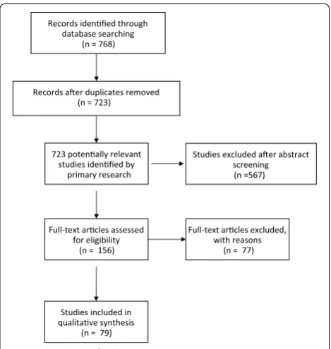
Records idenfied through database searching
(n = 768)
Records aer duplicates removed (n = 723)
Full-text arcles assessed for eligibility
(n = 156)
Full-text arcles excluded, with reasons
(n = 77)
Studies included in qualitave synthesis
(n = 79)
Studies excluded aer abstract screening
(n =567) 723 potenally relevant
studies idenfied by primary research
The literature review exclusively included studies of adults and publications in the English or German language. All references from the publications used in this study were examined for additionally relevant publications.
We identified 723 studies that met our search criteria. To perform statistically valid analyses, we only included studies with a minimum of 26 patients (Fig. 1).
Furthermore, only studies that used the definition cri-teria [22] of the Musculoskeletal Infection Society (MSIS) [23], Infectious Diseases Society of America (IDSA) [24] and International Consensus Meeting [25] (Table 2) were included.
A total of 79 studies were included, and the data were extracted from the studies. Subsequently, sensitivities, spe-cificities, positive and negative likelihood ratios and posi-tive and negaposi-tive predicposi-tive values were calculated from the extracted data if they were not stated in the publication.
The evidence levels of the selected studies were taken into account, fulfilling the formal requirement of the International Organization for Standardization (ISO) for development of algorithms. The creation of the algo-rithm was performed according to ISO norm 5807 Modi-fication ITU-I, initially designed for telecommunication defaults, to ensure explicit decision-making criteria for a logical and standardized procedure. The ISO 5807 norm defines the use of a different symbol for the single opera-tion to create an operaopera-tion plan that has only one input and output [26]. An algorithm that meets the ISO 5807 criteria is composed of process and decision symbols that differ from the symbols for the initial criteria and end-points. Checklists are introduced to reduce the number of decision symbols. For more practical reasons, the algo-rithm should not exceed a single page in length.
Results
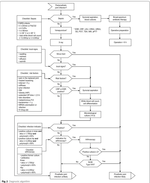
The algorithm was a composition of evidence-based procedures developed in our clinic that fulfilled the ISO 5807. Studies were integrated dependent of their LoE in a logical, structured, priority-orientated way in the algo-rithm. Checklists are located on the left side, the vertical flow represents the main diagnostic criteria and treat-ment, and the horizontal flow represents the supportive criteria [27] (Fig. 2). For practical reasons, the decisions symbol has been modified to a binary-decision hexagon.
All diagnostic aspects of the algorithm and the under-lying literature are specified below.
Risk factors checklist
In total, 31 included studies (patients: n = 312.946; LoE
I: n = 2, LoE II: n = 13, LoE III: n = 16) discussed the
risk factors for a PJI (Table 3). According to a study by Virolainen, pain in the index joint exhibits a specificity of 100% in patients with PJI. Various consensus recom-mendations and expert opinions consider a limited range of motion in the total joint an indicator for PJI [25]. The risk factors according to studies with a class of recom-mendation of I (CoR I) are an extended operation time (n = 142.120) [28–32], obesity (n = 116,682) [33–41],
malnutrition (n = 678) [42], diabetes (n = 72,778) [35, 39,
40, 43], immunosuppression (n = 86,675) [32, 34, 35, 37,
41, 44, 45], Prior infection of the joint [44], prior infec-tion [44], early implant failure [46], early implant loos-ening [46, 47] and superficial surgical site infections [32, 48, 49]. Other risk factors have been associated with PJI, although the related studies do not provide strong evi-dence (CoR II–III); these factors include asymptomatic bacteriuria [50] and tooth interventions, oral surgery and colonoscopies, which provide a crucial risk factor for PJI
Table 2 Definition of prosthetic joint infection
MSIS IDSA International consensus Main
criteria Supportive criteria Main criteria Supportive criteria Main criteria Supportive criteria
Sinus tract o o o
Identical microorganisms isolated form 2
or more cultures o o o
Purulence surrounding the prosthesis o o
Inflammation in histological examination
of prosthetic tissue o o o
Single positive culture o o
Single positive culture with virulent
microorganism o
Elevated synovial fluid leukocyte count o o
Elevated synovial fluid neutrophil percentage o o
Periprosthetic joint infection?
Checklist: Sepsis
2 SIRS-criteria: • rr ≥ 20/min or PaCO2 ≤ 4.3 kPa • h ≥ 90/min • t ≤ 36° C or ≥ 38° C • total white blood cell count ≥ 12,000/µl or ≤ 4,000/µl
Sepsis
No
Venepuncture*
Yes Synovial aspiration, blood cultures
* ESR, CRP, LEU, CREA, UREA, SO, POT, TSH, INR, aPTT
Broad-spectrum antibiotic therapy
Operative preparation
X-ray
Sinus tract
No
local signs? Yes
Yes
Operation < 6 h
Checklist : risk factors
• pain in the replaced joint • implant loosening • interval < 5 y • stiffness • prior infection • SSI • obesity (HIP) • extended OP-time > 2.5 h • skin disorders • metachronous PJI • bacteremia < 1 y • MRSA-colonisation or infection
• IV drug use
No
Risk factors?
No
CRP or ESR positive? No
Yes
Yes
Synovial aspiration
White blood cell count, cell differentiation
Microbiological culture (14 d)
Checklist: infection indicator Positive? Yes
• positive culture in knee and
wbcc ≥ 1,700/µl and
polymorph ≥ 65% • positive culture hip and
wbcc ≥ 4,200/µl and
polymorph ≥ 80%
Checklist: arthroscopy • positive former culture • antibiotics Knee: • wbcc ≥ 1,700/µl • polymorph ≥ 65% Hip:
• wbcc ≥ 4,200/µl • polymorph ≥ 80%
No
Indication for arthroscopy?
No
Prosthetic joint infection unlikely
Yes
No
Arthroscopy
Positive culture ≥ 2?
No
SLIM Type II/III?
Yes
Yes
Prosthetic joint infection likely Checklist: local signs
• swelling • redness • effusion • warmth
because of relevant bacteraemia [51, 52]. Likewise, skin disorders in the surgical area during implantation (pso-riasis, chronic venous stasis, skin ulcers, lymphedema) increase the risk of implant-associated infection [41, 53].
Sepsis checklist
As a first step, sepsis and septic shock are ruled out. Con-cerning sepsis, we included 9 studies discussing diagnos-tic parameters and treatment options for highly acute
PJIs. Two LoE I studies, three LoE II studies and six LoE III studies were included. To identify sepsis, we used the diagnostic criteria published by Llewelyn and Dellinger (CoR III) [54–56]. Prior to the initiation of a calculated antibiotic therapy, a synovial aspiration (CoR III) for sub-sequent examination of cell counts and differentials and a microbiologic workup to identify the causative pathogen should be performed [55, 57, 58]. Additionally, blood cul-tures should be obtained (CoR I) [59, 60]. Subsequently, early surgical focus management can significantly reduce the mortality rate (CoR II) [61, 62].
Physical exam
The highest LoE (CoR III) for physical examination is found in a study by Teller et al. [63]. They report a sen-sitivity of 18.95% (CI 0.05–0.4) and a specificity of 100% (CI 0.98–1.0) for the identification of PJI based on local signs of inflammation, such as warmth, effusion, redness and swelling of the corresponding joint [63]. For fever, a sensitivity of 9% (CI 95 0.03–0.21) and a specificity of 99% (CI 95 0.98–1.00) were reported. These data contrib-ute to a negative predictive value of 0.89 and a negative likelihood ratio of 0.82 (CI 0.66–0.98) for local signs of inflammation (Table 4). Studies with higher levels of evi-dence for clinical signs of inflammation have not yet been published.
Inflammatory markers
Decision paths for the CRP and ESR were derived from six studies with a LoE of I, resulting in a CoR I. In this context, Bottner et al. showed significantly increased pre-operative CRP and ESR levels in patients with PJI com-pared with cases with aseptic knee and hip revisions. Considering a cut-off value of 1.5 mg/dl for the CRP, the authors reported a sensitivity of 0.95 (95% CI 0.86–1.0) and a specificity of 0.91 (0.84–0.99). The ESR showed a lower sensitivity of 0.81 (0.64–0.98) and a lower speci-ficity of 0.89 (0.82–0.97) compared with the CRP given a cut-off value of 32 mm/h [64]. In their series of total knee arthroplasties, Valle Della et al. reported sensitivi-ties of 0.95 (0.89–1.0) and 0.9 (0.81–0.99) and specifici-ties of 0.75 (0.64–0.87) and 0.66 (0.53–0.79) for the CRP and ESR, respectively [65]. Greidanus et al. observed a lower sensitivity of 0.82 (0.71–0.95) and a higher specificity of 0.88 (0.76–0.9) for the ESR, with a cut-off value of 30 mm/h. The CRP showed a sensitivity of 0.93 (0.86–1.0) and a specificity of 0.83 (0.76–0.9) given a cut-off value of 1.0 mg/dl [66]. In the 1980s, Kamme et al. determined the ESR and reported a sensitivity of 0.89 (0.8–0.95) and a specificity of 0.73 (0.54–0.9) [67]. Schin-sky et al. showed high sensitivity [ESR: 0.96 (0.91–1.0); CRP: 0.95 (0.89–1.0)] and a low specificity [ESR: 0.39 (0.31–0.47); CRP: 0.71 (0.94–1.0)] for the ESR and CRP Table 3 Riskfactors for prosthetic joint infections
Author Year LoE (n = x) Risk factor CoR
Huotari et al. 2007 LoE II n= 8201 Smarbrekke et al. 2004 LoE II n= 31,750
Kurtz et al. 2010 LoE II n= 69,663 Extended time CoR I Uckay et al. 2009 LoE I n= 6001
Berbari et al. 1998 LoE II n= 26,505 Dowsey et al. 2009 LoE III n= 1214 Peersman et al. 2001 LoE III n= 6120 Lübbeke et al. 2007 LoE II n= 2495 Dowsey et al. 2008 LoE III n= 1207
Pulido et al. 2008 LoE III n= 9245 Obesity CoR I Namba et al. 2012 LoE II n= 30,491
Namba et al. 2013 LoE II n= 56,216 Malinzak et al. 2009 LoE III n= 8494 Peel et al. 2011 LoE III n= 1200
Berbari et al. 2012 LoE III n= 678 Malnutrition CoR III Namba et al. 2013 LoE II n= 56,216
Malinzak et al. 2009 LoE III n= 8494 Diabetes CoR I Peersman et al. 2001 LoE III n= 6120
Mraovic et al. 2011 LoE III n= 1948 Dowsey et al. 2008 LoE III n= 1207 Peersman et al. 2001 LoE III n= 6120
Jämsen et al. 2009 LoE II n= 43,149 Immunsuppres‑
sion CoR I
Pulido et al. 2008 LoE II n= 9245 Peel et al. 2011 LoE III n= 63 Berbari et al. 1998 LoE II n= 26,505 Bongratz et al. 2008 LoE III n= 462
Jämsen et al. 2009 LoE II n= 43,149 Prior infection CoR II Aslam et al. 2010 LoE III n= 126
Coelho‑Prabhu
et al. 2013 LoE III
n= 678 Bacteremia CoR III
Murdoch et al. 2001 LoE III n= 80
Murray et al. 1991 Level III n= 159 Metachronous infections CoR III Luessenhop
et al. 1996 Level III n= 145
Portillo et al. 2013 LoE I n= 116 Implant loosen‑
ing CoR I
[68]. In contrast, Savorino et al. reported a low sensitiv-ity [ESR: 0.6 (0.3–0.9); CRP 0.38 (0.14–0.61)] but a higher specificity [ESR: 0.94 (0.82–1.0); CRP: 0.7(0.42–0.98)] [69]. Recently, Fink et al. calculated a sensitivity of 0.73 (0.59–0.86) and a specificity of 0.81 (0.73–0.88) for the CRP in their series of total knee arthroplasties [70]. Posi-tive and negaPosi-tive predicPosi-tive values and posiPosi-tive and nega-tive likelihoods were calculated and are shown in Table 5. Inflammatory markers such as IL-6, PCT and TNF-alpha have also been the focus of clinical trials; however, there is no Level I or Level II study indicating their superior diagnostic value [71–73].
Sinus tract
According to the criteria of the MSIS, the ISDA and the International Consensus Meeting, a sinus tract commu-nicating with the prosthesis is a criterion for the presence of a PJI [23, 25, 74]. Two studies dealing with micro-biological cultures of patients with a sinus tracts were included; however, a pathogen was not identified in all cases (Table 4). Bogut et al. calculated a sensitivity of 0.82 (0.66–0.98) and a specificity of 1.0 (1.0–1.0; LoE II) [75]. Trampus et al. identified a positive microbiological cul-ture in all cases after sonication and described a sensitiv-ity of 1.0 and a specificsensitiv-ity of 1.0 (LoE I) [76].
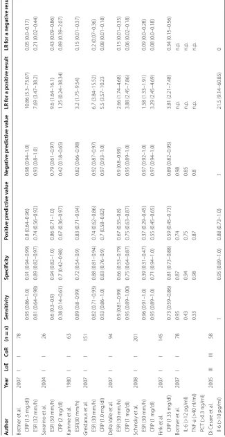
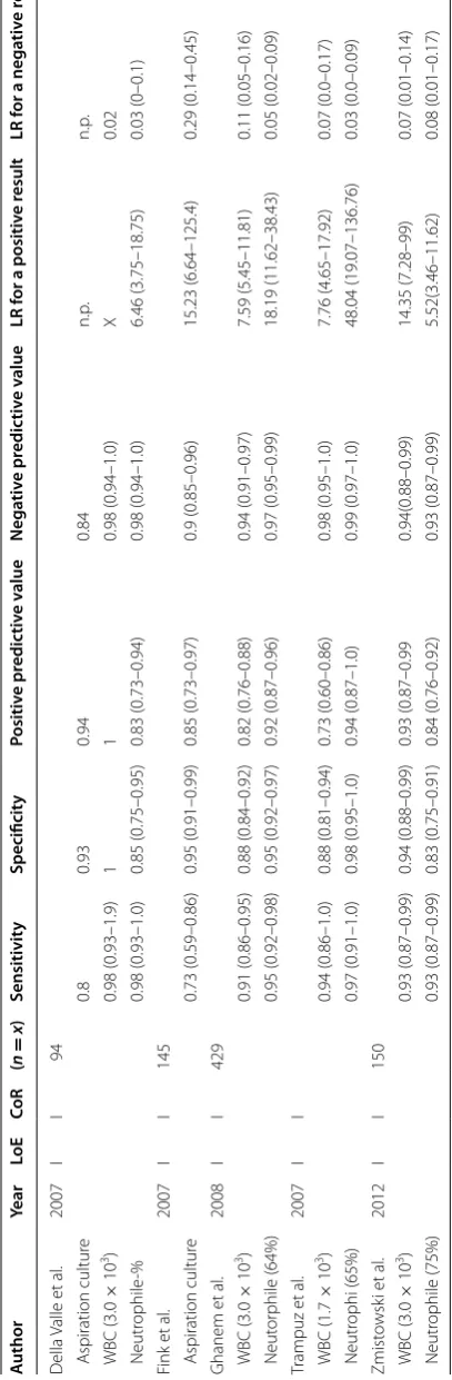
Joint aspiration (knee)
For tentative PJI of the knee, five studies (5× LoE I) addressing percutaneous aspiration of synovial fluid were included. Based on an exclusive bacteriologic culture analysis, Fink et al. reported a sensitivity of 0.73 (0.59– 0.86) and a specificity of 0.95 (0.91–0.99) [70]. Della Valle et al. reported a similar result for microbiological cul-tures (sensitivity 0.8 (95% CI n.p.); specificity 0.93 (95% CI n.p.) [65]. Four authors examined the synovial fluid, considering white blood cell counts (WBC) and cell dif-ferentiation (Neutrophil-%). Trampuz et al. observed a sensitivity of 0.94 (0.86–1.0) for WBC and of 0.97 (0.91– 1.0) for Neutrophil-%, with specificities of 0.88 (0.81– 0.94) and 0.98 (0.95–1.0), respectively [77]. Della Valle et al. showed comparable results: WBC sensitivity 0.91 (0.86–0.95)/specificity 1; Neutrophil-% sensitivity 0.98 (0.93–1.0) and specificity 0.85 (0.75–0.95). Ghanem et al. showed a sensitivity for WBC of 0.91 (0.86–0.95) and a
specificity of 0.88 (0.84–0.92); for Neutrophil-%, they found a sensitivity of 0.95 (0.92–0.98) and a specificity of 0.95 (0.92–0.97). Zmistowski et al. confirmed the results in their series, reporting a sensitivities of 0.93 (0.87–0.99) for WBC and Neutrophil-% and specificities of 0.94 (0.88–0.99) and 0.83 (0.75–0.91), respectively. Positive and negative predictive values and positive and negative likelihoods were calculated for all studies and are shown in Table 6.
Joint aspiration (hip)
The diagnostic value of percutaneous synovial aspiration in total hip arthroplasties was addressed in six studies cor-responding to a LoE of I. Four studies addressed microbi-ological cultures, and two studies addressed the WBC and Neutrophil-% of the synovial aspiration (Table 7).
Mulcahy et al. reported a sensitivity of 0.69 (0.46–0.91) and a specificity of 0.91 (0.83–0.99) for microbiologi-cal culture [78]. Similar results with a sensitivity of 0.44 (0.12–0.77) and a specificity of 0.91 (0.81–1.0) were pub-lished by Malhotra et al. [79], and Barrack et al. reported a sensitivity of 0.6 (0.3–0.9) and a specificity of 0.88 (0.84–0.92) [80]. Williams et al. reported a higher sensi-tivity of 0.8 (0.71–0.9) and a specificity of 0.94 (0.9–0.97) [81], while Schinsky et al. published a sensitivity of 0.84 (0.74–0.93) and specificity of 0.93 (0.89–0.97) for cell count analysis and a sensitivity of 0.82 (0.72–0.92) and specificity 0.83 (0.77–0.89) [68]. The results confirm the series of Dinneen et al., who reported a sensitivity of 0.89 (0.783–0.997) and specificity of 0.91 (0.827–0.99); for WBC, the values were 0.89 (0.79–0.99) and 0.86 (0.76– 0.97), respectively [82].
Other synovial fluid markers, such as synovial CRP and synovial IL-6, and antimicrobial peptides, such as alpha-defensin, are undergoing clinical trials [6, 83–85]. How-ever, there is no Level I or Level II study indicating their superior diagnostic value.
Synovial biopsy histological workup
Overall, seven LoE I studies including 822 patients (5×
frozen sections/2× fixed sections) addressing synovial biopsy and histological workup and one LoE III study establishing a histopathological classification of the periprosthetic membrane were included (Table 8). Banit Table 4 Physical Exam
Author Year LoE CoR (n = x) Sensitivity Specificity Positive predictive value
Negative predictive value
LR for a positive result
LR for a negative result
Teller et al. 2000 III III 166
Local signs 0.18 (0.02–0.34) 1 1 0.5 (0.34–0.66) n.p. 0.82 (0.66–0.98)
Table
6
V
alue of join
t aspir
ation in the diagnosis of inf
ec
ted t
otal k
nee ar
thr
oplast
y
A
uthor
Year
Lo
E
Co
R
(
n
=
x
)
Sensitivit
y
Specificit
y
Positiv
e pr
edic
tiv
e v
alue
Nega
tiv
e pr
edic
tiv
e v
alue
LR f
or a positiv
e r
esult
LR f
or a nega
tiv
e r
esult
D
ella
Valle et
al
.
2007
I
I
94
A
spiration cultur
e
0.8
0.93
0.94
0.84
n.p
.
n.p
.
WBC
(3.0
×
10
3)
0.98 (0.93–1.9)
1
1
0.98 (0.94–1.0)
X
0.02
Neutr
ophile
‑%
0.98 (0.93–1.0)
0.85 (0.75–0.95)
0.83 (0.73–0.94)
0.98 (0.94–1.0)
6.46 (3.75–18.75)
0.03 (0–0.1)
Fink et
al
.
2007
I
I
145
A
spiration cultur
e
0.73 (0.59–0.86)
0.95 (0.91–0.99)
0.85 (0.73–0.97)
0.9 (0.85–0.96)
15.23 (6.64–125.4)
0.29 (0.14–0.45)
Ghanem et
al
.
2008
I
I
429
WBC
(3.0
×
10
3)
0.91 (0.86–0.95)
0.88 (0.84–0.92)
0.82 (0.76–0.88)
0.94 (0.91–0.97)
7.59 (5.45–11.81)
0.11 (0.05–0.16)
Neut
or
phile (64%)
0.95 (0.92–0.98)
0.95 (0.92–0.97)
0.92 (0.87–0.96)
0.97 (0.95–0.99)
18.19 (11.62–38.43)
0.05 (0.02–0.09)
Trampuz et
al
.
2007
I
I
WBC
(1.7
×
10
3)
0.94 (0.86–1.0)
0.88 (0.81–0.94)
0.73 (0.60–0.86)
0.98 (0.95–1.0)
7.76 (4.65–17.92)
0.07 (0.0–0.17)
Neutr
ophi (65%)
0.97 (0.91–1.0)
0.98 (0.95–1.0)
0.94 (0.87–1.0)
0.99 (0.97–1.0)
48.04 (19.07–136.76)
0.03 (0.0–0.09)
Zmist
owsk
i et
al
.
2012
I
I
150
WBC
(3.0
×
10
3)
0.93 (0.87–0.99)
0.94 (0.88–0.99)
0.93 (0.87–0.99
0.94(0.88–0.99)
14.35 (7.28–99)
0.07 (0.01–0.14)
Neutr
ophile (75%)
0.93 (0.87–0.99)
0.83 (0.75–0.91)
0.84 (0.76–0.92)
0.93 (0.87–0.99)
5.52(3.46–11.62)
Table
7
V
alue of join
t aspir
ation in the diagnosis of inf
ec
ted t
otal hip ar
et al. showed a sensitivity of 0.45 (0.16–0.75) and speci-ficity of 0.92 (0.85–1.0) in their cohort. Borrego et al. reported a sensitivity of 0.5 (0.15–0.85), and a specificity of 1.00 in their series of 83 patients with THA and a sen-sitivity of 0.67 (0.48–0.86) and a specificity of 0.8 (0.7– 0.93) in their series of 63 patients with TKA [86].
Nunez et al. calculated a sensitivity of 0.86 (0.76–0.96) and a specificity of 0.87 (0.8–0.94) [87].
Banit et al. observed a sensitivity of 1.0 and a specificity of 0.96 (0.9–1.0) for TKA and a sensitivity of 0.45 (0.16– 0.75) and a specificity of 0.92 (0.85–1.0) for THA. Over-all, the authors reported a sensitivity of 0.67 (0.48–0.86) and a specificity of 0.95 (0.91–1.0) [88]. In their series of 105 patients with painful TKA, Valle Della et al. observed a sensitivity of 0.88 (0.78–0.98) and a specificity of 0.96 (0.91–1.0). For THA, Schinsky et al. reported a sensitiv-ity of 0.73 (0.61–0.84) and a specificsensitiv-ity of 0.94 (0.9–0.98) [65, 68].
Regarding fixed sections, Fink et al. showed a sensi-tivity of 1.0 and a specificity of 0.98 (0.95–1.0) for TKA (145) and a sensitivity of 0.62 (0.48–0.76) and a specificity of 1.0 for THA (100) [70, 89].
Synovial biopsy microbiological workup
In their series of THAs, Fink et al. showed a sensitivity of 0.73 (0.6–0.86) and a specificity of 0.98 for micro-biological culture of biopsies (0.95–1.0). The combina-tion of histological biopsy and microbiological culture increased the sensitivity to 0.82 (0.71–0.93) [90]. In their series of TKAs, they reported a sensitivity of 0.78 (0.65–0.9) and a specificity of 0.98 (0.95–1.0); when combined with histological analysis, the sensitivity increased to 1, with equal specificity. Regarding the recommended number of microbiological cultures, the mathematical model of Atkins et al. was used: at least 5 or 6 biopsy cultures should be taken [91]. Marin et al. showed a sensitivity of 0.87 (0.66–0.94) and a specificity of 0.67 (0.56–0.76) for one positive micro-biological culture using that model; when three posi-tive cultures were used, the sensitivity decreased to 0.46 (0.32–0.61), while the specificity increased to 0.98 (0.93–0.99) [92].
Discussion
Although the diagnosis of PJIs prior to revision surgery is of paramount importance for further treatment, it can be challenging, and a well-structured diagnostic approach is necessary. A PJI diagnosis results in substantial changes in the therapeutic procedure [3]. Thus, an evidence-based and priority-orientated algorithm (Fig. 2) can provide an incremental and easy-to-use guideline for non-specialists and less-experienced orthopedic surgeons.
AAOS guidelines strongly recommend determining the ESR and CRP [93]. According to the included studies, sensitivities vary from 81 to 93% for the ESR and from 73 to 95% for the CRP [93]. In a recent meta-analysis by Ber-bari and colleagues that included 30 studies with a total of 3909 patients, pooled sensitivities of 75% for the ESR and 88% for the CRP were reported [94]. The specifici-ties were 70 and 74%, respectively. Despite the relatively high sensitivity of the CRP, its specificity remains unsat-isfactory, confirming the observations of McArthur et al. who identified a considerable subset of patients with PJI and negative serology within their series of 414 infected THAs. In contrast, the AAOS guidelines recommend percutaneous aspiration only in case of altered ESR and CRP levels and thus exclude seronegative patients from this procedure [93]. In these cases, one-stage revision surgery without adequate antibiotic treatment may be performed, inevitably resulting in new prosthetic failure and PJI persistence. In our algorithm, the decision to per-form joint aspiration is based on ESR and CRP levels and on the radiological findings and medical history (risk fac-tors) of the patient.
Large multicenter LoE I studies were able to define some risk factors. In particular, potential intraoperative contamination and the immune system of the patients were determined to have an important role. Namba et al. showed in a large multicenter study that an extended operation time leads to an increase of PJIs. An additional 15 min of operation time was determined to increase the risk by up to 9% [39]. This relationship is explained by increased time for potential intraoperative microbial con-tamination. However, the increased risk of PJI in immu-nocompromised patients, such as those with rheumatoid arthritis and/or diabetes, has also been proven. The most recent studies suggest that even asymptomatic bacteriu-ria is an independent risk factor for PJI; the authors indi-cated that an immunocompromised status puts patients at risk for colonization with Gram-negative microorgan-isms [50]. Early implant loosening (<5 years) without evi-dence of mechanical failure or progressive radiolucency adjacent to the implant must be considered a decisive risk factor for a low-grade PJI. As proposed by Lachie-wicz et al., premature implant loosening and the presence of the previously described risk factors require further diagnostic procedures [47]. In this context, Portillo et al. were able to demonstrate a significantly longer period between primary implantation and diagnosed aseptic loosening (7.8 years) compared with septic implant loos-ening (2 years; CoR I) [46].
complications and economic issues. Although iatrogenic complications in the context of synovial aspiration are considered rare, Murray et al. reported a complication rate of 5.1% (0.2–10%), including hematoma, infections and lesions on nerve structures after synovial aspiration of the hip [95]. Barrack et al. showed a 1% (0.1–2.2%) rate of infections after synovial aspiration of the hip [96]. This benefit–risk assessment is of major importance to mini-mize the risk of infection for the patient and thus avoid false-positive results leading to overtreatment [97].
However, the diagnostic value of synovial aspiration and subsequent microbiological workup is controversial according to recent literature. Sensitivities vary between 12 and 89%, with specificities between 50 and 100% for synovial aspiration of hip joints [68, 78, 79, 81, 82, 96, 98, 99]. Similar results are available for TKA [65, 70, 77, 100, 101]. However, extended synovial analysis combin-ing microbiological culture with WBC and neutrophil-% is the gold standard for synovial aspiration investigation. The sensitivity, specificity, positive/negative predictive value and positive/negative likelihood ratio for the WBC and neutrophil-% are given in Tables 6 and 7. Several stud-ies have examined the optimal cut-off values for WBC and neutrophil-%. Trampuz et al. suggest 1.7 × 10³/µl (WBC) and 65% (neutrophil-%), and Zmistowski et al. and Della Valle report quite similar results, using higher cut-off val-ues of 3.0 × 10³ for WBC and 75% neutrophils [65, 101]. However, cut-off values calculated using receiver-operat-ing characteristics are linked to the microbiological strains that cause the PJI. In our algorithm, we used the lower cut-off values that Trampuz et al. and Schinsky et al. iden-tified to ensure that we detected the low-grade infections caused by slow-growing and low virulence strains, such as coagulase-negative staphylococci or Proprionibacterium acnes, which generate a low immune reaction. Considering the defining criteria for PJI (Table 2), 2 or more separate synovial fluid samples should be obtained from the index joint. However, this main criterion is usually not met with routine synovial aspiration. In the daily clinical routine, the additional use of blood culture bottles, as proposed by Minassian et al. [102], to obtain two separate microbiologi-cal cultures should thus be encouraged. Although the data on diagnostic value are discordant, the causative patho-gen and its antibiotic sensitivity pattern can be identified via synovial aspiration and microbiological examination. This information, in turn, is of great importance for preop-eratively planning the surgical strategy and the antibiotic regimen.
Unfortunately, a causative pathogen can only be iden-tified in approximately 44% [79]–80% [81] of cases, reflecting the heterogeneous diagnostic value of syno-vial aspiration. Among the factors influencing micro-biological results, the length of the incubation period is
crucial because the bacteria that cause PJIs occur only in a very low number in the biofilm and often are in a ses-sile form that is very slow growing [103, 104]. Accord-ingly, in many of the aforementioned studies, the length of microbiological incubation was only 48 h or was not specifically disclosed. Furthermore, the omission of anti-biotic treatment termination at least 2 weeks prior to the joint aspiration can lead to false-negative microbiological results and thus to maltreatment [105].
Other synovial fluid markers, such as alpha-defensin, show promising results, with a sensitivity of 100% [4, 85], but they lack the evidence and independent studies to support their use. Similarly, the leukocyte esterase test requires further evidence to support its role in diagnos-ing PJI [5, 106]. According to the Plan-Do-Check-Act principle, constant improvement of the algorithm by reintegrating actual evidence-based literature at half-year intervals is intended. If new diagnostic procedures fulfill the LoR I criteria, they will be included in the algorithm.
As a further, more invasive diagnostic step, arthro-scopic synovial biopsy has been implemented in our algorithm. As itemized in the “Arthroscopy” checkbox (Fig. 2), increased WBC or neutrophil percentage but negative microbiological assessment of the aspirate, con-tinued antibiotic treatment and history of PJI are indica-tions for synovial biopsy according to our algorithm.
In this context, recent studies by Williams et al. showed equal results for aspiration and tissue biopsy with sole microbiological examination [81]. These results underline the importance of concurrent histological and microbio-logical workup of the biopsy specimens, as confirmed by Malhotra and Morgan in their series of 41 THAs [79]. The authors reported a sensitivity of 80% and a specificity of 100% for synovial biopsy compared with a sensitivity of 44% and a specificity of 91% for synovial fluid aspiration. Likewise, Fink et al. reported synovial biopsy sensitivities of 100 and 87% for TKA and THA, respectively [70, 89] Specificity was 98% for both TKA and THA. According to the authors, the underlying hypothesis for the discrepancy of results between hip and knee joints was that biopsy samples can be obtained at many more places adjacent to the prosthesis in the knee compared with hip joints, where only the head and neck of the prosthesis and the inlay of the acetabular cup are easily accessible [89].
is essential to maximize the diagnostic yield within the diagnostic cascade and while minimizing the potentially harmful effects for the patient.
Conclusions
The diagnostic algorithm presented in this study is derived from high-quality studies in the field of PJI and provides a well-structured diagnostic approach in form of a detailed and transparent SOP. These incremental and easy-to-use guidelines facilitate consistently high and examiner-independent process quality in terms of PJI treatment and provide a basis for scientific analyses.
Abbreviations
PJI: prosthetic joint infection; THA: total hip arthroplasty; TKA: total knee arthroplasty; EAST: Eastern Association of the Surgery of Trauma; SOP: standard operating procedure; ESR: erythrocyte sedimentation rate; CRP: c‑reactive protein; GRADE: The Grading of Recommendation Assessment, Development and Evalutation; STARD: Standards for Reporting of Diagnostic Accuracy; LoE: level of evidence; CoR: class of recommendation; IDSA: Infectious Deceases Society of America; MSIS: Musculoskeletal Infection Society; ISO: International Organization for Standardization; IL‑6: interleukin‑6; PCT: procalcitonin; AAOS: American Academy of Orthopaedic Surgeons.
Authors’ contributions
Conception and designs: HM, FP, JS,RvER, KGK, UL. Generation, collection assembly, analysis and interpretation: HM, FP, KGK, UL, FL, AT, SK, NH. Drafting and revising the manuscript: HM, FP, KGK, JS. Approval of the final version of the manuscript: HM, FP, RvER, JS, KGK. All authors read and approved the final manuscript.
Author details
1 Department of Orthopaedic Surgery, Klinikum rechts der Isar, Technische Universität München, Ismaninger Str. 22, 81675 Munich, Germany. 2 Depart‑ ment of Trauma Surgery, Klinikum rechts der Isar, Technische Universität München, Ismaninger Str. 22, 81675 Munich, Germany.
Acknowledgements
None.
Competing interests
All authors declare that they have no competing interests.
Availability of data and materials
All data are available in the included figures, tables and bibliography.
Ethics approval and consent to participate
No approval by an institutional review board and no consent to participate were required.
Funding
The present study was funded by the authors institution. Received: 1 September 2016 Accepted: 24 February 2017
References
1. Kurtz S. Projections of primary and revision hip and knee arthro‑ plasty in the United States from 2005 to 2030. J Bone Joint Surg Am. 2007;89:780.
2. Urquhart DM, Hanna FS, Brennan SL, Wluka AE, Leder K, Cameron PA, et al. Incidence and risk factors for deep surgical site infection after pri‑ mary total hip arthroplasty: a systematic review. J Arthroplasty Elsevier. 2010;25:1216–22.
3. Zimmerli W, Trampuz A. Prosthetic‑joint infections. N Engl J Med. 2004;351:1645–54.
4. Deirmengian C, Kardos K, Kilmartin P, Cameron A, Schiller K, Parvizi J. Diagnosing periprosthetic joint infection: has the era of the biomarker arrived? Clin Orthop Relat Res. 2014;472:3254–62.
5. Wetters NG, Berend KR, Lombardi AV, Morris MJ, Tucker TL, Della Valle CJ. Leukocyte esterase reagent strips for the rapid diagnosis of peripros‑ thetic joint infection. J Arthroplast. 2012;27(8):8–11 (Elsevier Inc). 6. Gollwitzer H, Dombrowski Y, Prodinger PM, Peric M, Summer B, Hap‑
felmeier A, et al. Antimicrobial peptides and proinflammatory cytokines in periprosthetic joint infection. J Bone Joint Surg Am. 2013;95:644. 7. Whiting P, Rutjes AWS, Reitsma JB, Bossuyt PMM, Kleijnen J. The
development of QUADAS: a tool for the quality assessment of studies of diagnostic accuracy included in systematic reviews. BMC Med Res Methodol. 2003;3:25 (BioMed Central Ltd).
8. Bossuyt PM, Reitsma JB, Bruns DE, Gatsonis CA, Glasziou PP, Irwig LM, et al. The STARD statement for reporting studies of diagnostic accuracy: explanation and elaboration. Clin Chem. 2003;49:7–18.
9. Bossuyt PM, Reitsma JB, Bruns DE. Towards complete and accurate reporting of studies of diagnostic accuracy: the STARD initiative. Clin Chem Lab Med. 2003;41:68–73.
10. Cook DJ, Mulrow CD, Haynes RB. Systematic reviews: synthesis of best evidence for clinical decisions. Ann Intern Med. 1997;126:376–80. 11. Moher D, Liberati A, Tetzlaff J, Altman DG, PRISMA Group. Preferred
reporting items for systematic reviews and meta‑analyses: the PRISMA statement. PLoS Med. 2009;6:e1000097.
12. Guyatt G, Oxman AD, Akl EA, Kunz R, Vist G, Brozek J, et al. GRADE guidelines: 1. Introduction‑GRADE evidence profiles and summary of findings tables. J Clin Epidemiol. 2011;64:383–94 (Elsevier Inc). 13. Guyatt GH, Oxman AD, Kunz R, Atkins D, Brozek J, Vist G, et al. GRADE
guidelines: 2. Framing the question and deciding on important out‑ comes. J Clin Epidemiol. 2011;64:395–400 (Elsevier Inc).
14. Guyatt GH, Oxman AD, Vist G, Kunz R, Brozek J, Alonso‑Coello P, et al. GRADE guidelines: 4. Rating the quality of evidence‑study limitations (risk of bias). J Clin Epidemiol. 2011;64:407–15 (Elsevier Inc). 15. Guyatt GH, Oxman AD, Montori V, Vist G, Kunz R, Brozek J, et al. GRADE
guidelines: 5. Rating the quality of evidence‑publication bias. J Clin Epidemiol. 2011;64:1277–82 (Elsevier Inc).
16. Guyatt GH, Oxman AD, Kunz R, Brozek J, Alonso‑Coello P, Rind D, et al. GRADE guidelines 6. Rating the quality of evidence‑imprecision. J Clin Epidemiol. 2011;64:1283–93 (Elsevier Inc).
17. Guyatt GH, Oxman AD, Kunz R, Woodcock J, Brozek J, Helfand M, et al. GRADE guidelines: 8. Rating the quality of evidence‑indirectness. J Clin Epidemiol. 2011;64:1303–10 (Elsevier Inc).
18. Dellinger EP, Gross PA, Barrett TL, Krause PJ, Martone WJ, McGowan JE, et al. Quality standard for antimicrobial prophylaxis in surgical procedures. Infectious diseases Society of America. Clin Infect Dis. 1994;18:422–7.
19. Guyatt GH, Oxman AD, Sultan S, Glasziou P, Akl EA, Alonso‑Coello P, et al. GRADE guidelines: 9. Rating up the quality of evidence. J Clin Epidemiol. 2011;64:1311–6 (Elsevier Inc).
20. Guyatt GH, Oxman AD, Schünemann HJ, Tugwell P, Knottnerus A. GRADE guidelines: a new series of articles in the journal of clinical epidemiology. J Clin Epidemiol. 2011;64:380–2.
21. Balshem H, Helfand M, Schünemann HJ, Oxman AD, Kunz R, Brozek J, et al. GRADE guidelines: 3. Rating the quality of evidence. J Clin Epide‑ miol. 2011;64:401–6 (Elsevier Inc).
22. Oussedik S, Gould K, Stockley I. Defining peri‑prosthetic infection. J Bone Joint Surg Br. 2012;94:1455–6.
23. Society TWCBTMI. New definition for periprosthetic joint infection. J Arthroplast. 2011;26:1136–8 (Elsevier Inc).
24. Osmon DR, Berbari EF, Berendt AR, Lew D, Zimmerli W, Steckelberg JM, et al. Diagnosis and management of prosthetic joint infection: clinical practice guidelines by the Infectious Diseases Society of America. Clin Infect Dis. Oxford University Press; 2013. pp. e1–e25.
25. Parvizi J, Gehrke T, Chen AF. Proceedings of the international consensus on periprosthetic joint infection. Bone Joint J. 2013;95:1450–2. 26. ISO G. International Organisation for Standarisation (1985) ISO 5807.
1985 Aug.
clinical medicine and quality management. Langenbecks Arch Surg. 2010;396:31–40.
28. Huotari K, Lyytikäinen O, Seitsalo S. Patient outcomes after simultane‑ ous bilateral total hip and knee joint replacements. J Hosp Infect. 2007;65:219–25.
29. Småbrekke A, Espehaug B, Havelin LI, Furnes O. Operating time and survival of primary total hip replacements: an analysis of 31,745 primary cemented and uncemented total hip replacements from local hospitals reported to the Norwegian Arthroplasty Register 1987–2001. Acta Orthop Scand. 2004;75:524–32.
30. Kurtz SM, Ong KL, Lau E, Bozic KJ, Berry D, Parvizi J. Prosthetic joint infection risk after TKA in the medicare population. Clin Orthop Relat Res. 2009;468:52–6 (Springer-Verlag).
31. Uçkay I, Lübbeke A, Emonet S, Tovmirzaeva L, Stern R, Ferry T, et al. Low incidence of haematogenous seeding to total hip and knee prostheses in patients with remote infections. J Infect. 2009;59:337–45 (Elsevier Ltd). 32. Berbari EF, Hanssen AD, Duffy MC, Steckelberg JM, Ilstrup DM, Harmsen
WS, et al. Risk factors for prosthetic joint infection: case‑control study. Clin Infect Dis. 1998;27:1247–54.
33. Dowsey MM, Choong PFM. Obese diabetic patients are at substan‑ tial risk for deep infection after primary TKA. Clin Orthop Relat Res. 2008;467:1577–81.
34. Dowsey MM, Choong PFM. Obesity is a major risk factor for pros‑ thetic infection after primary hip arthroplasty. Clin Orthop Relat Res. 2008;466:153–8.
35. Peersman G, Laskin R, Davis J, Peterson M. Infection in total knee replacement: a retrospective review of 6489 total knee replacements. Clin Orthop Relat Res. 2001;392:15–23.
36. Lübbeke A, Stern R, Garavaglia G, Zurcher L, Hoffmeyer P. Differences in outcomes of obese women and men undergoing primary total hip arthroplasty. Arthritis Rheum. 2007;57:327–34.
37. Pulido L, Ghanem E, Joshi A, Purtill JJ, Parvizi J. Periprosthetic joint infec‑ tion: the incidence, timing, and predisposing factors. Clin Orthop Relat Res. 2008;466:1710–5.
38. Namba RS, Inacio M, Paxton EW. Risk factors associated with surgical site infection in 30,491 primary total hip replacements. J Bone Joint Surg Br. 2012;94:1330–8.
39. Namba RS, Inacio MCS, Paxton EW. risk factors associated with deep surgical site infections after primary total knee arthroplasty. J Bone Joint Surg Am. 2013;95:775–8.
40. Malinzak RA, Ritter MA, Berend ME, Meding JB, Olberding EM, Davis KE. Morbidly obese, diabetic, younger, and unilateral joint arthroplasty patients have elevated total joint arthroplasty infection rates. J Arthro‑ plast. 2009;24(6):84–8.
41. Peel TN, Dowsey MM, Daffy JR, Stanley PA, Choong PFM, Buising KL. Risk factors for prosthetic hip and knee infections according to arthroplasty site. J Hosp Infect. 2011;79:129–33.
42. Berbari EF, Osmon DR, Lahr B, Eckel‑Passow JE, Tsaras G, Hanssen AD, et al. The mayo prosthetic joint infection risk score: implication for surgi‑ cal site infection reporting and risk stratification. Infect Control Hosp Epidemiol. 2012;33:774–81.
43. Mraovic B, Suh D, Jacovides C, Parvizi J. Perioperative hyperglycemia and postoperative infection after lower limb arthroplasty. J Diabetes Sci Technol. 2011;5:412–8.
44. Jämsen E. Risk factors for infection after knee arthroplasty. J Bone Joint Surg Am. 2009;91:38.
45. Bongartz T, Halligan CS, Osmon DR, Reinalda MS, Bamlet WR, Crowson CS, et al. Incidence and risk factors of prosthetic joint infection after total hip or knee replacement in patients with rheumatoid arthritis. Arthritis Rheum. 2008;59:1713–20.
46. Portillo ME, Salvadó M, Alier A, Sorli L, Martínez S, Horcajada JP, et al. Prosthesis failure within 2 years of implantation is highly predictive of infection. Clin Orthop Relat Res. 2013;471:3672–8.
47. Lachiewicz PF, Rogers GD, Thomason HC. Aspiration of the hip joint before revision total hip arthroplasty. Clinical and laboratory factors influencing attainment of a positive culture. J Bone Joint Surg Am. 1996;78:749–54.
48. Carroll K, Dowsey M, Choong P, Peel T. Risk factors for superficial wound complications in hip and knee arthroplasty. Clin Microbiol Infect. 2014;20:130–5.
49. Wymenga AB, van Horn JR, Theeuwes A, Muytjens HL, Slooff TJ. Perioperative factors associated with septic arthritis after arthroplasty. Prospective multicenter study of 362 knee and 2651 hip operations. Acta Orthop Scand. 1992;63:665–71.
50. Sousa R, Muñoz‑Mahamud E, Quayle J, Dias da Costa L, Casals C, Scott P, et al. Is asymptomatic bacteriuria a risk factor for prosthetic joint infec‑ tion? Clin Infect Dis. 2014;59:41–7 (Oxford University Press). 51. Murdoch DR, Roberts SA, Fowler VG Jr, Shah MA, Taylor SL, Morris AJ,
et al. Infection of orthopedic prostheses after Staphylococcus aureus bacteremia. Clin Infect Dis. 2001;32:647–9.
52. Coelho‑Prabhu N, Oxentenko AS, Osmon DR, Baron TH, Hanssen AD, Wilson WR, et al. Increased risk of prosthetic joint infection associated with esophago‑gastro‑duodenoscopy with biopsy. SORT. 2013;84:82–6. 53. Mishriki SF, Law D, Jeffery PJ. Factors affecting the incidence of postop‑
erative wound infection. J Hosp Infect. 1990;16:223–30.
54. Dellinger RP, Levy MM, Rhodes A, Annane D, Gerlach H, Opal SM, et al. Surviving sepsis campaign: international guidelines for management of severe sepsis and septic shock: 2014. Crit Care Med. 2012;2013:580–637. 55. Llewelyn M, Cohen J. International sepsis forum. Diagnosis of infection
in sepsis. Intensive Care Med. 2001;27:S10–32.
56. Reinhart K, Brunkhorst FM, Bone HG, Bardutzky J, Dempfle CE, Kern W, et al. Prävention, diagnose, therapie und Nachsorge der Sepsis. Der Anaesthesist. 2010;59:1–68.
57. Brook I, Frazier EH. Aerobic and anaerobic microbiology of retroperito‑ neal abscesses. Clin Infect Dis. 1998;26:938–41.
58. Nichols RL, Smith JW. Wound and intraabdominal infections: micro‑ biological considerations and approaches to treatment. Clin Infect Dis. 1993;16(Suppl 4):S266–72.
59. Bates DW, Cook EF, Goldman L, Lee TH. Predicting bacteremia in hospitalized patients. A prospectively validated model. Ann Intern Med. 1990;113:495–500.
60. Smith‑Elekes S, Weinstein MP. Blood cultures. Infect Dis Clin North Am. 1993;7:221–34.
61. Koperna T, Schulz F. Relaparotomy in peritonitis: prognosis and treat‑ ment of patients with persisting intraabdominal infection. World J Surg. 2000;24:32–7.
62. Sia IG, Berbari EF, Karchmer AW. Prosthetic joint infections. Infect Dis Clin North Am. 2005;19:885–914.
63. Teller RE, Christie MJ, Martin W, Nance EP, Haas DW. Sequential indium‑ labeled leukocyte and bone scans to diagnose prosthetic joint infec‑ tion. Clin Orthop Relat Res. 2000;373:241–7.
64. Bottner F, Wegner A, Winkelmann W, Becker K, Erren M, Götze C. Interleukin‑6, procalcitonin and TNF‑alpha: markers of peri‑prosthetic infection following total joint replacement. J Bone Joint Surg Br. 2007;89:94–9.
65. Valle Della CJ, Sporer SM, Jacobs JJ, Berger RA, Rosenberg AG, Paprosky WG. Preoperative testing for sepsis before revision total knee arthro‑ plasty. J Arthroplast. 2007;22:90–3.
66. Greidanus NV, Masri BA, Garbuz DS, Wilson SD, McAlinden MG, Xu M, et al. Use of erythrocyte sedimentation rate and C‑reactive protein level to diagnose infection before revision total knee arthroplasty. A prospec‑ tive evaluation. J Bone Joint Surg Am. 2007;89:1409–16.
67. Kamme C, Lindberg L. Aerobic and anaerobic bacteria in deep infections after total hip arthroplasty: differential diagnosis between infectious and non‑infectious loosening. Clin Orthop Relat Res. 1981;154:201–7.
68. Schinsky MF. Perioperative testing for joint infection in patients under‑ going revision total hip arthroplasty. J Bone Joint Surg Am. 2008;90:1869. 69. Savarino L, Baldini N, Tarabusi C, Pellacani A, Giunti A. Diagnosis of infec‑ tion after total hip replacement. J Biomed Mater Res. 2004;70:139–45. 70. Fink B, Makowiak C, Fuerst M, Berger I, Schäfer P, Frommelt L. The value
of synovial biopsy, joint aspiration and C‑reactive protein in the diagno‑ sis of late peri‑prosthetic infection of total knee replacements. J Bone Joint Surg Br. 2008;90:874–8.
71. Di Cesare PE, Chang E, Preston CF, Liu C‑J. Serum interleukin‑6 as a marker of periprosthetic infection following total hip and knee arthro‑ plasty. J Bone Joint Surg Am. 2005;87:1921–7.
• We accept pre-submission inquiries
• Our selector tool helps you to find the most relevant journal
• We provide round the clock customer support
• Convenient online submission
• Thorough peer review
• Inclusion in PubMed and all major indexing services
• Maximum visibility for your research
Submit your manuscript at www.biomedcentral.com/submit
Submit your next manuscript to BioMed Central
and we will help you at every step:
73. Worthington T, Dunlop D, Casey A. Serum procalcitonin, interleukin‑6, soluble intercellular adhesin molecule‑1 and IgG to shortchain exocel‑ lular lipoteichoic acid as predictors of infection in total joint prosthesis revision. Br J Biomed Sci. 2010;67:71–6.
74. Osmon DR, Berbari EF, Berendt AR, Lew D, Zimmerli W, Steckelberg JM, et al. Executive summary: diagnosis and management of prosthetic joint infection: clinical practice guidelines by the infectious diseases society of America. Clin Infect Dis. 2012;56:1–10.
75. Bogut A, Niedźwiadek J, Kozioł‑Montewka M, Strzelec‑Nowak D, Blacha J, Mazurkiewicz T, et al. Sonication as a diagnostic approach used to investigate the infectious etiology of prosthetic hip joint loosening. Pol J Microbiol. 2014;63:299–306.
76. Trampuz A, Piper KE, Jacobson MJ, Hanssen AD, Unni KK, Osmon DR, et al. Sonication of removed hip and knee prostheses for diagnosis of infection. N Engl J Med. 2007;357:654–63.
77. Trampuz A, Hanssen AD, Osmon DR, Mandrekar J, Steckelberg JM, Patel R. Synovial fluid leukocyte count and differential for the diagnosis of prosthetic knee infection. AJM. 2004;117:556–62.
78. Mulcahy DM, Fenelon GC, McInerney DP. Aspiration arthrography of the hip joint. Its uses and limitations in revision hip surgery. J Arthroplast. 1996;11:64–8.
79. Malhotra R, Morgan D. Role of core biopsy in diagnosing infection before revision hip arthroplasty. J Arthroplast. 2004;19:78–87. 80. Barrack RL, Jennings RW, Wolfe MW, Bertot AJ. The coventry award. The
value of preoperative aspiration before total knee revision. Clin Orthop Relat Res. 1997;345:8–16.
81. Williams JL, Norman P, Stockley I. The value of hip aspiration versus tissue biopsy in diagnosing infection before exchange hip arthroplasty surgery. J Arthroplast. 2004;19:582–6.
82. Dinneen A, Guyot A, Clements J. Synovial fluid white cell and differ‑ ential count in the diagnosis or exclusion of prosthetic joint infection. Bone Joint J. 2013;95:554–7.
83. Jacovides CL, Parvizi J, Adeli B, Am Jung K. Molecular markers for diag‑ nosis of periprosthetic joint infection. J Arthroplast. 2011;26:99–103. 84. Parvizi J, McKenzie JC, Cashman JP. Diagnosis of periprosthetic joint infection using synovial C‑reactive protein. J Arthroplast. 2012;27:12–6. 85. Deirmengian C, Kardos K, Kilmartin P, Cameron A, Schiller K, Parvizi J.
Combined measurement of synovial fluid‑defensin and C‑reactive pro‑ tein levels: highly accurate for diagnosing periprosthetic joint infection. J Bone Joint Surg Am. 2014;96:1439–45.
86. Francés Borrego A, Martínez FM, Cebrian Parra JL, Grañeda DS, Crespo RG, López‑Durán Stern L. Diagnosis of infection in hip and knee revi‑ sion surgery: intraoperative frozen section analysis. Int Orthop SICO. 2006;31:33–7.
87. Nuñez LV, Buttaro MA, Morandi A, Pusso R, Piccaluga F. Frozen sections of samples taken intraoperatively for diagnosis of infection in revision hip surgery. SORT. 2007;78:226–30.
88. Banit DM, Kaufer H, Hartford JM. Intraoperative frozen section analysis in revision total joint arthroplasty. Clin Orthop Relat Res. 2002;401:230–8.
89. Fink B, Gebhard A, Fuerst M, Berger I, Schäfer P. High diagnostic value of synovial biopsy in periprosthetic joint infection of the hip. Clin Orthop Relat Res. 2013;471:956–64.
90. Fink B, Gebhard A, Fuerst M, Berger I, Schäfer P. High diagnostic value of synovial biopsy in periprosthetic joint infection of the hip. Clin Orthop Relat Res. 2012;471:956–64.
91. Atkins BL, Athanasou N, Deeks JJ, Crook DW, Simpson H, Peto TE, et al. Prospective evaluation of criteria for microbiological diagnosis of prosthetic‑joint infection at revision arthroplasty. The OSIRIS Collabora‑ tive Study Group. J Clin Microbiol. 1998;36:2932–9.
92. Marin M, Garcia‑Lechuz JM, Alonso P, Villanueva M, Alcala L, Gimeno M, et al. Role of universal 16S rRNA gene PCR and sequencing in diagnosis of prosthetic Joint Infection. J Clin Microbiol. 2012;50:583–9.
93. Della Valle C, Parvizi J, Bauer TW, Dicesare PE, Evans RP, Segreti J, Spangehl M, Watters WC, III, Keith M, Turkelson CM, Wies JL, Sluka P, Hitchcock K. Diagnosis of periprosthetic joint infections of the hip and knee. J Am Acad Orthop Surg. 2010;18:760–70.
94. Berbari E, Mabry T, Tsaras G, Spangehl M, Erwin PJ, Murad MH, et al. Inflammatory blood laboratory levels as markers of prosthetic joint infection: a systematic review and meta‑analysis. J Bone Joint Surg. 2010;92:2102–9.
95. Murray RP, Bourne MH, Fitzgerald RH. Metachronous infections in patients who have had more than one total joint arthroplasty. J Bone Joint Surg Am. 1991;73:1469–74.
96. Barrack RL, Harris WH. The value of aspiration of the hip joint before revision total hip arthroplasty. J Bone Joint Surg Am. 1993;75:66–76. 97. Yee DKH, Chiu KY, Yan CH, Ng FY. Review article: joint aspiration for
diagnosis of periprosthetic infection. J Orthop Surg (Hong Kong). 2013;21:236–40.
98. Detection of occult infection following total joint arthroplasty using sequential technetium‑99 m HDP bone scintigraphy and indium‑111 WBC imaging. 1988;29:1347–53. http://eutils.ncbi.nlm.nih.gov/entrez/ eutils/elink.fcgi?dbfrom=pubmed&id=3404252&retmode=ref&cmd= prlinks.
99. Spangehl MJ, Masri BA, O’connell JX, Duncan CP. Prospective analysis of preoperative and intraoperative investigations for the diagnosis of infection at the sites of two hundred and two revision total hip arthro‑ plasties. J Bone Joint Surg Am. 1999;81(5):672–83.
100. Ghanem E, Parvizi J, Burnett RSJ, Sharkey PF, Keshavarzi N, Aggarwal A, et al. Cell count and differential of aspirated fluid in the diagnosis of infection at the site of total knee arthroplasty. J Bone Joint Surg. 2008;90:1637–43.
101. Zmistowski B, Restrepo C, Huang R, Hozack WJ, Parvizi J. Periprosthetic joint infection diagnosis. J Arthroplast. 2012;27:1589–93.
102. Minassian AM, Newnham R, Kalimeris E, Bejon P, Atkins BL, Bowler IC. Use of an automated blood culture system (BD BACTEC™) for diagnosis of prosthetic joint infections: easy and fast. BMC Infect Dis. 2014;14:1–7. 103. Is “aseptic” loosening of the prosthetic cup after total hip replacement
due to nonculturable bacterial pathogens in patients with low‑grade infection? Oxford University Press; 2004;39:1599–603. http://cid.oxford‑ journals.org/lookup/doi/10.1086/425303.
104. Schäfer P, Fink B, Sandow D, Margull A, Berger I, Frommelt L. Prolonged bacterial culture to identify late periprosthetic joint infection: a promis‑ ing strategy. Clin Infect Dis. 2008;47:1403–9 (Oxford University
Press).
105. Saleh KJ, Clark CR, Sharkey PF, Goldberg VM, Rand JA, Brown GA. Modes of failure and preoperative evaluation. J Bone Joint Surg Am. 2003;85(Suppl 1):S21–5.
106. Diagnosis of periprosthetic joint infection: the utility of a simple yet unappreciated enzyme. Am Orthop Assoc; 2011;93:2242–8. http://jbjs. org/cgi/doi/10.2106/JBJS.J.01413.