R E S E A R C H
Open Access
Application of RNA-Seq transcriptome analysis:
CD151 is an Invasion/Migration target in all
stages of epithelial ovarian cancer
Rebecca A Mosig
1, Li Lin
1, Emir Senturk
1, Hardik Shah
1, Fei Huang
1, Peter Schlosshauer
2, Samantha Cohen
3,
Robert Fruscio
4, Sergio Marchini
5, Maurizio D
’
Incalci
5,6, Ravi Sachidanandam
1, Peter Dottino
3and
John A Martignetti
1*Abstract
Background:RNA-Seq allows a theoretically unbiased analysis of both genome-wide transcription levels and mutation status of a tumor. Using this technique we sought to identify novel candidate therapeutic targets expressed in epithelial ovarian cancer (EOC).
Methods:Specifically, we sought candidate invasion/migration targets based on expression levels across all tumors, novelty of expression in EOC, and known function. RNA-Seq analysis revealed the high expression of CD151, a transmembrane protein, across all stages of EOC. Expression was confirmed at both the mRNA and protein levels using RT-PCR and immunohistochemical staining, respectively.
Results:In both EOC tumors and normal ovarian surface epithelial cells we demonstrated CD151 to be localized to the membrane and cell-cell junctions in patient-derived and established EOC cell lines. We next evaluated its role in EOC dissemination using two ovarian cancer-derived cell lines with differential levels of CD151 expression. Targeted antibody-mediated and siRNA inhibition or loss of CD151 in SKOV3 and OVCAR5 cell lines effectively inhibited their migration and invasion.
Conclusion:Taken together, these findings provide the first proof-of-principle demonstration for a next generation sequencing approach to identifying candidate therapeutic targets and reveal CD151 to play a role in EOC
dissemination.
Keywords:CD151, Epithelial Ovarian Cancer, Invasion, Migration, Metastasis, RNA-Seq
Background
Epithelial ovarian cancer (EOC) is the most common cause of gynecologic cancer death and the fifth most lethal cancer among women [1]. Despite a relatively low occurrence rate (1 in 72) compared to other female can-cers, the low 5-year survival rate of ~40% translates to greater than 14,000 yearly deaths from ovarian cancer in the United States [1]. One main contributor to the low survival rate is the late stage at which EOC is usually detected: upwards of 80% of EOC is discovered after
localized spread. When detected early, the EOC 5-year survival rate is ~90% [2].
Beyond earlier diagnosis and detection, the identifica-tion of novel therapeutic targets or approaches to over-come chemoresistance is necessary to treat late stage or recurrent disease that will occur even with more sensi-tive and specific screening and detection methods. Since the introduction of platinum-based drugs as first line chemotherapy in the early 1980’s followed by the addi-tion of taxane containing agents in the mid-1990’s, there has been little change to the first line treatment of EOC [3]. Novel administration methods, such as intraperito-neal therapy, and dosing, such as dose-dense taxol, have yielded slight improvements in progression-free survival and overall survival [3]. Molecularly targeted therapies
* Correspondence: [email protected]
1
Department of Genetics and Genomic Sciences, Mount Sinai School of Medicine, New York, NY, USA
Full list of author information is available at the end of the article
to treat recurrent and/or chemoresistant disease show some promise but large conclusive trials have not been completed [3]. Therefore, the need for new targets and drugs remains high [1].
Next Generation Sequencing technology is now allowing for the thorough and unbiased profiling of a number of cancer genomes and transcriptomes [4-7]. Analysis of mutational profiles, copy number variations, and expression profiles have yielded insights into commonly affected genes and pathways important for carcinogenesis in a number of cancers including melanoma, pancreatic, lung, and breast cancers [4-7]. Theoretically, by applying RNA-Seq technol-ogy to ovarian cancers relevant pathways and molecules for therapeutic intervention should be identifiable.
CD151 is an integral membrane protein and member of the tetraspanin family. It associates with integrins and other transmembrane proteins tetraspanin-enriched micro-domains, thereby playing a role in cell-matrix or cell- cell attachment [8,9]. Migration signaling pathways including PI3K, FAK, and Rho/Src mediate cell behavior in response to CD151 interactions [10-18]. Functionally, a CD151-tar-geting antibody was shown to inhibit cell migration in an in vivobreast cancer xenograft model and contain breast cancer cells within a single contiguous tumor [19].
Using a highly clinically annotated sample set of EOC, representing both early and late stage tumors, we inter-rogated global expression patterns by RNA-Seq. We identified the transmembrane protein CD151 as being overexpressed in all tumor samples and then demon-strated it to be a potential target to inhibit metastasis and dissemination of ovarian cancer.
Methods
Patients and Specimen Collection
EOC tumor samples and ascites cells were collected from MSSM and San Gerardo Hospital patients at the time of surgery under their respective IRB-approved protocols, as previously described [20]. Samples were divided in the operating room and a portion sent for pathology confirmation and staging. A portion was flash frozen for subsequent RNA and protein analysis, and another portion used for generating patient-derived cell lines. For the RNA-Seq “discovery set”, 16 papillary serous tumor samples representative of all stages of the disease (3 stage I/II, 8 stage III, 1 stage IV, 2 peritoneal metastatic lesions, and 2 recurrent tumors) and two borderline serous tumors were col-lected and analyzed. An additional set of 25 tumors (6 stage I/II, 7 stage III/IV, 8 peritoneal metastatic lesions, and 4 recurrent tumors) were used as a“validation set”.
RNA extraction
RNA was extracted from frozen tissue using QIAzol according to manufacturer’s instructions (Qiagen,
Valencia, California). Briefly, tissue was homogenized in QIAzol on ice. Chloroform was added, mixed and cen-trifuged to allow for separation and removal of the aqu-eous layer. RNA was precipitated in isopropanol overnight at -20°C. The suspension was centrifuged to pellet the RNA, washed with 75% ethanol and then resuspended in RNAase-free water. RNA integrity num-bers (RINs) were analyzed using the Agilent Bioanalyzer and only RNA with a RIN of > 8.0 was submitted for next-generation sequencing.
RNA-Seq
Epithelial ovarian cancer transcriptomes were prepared for paired-end sequencing on the Illumina GAII plat-form using the manufacturer’s protocols and with a sec-ond size selection step to reduce ligation artifacts. Reads were aligned using Eland32 (provided with the Illumina sequencing platform). Expression levels were quantified by running ERANGE v. 3.0.2. [21]. For each gene, ERANGE reported the number of mapped reads per kilobase of exon per million mapped reads (RPKM).
Quantitative Real-time Reverse Transcription PCR
RNA-Seq data was confirmed by quantitative real-time PCR. One microgram of RNA was reverse transcribed to cDNA using the BioRad Iscript system (Biorad, Her-cules, California). Quantitative real-time PCR was per-formed on an ABI PRISM 7900 HT sequence detection system (Applied Biosystems, Carlsbad, California). Cycle number values were normalized against two housekeep-ing genes, B2 M and GAPDH. Data shown is the aver-age of three separate experiments, each performed in triplicate. The CD151 primers used were CD151 Fwd: 5’- AGACAGCTGCTGCAAGAC-3’and CD151 Rev: 5’ -TGGATGAAGGTCTCCAACT-3’.
Immunohistochemistry and Fluorescent Immunocytochemistry
Four-micrometer thick tumor sections were stained with a-CD151 antibody (Cat # NCL-CD151, Leica, Wetzlar, Germany) and the R&D mouse cell and tissue DAB staining kit and counterstained with Hematoxylin. A murine IgG1 isotype control antibody (Clone 11711, R&D systems MAB002) was used as a negative experi-mental control.
Generation of Low Passage Number Ascites Cell Lines and Cell Culture
Ascites fluid was centrifuged for 10 minutes at low speed at 4°C to pellet the cellular fraction. Cells were resuspended in DMEM containing 10% FBS and Penicil-lin-Streptomycin and allowed to adhere. Media was changed daily until cells reached confluence at which time they were passaged. RNA and immunostaining pro-cedures were performed using only 2nd or 3rd passage cells.
SiRNA Transfection
SMARTPool siRNA targeting human CD151 (Dharma-con, Lafayette, Colorado) was transfected into SKOV3 or OVCAR5 cells using Lipofectamine as described pre-viously [22]. Knockdown was confirmed at the RNA level using qRT-PCR and at the protein level using IHC as described above.
Migration and Invasion Assays
Migration and invasion experiments were performed according to the manufacturer’s recommended protocol (BD Biosciences, Franklin Lakes, New Jersey). Briefly, SKOV3 or OVCAR5 cells were resuspended in serum free media with or without mousea-CD151 or control mousea-V5 antibody (Invitrogen, Carlsbad, California) in the upper chamber of modified Boyden chamber transwells. Bottom chambers were filled with media containing 10% FBS as a chemoattractant. Cells were allowed to migrate or invade through matrigel for 24 hours followed by calcein dye staining and visualization using a fluorescent detector. Results shown are the averages of 3 separate experiments performed in tripli-cate. Statistical significance was measured using the Stu-dent’s T test with p < 0.05 considered significant.
Results
CD151 is expressed across all stages of EOC
A total of16 papillary serousepithelial ovarian tumors, representing early- and late-stage disease and metastatic nodules and recurrences, and two borderline ovarian tumors were selected for RNA-Seq in our “discovery set” of samples. Data for ~10,000 transcripts with an average expression coverage level greater than one across all samples was achieved. Coverage is a represen-tation of the number of sequence reads mapping to the exonic regions of a gene adjusted for the overall tran-script length. Our search for candidates was defined by: 1) representation in all samples, 2) high transcript abun-dance (top 5%), 3) lack of previous identification in EOC, and 4) potential functionality as a treatment tar-get. Based on these search criteria, CD151 became a high-ranking candidate including the fact that it ranked as high as the top 2% of genes expressed in our samples
(Table 1). The ability of CD151 to affect cell dissociation and migration in other tumor models and our novel dis-covery of its expression in EOC made CD151 a good proof-of-principle candidate for further study.
To confirm these RNA-Seq results, we analyzed CD151 expression using quantitative real-time RT-PCR (qRT-PCR) in each of the discovery set of tumors that had been sequenced (Table 2). In addition to these 18 samples, 25 additional papillary serous tumors, not used for RNA-Seq, were then analyzed as a “validation set”. While RNA-Seq expression levels and the qRT-PCR-measured expression levels did not always correlate precisely, all tumors expressed CD151 in readily detect-able amounts. One explanation for the variation may be that the real-time values are normalized to B2M and GAPDH, whose expression may vary between tumor samples [23]. In fact, in our RNA- Seq sample set, the B2M and GAPDH transcript levels also varied markedly. B2M coverage levels ranged greater than 30 fold between samples and GAPDH levels ranged greater than 10 fold (data not shown).
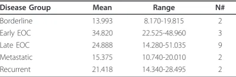
Overall, CD151 was expressed across all stages and no differences were noted with either increasing stage or a particular subtype (Table 1). One trend of note, given the relatively small sample set in this study, was that primary tumors, regardless of stage, on average expressed higher levels of CD151 than either recurrent tumors or metastatic lesions presenting at time of pri-mary debulking surgery (27.4 v 18.4, respectively, p = 0.11). Borderline tumors possessed the lowest average coverage values (14.0).
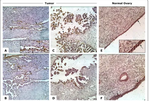
CD151 is expressed in EOC tumors and normal ovary surface epithelial cells
Having identified overexpression on the RNA level, we next evaluated the expression of CD151 protein in tumors and normal ovary tissues using immunohisto-chemistry. Previous reports revealed that CD151 loca-lizes to either the cell membrane or within the cytoplasm of cells in a context specific manner [9,24-26]. Similar to both breast and colorectal cancers [10,12,27-29], CD151 staining in ovarian tumors was seen to be both membranous and cytoplasmic (Figure 1). CD151 was also present in normal ovarian surface
Table 1 RNA-Seq tumor coverage values
Disease Group Mean Range N#
Borderline 13.993 8.170-19.815 2
Early EOC 34.820 22.525-48.960 3
Late EOC 24.888 14.280-51.035 9
Metastatic 15.375 10.740-20.010 2
epithelial cells where it was primarily expressed on the membrane (Figure 1).
CD151 is expressed in ascites-derived and EOC cell lines and immortalized ovarian surface epithelial cells (IOSE) and is localized to cell-cell junctions
The known role of CD151 in cell-cell attachment and its potential role in invasion/migration in ovarian cancer led us next to examine the expression of CD151 in ovar-ian-derived cell lines. These included ascites-derived lines that we had established from patients with ovarian
cancer, commercially available EOC cell lines (A2780, OVCAR3, OVCAR5 and SKOV3), and a number of immortalized ovarian surface epithelial cell lines (IOSE, IOSE397 and IOSE527). Quantitative RT-PCR revealed that all ascites-derived cell lines and ovarian cancer cells expressed CD151 message (Figure 2). In accord with our finding that ovarian surface epithelial cells express CD151 (Figure 1), IOSE also express CD151. The estab-lished ovarian cell lines expressed similar levels of CD151 compared to the primary ascites-derived lines and the immortalized OSE lines (Figure 2).
Table 2 qRT-PCR tumor expression values
———Discovery—— ————Validation———
Disease Group Mean Range N# Mean Range N#
Borderline 0.038 0.019-0.056 2 - -
-Early EOC 0.257 0.046-0.495 3 0.050 0.014-0.090 6
Late EOC 0.404 0.003-2.45 9 1.022 0.085-3.410 7
Metastatic 0.708 0.497-0.920 2 1.160 0.039-1.231 8
Recurrent 0.122 0.005-0.239 2 0.365 0.031-3.605 4
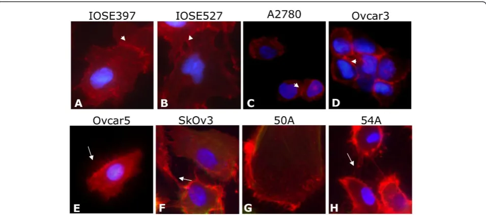
Immunocytochemical fluorescent staining of primary ovarian ascites cell lines and EOC and IOSE cell lines revealed that all patient-derived cell lines expressed CD151 at the protein level (Figure 3). Furthermore, CD151 localized not only to the cell membrane but intriguingly was also expressed at very high concentra-tions at cell-cell attachment points and cell membrane
extensions between cells (Figure 3). We are unaware of this association being previously described in EOC derived cell lines, although it has been reported pre-viously in breast and epidermal carcinoma cell lines [12,16,30,31]. Nonetheless, this is in agreement with the postulated role of CD151 in mediating cell attachment and migration.
Figure 2CD151 qRT-PCR mRNA expression levels in patient-derived ascites cell lines. Patient-derived ascites cell lines, immortalized OSE, and EOC cell lines express CD151.
Migration and invasion of ovarian cancer cell lines is blocked by inhibition of CD151
The postulated role of CD151 in cell migration and tumor spread led us to test if blocking CD151 expres-sion or function could inhibit ovarian cell migration and invasion. Therefore, for these experiments we used either siRNA- mediated knockdown or a specific CD151 antibody. We chose the SKOV3 (lower expression) and OVCAR5 (higher expression) cell lines for their differen-tial RNA expression of CD151 (Figure 2). Using a pool of siRNA oligonucleotides targeting CD151, titration experiments revealed optimal knockdown of CD151 mRNA (90% knockdown) and protein occurred 96 hours after transfection (Figure 4a-c). Both invasion and migration of siCD151- transfected SKOV3 cells over 24 hours were significantly reduced following inhibition of CD151 (Figure 4d-e). We also tested the inhibition of invasion and migration in the higher expressing
OVCAR5 cell line. While siRNA- mediated inhibition of CD151 in OVCAR5 cells significantly reduced their migration, we found no evidence of an effect on inva-sion (Figure 4d-e).
A second method of CD151 inhibition, antibody-mediated, also significantly inhibited the ability of SKOV3 cells to invade and migrate (Figure 4d-e). In the CD151 higher expressing OVCAR5 cells, antibody-mediated blockade, at the concentrations used, had only an ~ 20% decrease in invasion but no measurable effect on migration (Figure 4d-e).
Discussion
These studies are the first to demonstrate that the tetra-spanin CD151 is expressed in epithelial ovarian cancers and that its expression is independent of stage or histo-logical subtype. We analyzed EOC tumor expression levels of all RNA transcripts in an unbiased manner
through the use of RNA-Seq with the goal of identifying possible novel therapeutic targets. From this analysis of 18 whole transcriptomes we identified high CD151 expression across all stages and subtypes of EOC, including borderline ovarian tumors. We then con-firmed expression of CD151 in this discovery set and a second set of tumor tissues using qRT-PCR and immu-nohistochemistry. The previously described role of CD151 as a possible anti-metastatic target led us to further examine the role of CD151 in ovarian cancer and its migration and invasion in culture.
Immunohistochemical staining of EOC tumors and normal ovary showed CD151 expression in tumor cells as well as normal OSE cells. Immunocytochemistry of cells from patient ascites fluid showed CD151 to be localized to the cell periphery and highly concentrated at cell-cell junction points and along outstretched cellu-lar elongations. Finally, functional studies showed that SKOV3 and OVCAR5 cell invasion and migration are differentially inhibited bya-CD151 antibody or siRNA-mediated knockdown of CD151. It appears from these experiments that the level of CD151 expression may affect our ability to inhibit invasion and migration and therefore CD151 blockade may need to be titrated to achieve the most robust inhibition.
CD151 has previously been postulated to play impor-tant roles in a number of cancers including colorectal and breast cancers but no studies had previously identi-fied or examined its expression and function in ovarian cancer. In colorectal cancer CD151 is differentially expressed between normal (high expression), primary (low expression), and metastatic (high expression) tissue [28]. It is proposed that these changes are part of a hypoxic response that drives cell detachment and migra-tion [28]. In our own discovery sample set of EOC tumors, while not reaching statistical significance, we did find an interesting trend of higher CD151 expression in primary ovarian tumors and relatively lower expres-sion in disseminated metastatic or recurrent disease (Tables 1 and 2). It is possible that this is due to differ-ent environmdiffer-ental effects in secondary sites, such as hypoxia or surrounding cell types. This hypothesis will need to be expanded upon by examination of a larger collection of samples.
The expression of tetraspanin CD151 on the cell sur-face of EOC cells may play a role in the spread of these cells to other organs in the peritoneal cavity. In xeno-graft tumor models, antibody-mediated inhibition of CD151 has been shown to hinder the spread of meta-static tumors, reinforcing the idea that CD151 is func-tionally important for either cell detachment from a tumor or migration away from that tumor [19,32]. Intri-guingly, in breast cancer models comparing CD151-expressing cells against CD151-ablated cells, tumor
growth was delayed in the absense of CD151 [12]. In immunohistochemical studies of breast cancer, staining varied greatly in both intensity and cellular localization, which may reflect the heterogeneity of the tumor cells’ environment or activity [33]. CD151 staining intensity and localization in normal OSE and ovarian tumor cells also varies. CD151 protein was localized to both the membrane and the cytoplasm of cancer cells (Figure 1). In contrast, in normal ovarian surface CD151 localized mostly to the cell membrane. It is possible that the internalization of CD151 into cytoplasmic endosomes may reduce its ability to cooperate with other binding partners and cells, allowing detachment from the pri-mary tumor [25]. On ascites-derived patient cell lines, IOSE, and EOC cell lines, CD151 not only localized to the membrane but more specifically to the cell-cell junc-tions and along cell membrane extensions (Figure 3). Althoughin vitrocellular localization suggests no differ-ence between cancerous cell lines and OSE cells, this may due to distinct conditions in cell culture such as attachment to plastic and tightly controlled O2/CO2 balance.
Expression of CD151 in EOC tumors, the known involvement of CD151 in cell migration and invasion, and thein vivo ability ofaCD151 antibody to contain breast cancer tumors to single nodules and eliminate tumor spread suggested that CD151 may also represent a promising and relevant candidate for therapeutic tar-geting in ovarian cancer. We show that in the ovarian cancer cell lines SKOV3 and OVCAR5, CD151 is a functionally important molecule whose silencing or blockade impeded cell migration or invasion to different levels dependent on level of CD151 expression. A role for CD151 in cell migration and invasion is consistent through many cancer types including breast, prostate, colorectal, and pancreatic cancers [15,19,32,34-36]. Ulti-mately, the value of CD151 as a therapeutic target in EOC will need to be demonstrated inin vivostudies.
Abbreviations
EOC: epithelial ovarian cancer; RIN: RNA integrity number; IOSE: immortalized ovarian surface epithelial.
Author details
1
Department of Genetics and Genomic Sciences, Mount Sinai School of Medicine, New York, NY, USA.2Department of Pathology, Mount Sinai
School of Medicine, New York, NY, USA.3Division of Gynecologic Oncology, Mount Sinai School of Medicine, New York, NY, USA.4San Gerardo Hospital,
University of Milano-Bicocca, Monza, Italy.5Department of Oncology, Instituto“Mario Negri”, Milano, Italy.6Mario Negri Gynecological Oncology
Group (MaNGO), Milano, Italy.
Authors’contributions
staining analysis. SC supplied samples and clinical information. RF, SM, and MD supplied samples and clinical information and participated in study design. RS provided bioinformatics support and analysis as well as data interpretation. PD supplied samples and clinical information and participated in study design. JAM participated in overall study design, sample selection, sequencing analysis, and preparation of the manuscript. All Authors reviewed and approved the final version of the manuscript.
Competing interests
The authors declare that they have no competing interests.
Received: 21 November 2011 Accepted: 24 January 2012 Published: 24 January 2012
References
1. Altekruse SF, Kosary CL, Krapcho M, Neyman N, Aminou R, Waldron W, Ruhl J, Howlader N, Tatalovich Z, Cho H, Mariotto A, Eisner MP, Lewis DR, Cronin K, Chen HS, Feuer EJ, Stinchcomb DG, Edwards BK:SEER Cancer Statistics Review, 1975-2007 [Internet]Bethesda, MD: National Cancer Institute.
2. Cannistra SA:Cancer of the ovary.N Engl J Med2004,351:2519-29. 3. Guarneri V, Piacentini F, Barbieri E, Conte PF:Achievements and unmet
needs in the management of advanced ovarian cancer.Gynecol Oncol 2010,117:152-8.
4. Pleasance ED, Stephens PJ, O’Meara S, McBride DJ, Meynert A, Jones D, Lin ML, Beare D, Lau KW, Greenman C, Varela I, Nik-Zainal S, Davies HR, Ordonez GR, Mudie LJ, Latimer C, Edkins S, Stebbings L, Chen L, Jia M, Leroy C, Marshall J, Menzies A, Butler A, Teague JW, Mangion J, Sun YA, McLaughlin SF, Peckham HE, Tsung EF, Costa GL, Lee CC, Minna JD, Gazdar A, Birney E, Rhodes MD, McKernan KJ, Stratton MR, Futreal PA, Campbell PJ:A small-cell lung cancer genome with complex signatures of tobacco exposure.Nature2010,463:184-90.
5. Timmermann B, Kerick M, Roehr C, Fischer A, Isau M, Boerno ST, Wunderlich A, Barmeyer C, Seemann P, Koenig J, Lappe M, Kuss AW, Garshasbi M, Bertram L, Trappe K, Werber M, Herrmann BG, Zatloukal K, Lehrach H, Schweiger MR:Somatic mutation profiles of MSI and MSS colorectal cancer identified by whole exome next generation sequencing and bioinformatics analysis.PLoS One2010,5:e15661. 6. Pleasance ED, Cheetham RK, Stephens PJ, McBride DJ, Humphray SJ,
Greenman CD, Varela I, Lin ML, Ordonez GR, Bignell GR, Ye K, Alipaz J, Bauer MJ, Beare D, Butler A, Carter RJ, Chen L, Cox AJ, Edkins S, Kokko-Gonzales PI, Gormley NA, Grocock RJ, Haudenschild CD, Hims MM, James T, Jia M, Kingsbury Z, Leroy C, Marshall J, Menzies A, Mudie LJ, Ning Z, Royce T, Schulz-Trieglaff OB, Spiridou A, Stebbings LA, Szajkowski L, Teague J, Williamson D, Chin L, Ross MT, Campbell PJ, Bentley DR, Futreal PA, Stratton MR:A comprehensive catalogue of somatic mutations from a human cancer genome.Nature2010,463:191-6. 7. Bignell GR, Greenman CD, Davies H, Butler AP, Edkins S, Andrews JM,
Buck G, Chen L, Beare D, Latimer C, Widaa S, Hinton J, Fahey C, Fu B, Swamy S, Dalgliesh GL, Teh BT, Deloukas P, Yang F, Campbell PJ, Futreal PA, Stratton MR:Signatures of mutation and selection in the cancer genome.Nature2010,463:893-8.
8. Hasegawa M, Furuya M, Kasuya Y, Nishiyama M, Sugiura T, Nikaido T, Momota Y, Ichinose M, Kimura S:CD151 dynamics in carcinoma-stroma interaction: Integrin expression, adhesion strength and proteolytic activity.Lab Invest2007,87:882-92.
9. Liu L, He B, Liu WM, Zhou D, Cox JV, Zhang XA:Tetraspanin CD151 promotes cell migration by regulating integrin trafficking.J Biol Chem 2007,282:31631-42.
10. Yang XH, Flores LM, Li Q, Zhou P, Xu F, Krop IE, Hemler ME:Disruption of laminin-integrin-CD151-focal adhesion kinase axis sensitizes breast cancer cells to ErbB2 antagonists.Cancer Res2010,70:2256-63. 11. Zuo H, Liu Z, Liu X, Yang J, Liu T, Wen S, Zhang XA, Cianflone K, Wang D:
CD151 gene delivery after myocardial infarction promotes functional neovascularization and activates FAK signaling.Mol Med2009,15:307-15. 12. Yang XH, Richardson AL, Torres-Arzayus MI, Zhou P, Sharma C, Kazarov AR,
Andzelm MM, Strominger JL, Brown M, Hemler ME:CD151 accelerates breast cancer by regulating alpha 6 integrin function, signaling, and molecular organization.Cancer Res2008,68:3204-13.
13. Sawada S, Yoshimoto M, Odintsova E, Hotchin NA, Berditchevski F:The tetraspanin CD151 functions as a negative regulator in the adhesion-dependent activation of ras.J Biol Chem2003,278:26323-6. 14. Franco M, Muratori C, Corso S, Tenaglia E, Bertotti A, Capparuccia L,
Trusolino L, Comoglio PM, Tamagnone L:The tetraspanin CD151 is required for met-dependent signaling and tumor cell growth.J Biol Chem2010.
15. Zhu GH, Huang C, Qiu ZJ, Liu J, Zhang ZH, Zhao N, Feng ZZ, Lv XH: Expression and prognostic significance of CD151, c-met, and integrin alpha3/alpha6 in pancreatic ductal adenocarcinoma.Dig Dis Sci2010. 16. Johnson JL, Winterwood N, DeMali KA, Stipp CS:Tetraspanin CD151
regulates RhoA activation and the dynamic stability of carcinoma cell-cell contacts.J Cell Sci2009,122:2263-73.
17. Ke AW, Shi GM, Zhou J, Wu FZ, Ding ZB, Hu MY, Xu Y, Song ZJ, Wang ZJ, Wu JC, Bai DS, Li JC, Liu KD, Fan J:Role of overexpression of CD151 and/ or c-met in predicting prognosis of hepatocellular carcinoma.Hepatology 2009,49:491-503.
18. Yamada M, Sumida Y, Fujibayashi A, Fukaguchi K, Sanzen N, Nishiuchi R, Sekiguchi K:The tetraspanin CD151 regulates cell morphology and intracellular signaling on laminin-511.FEBS J2008,275:3335-51. 19. Zijlstra A, Lewis J, Degryse B, Stuhlmann H, Quigley JP:The inhibition of
tumor cell intravasation and subsequent metastasis via regulation of in vivo tumor cell motility by the tetraspanin CD151.Cancer Cell2008, 13:221-34.
20. Marchini S, Mariani P, Chiorino G, Marrazzo E, Bonomi R, Fruscio R, Clivio L, Garbi A, Torri V, Cinquini M, Dell’Anna T, Apolone G, Broggini M, D’Incalci M:Analysis of gene expression in early-stage ovarian cancer. Clin Cancer Res2008,14:7850-60.
21. Mortazavi A, Williams BA, McCue K, Schaeffer L, Wold B:Mapping and quantifying mammalian transcriptomes by RNA-seq.Nat Methods2008, 5:621-8.
22. Mosig RA, Dowling O, Difeo A, Ramirez MC, Parker IC, Abe E, Diouri J, Aqeel AA, Wylie JD, Oblander SA, Madri J, Bianco P, Apte SS, Zaidi M, Doty SB, Majeska RJ, Schaffler MB, Martignetti JA:Loss of MMP-2 disrupts skeletal and craniofacial development and results in decreased bone mineralization, joint erosion and defects in osteoblast and osteoclast growth.Hum Mol Genet2007,16:1113-23.
23. Tricarico C, Pinzani P, Bianchi S, Paglierani M, Distante V, Pazzagli M, Bustin SA, Orlando C:Quantitative real-time reverse transcription polymerase chain reaction: Normalization to rRNA or single housekeeping genes is inappropriate for human tissue biopsies.Anal Biochem2002,309:293-300.
24. Lineberry N, Su L, Soares L, Fathman CG:The single subunit transmembrane E3 ligase gene related to anergy in lymphocytes (GRAIL) captures and then ubiquitinates transmembrane proteins across the cell membrane.J Biol Chem2008,283:28497-505.
25. Rana S, Claas C, Kretz CC, Zoeller M:Activation-induced internalization differs for the tetraspanins CD9 and Tspan8: Impact on tumor cell motility.Int J Biochem Cell Biol2010.
26. Yang X, Claas C, Kraeft SK, Chen LB, Wang Z, Kreidberg JA, Hemler ME: Palmitoylation of tetraspanin proteins: Modulation of CD151 lateral interactions, subcellular distribution, and integrin-dependent cell morphology.Mol Biol Cell2002,13:767-81.
27. Klosek SK, Nakashiro K, Hara S, Goda H, Hasegawa H, Hamakawa H:CD151 regulates HGF-stimulated morphogenesis of human breast cancer cells. Biochem Biophys Res Commun2009,379:1097-100.
28. Chien CW, Lin SC, Lai YY, Lin BW, Lin SC, Lee JC, Tsai SJ:Regulation of CD151 by hypoxia controls cell adhesion and metastasis in colorectal cancer.Clin Cancer Res2008,14:8043-51.
29. Sauer G, Kurzeder C, Grundmann R, Kreienberg R, Zeillinger R, Deissler H: Expression of tetraspanin adaptor proteins below defined threshold values is associated with in vitro invasiveness of mammary carcinoma cells.Oncol Rep2003,10:405-10.
30. Sharma C, Yang XH, Hemler ME:DHHC2 affects palmitoylation, stability, and functions of tetraspanins CD9 and CD151.Mol Biol Cell2008, 19:3415-25.
31. Shigeta M, Sanzen N, Ozawa M, Gu J, Hasegawa H, Sekiguchi K:CD151 regulates epithelial cell-cell adhesion through PKC- and Cdc42-dependent actin cytoskeletal reorganization.J Cell Biol2003,163:165-76. 32. Testa JE, Brooks PC, Lin JM, Quigley JP:Eukaryotic expression cloning with
CD151) as an effector of human tumor cell migration and metastasis. Cancer Res1999,59:3812-20.
33. Sadej R, Romanska H, Baldwin G, Gkirtzimanaki K, Novitskaya V, Filer AD, Krcova Z, Kusinska R, Ehrmann J, Buckley CD, Kordek R, Potemski P, Eliopoulos AG, Lalani e, Berditchevski F:CD151 regulates tumorigenesis by modulating the communication between tumor cells and endothelium. Mol Cancer Res2009,7:787-98.
34. Gesierich S, Paret C, Hildebrand D, Weitz J, Zgraggen K, Schmitz-Winnenthal FH, Horejsi V, Yoshie O, Herlyn D, Ashman LK, Zoller M: Colocalization of the tetraspanins, CO-029 and CD151, with integrins in human pancreatic adenocarcinoma: Impact on cell motility.Clin Cancer Res2005,11:2840-52.
35. Funakoshi T, Tachibana I, Hoshida Y, Kimura H, Takeda Y, Kijima T, Nishino K, Goto H, Yoneda T, Kumagai T, Osaki T, Hayashi S, Aozasa K, Kawase I:Expression of tetraspanins in human lung cancer cells: Frequent downregulation of CD9 and its contribution to cell motility in small cell lung cancer.Oncogene2003,22:674-87.
36. Tokuhara T, Hasegawa H, Hattori N, Ishida H, Taki T, Tachibana S, Sasaki S, Miyake M:Clinical significance of CD151 gene expression in non-small cell lung cancer.Clin Cancer Res2001,7:4109-14.
doi:10.1186/1757-2215-5-4
Cite this article as:Mosiget al.:Application of RNA-Seq transcriptome analysis: CD151 is an Invasion/Migration target in all stages of epithelial ovarian cancer.Journal of Ovarian Research20125:4.
Submit your next manuscript to BioMed Central and take full advantage of:
• Convenient online submission
• Thorough peer review
• No space constraints or color figure charges
• Immediate publication on acceptance
• Inclusion in PubMed, CAS, Scopus and Google Scholar
• Research which is freely available for redistribution