M
AŁGORZATAJ
AREMKO1, 2, A
NNAS
ADAKIERSKA−C
HUDY1, T
ADEUSZD
OBOSZ1Genotyping
FOXA1
G559A Polymorphism
in Breast Cancer Archival Samples Using
the SNaPshot Technique*
Genotypowanie polimorfizmu
FOXA1
G559A
w archiwalnych preparatach raka gruczołu piersiowego
techniką SNaPshot
1Department of Forensic Medicine, Molecular Techniques Unit, Wroclaw Medical University, Poland 2Mount Sinai School of Medicine, Department of Genetic and Genomics Sciences, NY, USA
Adv Clin Exp Med 2009, 18, 1, 19–24 ISSN 1230−025X
ORIGINAL PAPERS
Abstract
Background.FOXA1, a forkhead family transcription factor, is expressed in breast cancer cells and plays an essential role in the regulation of approximately 50% of estrogen receptor alpha (ERα)−dependent genes. Recent studies of expression signatures of breast cancer types have indicated its potential use as a prognostic factor in luminal type A breast cancer (BC).
Objectives.To establish the frequencies of FOXA1 G559A polymorphisms in a Polish population of breast cancer patients diagnosed with early breast carcinoma and to test whether the SNaPshot methodology is useful for geno− typing DNA extracted from formalin−fixed paraffin−embedded tissues (FFPETs).
Material and Methods. DNA was extracted from 70 FFPET blocks and FOXA1 G559A polymorphism was suc− cessfully genotyped in 51 DNA samples using SNaPshot technology (Applied Biosystems).
Results. It is shown that the SNapShot technology based on the single−base primer extension reaction is suitable for genotyping DNA recovered from archived breast cancer tissues. The rate of successful amplifications was 94.4%. No genotypes deviated from Hardy−Weinberg equilibrium. The observed distribution of the FOXA1
G559A genotypes for this cohort of BC patients was GG 84%, GA 14%, and AA 2%.
Conclusion. SNaPshot is a credible method for the analysis of DNA extracted from FFPETs and it can be used in large−scale FOXA1 G559A analysis. Further study of this polymorphism and analysis of patients’ case histories may provide better insight into its utility as a putative marker of breast cancer prognosis (Adv Clin Exp Med 2009, 18, 1, 19–24).
Key words:FOXA1, luminal breast cancer, SNaPshot.
Streszczenie
Wprowadzenie.FOXA1 to czynnik transkrypcyjny z rodziny Forkhead, ekspresjonowany w komórkach raka pier− si, odgrywający kluczową rolę w regulacji około 50% genów zależnych od receptora estrogenowego alfa (ERα). Ostatnie badania niektórych typów raka gruczołu piersiowego wykazały, że FOXA1 może być potencjalnym czyn− nikiem prognostycznym w luminalnym raku typu A.
Cel pracy. Ustalenie częstości polimorfizmu FOXA1 G559A w populacji polskiej pacjentek ze zdiagnozowanym wczesnym rakiem piersi oraz określenie przydatności metody SNaPshot w genotypowaniu DNA pochodzącego z utrwalonego materiału, przechowywanego w postaci bloczków parafinowych.
Materiał i metody. DNA izolowano z 70 bloczków parafinowych, a genotypowanie udało się przeprowadzić z sukcesem tylko w 51 próbkach z użyciem metody SNaPshot (Applied Biosystems).
Wyniki.Wykazano, że technika SNaPshot oparta na reakcji wydłużania starterów o jedną parę zasad pozwala na genotypowanie DNA pochodzącego z archiwalnych tkanek raka piersi. Wskaźnik udanych amplifikacji wyniósł 94,4%. Stwierdzone genotypy były ilościowo zgodne z przewidywaniami wynikającymi z prawa Hardy’ego−We− inberga. Obserwowany rozkład genotypów w badanej grupie pacjentek wynosił: GG 84%, GA 14%, AA 2%.
© Copyright by Wroclaw Medical University
Breast cancer (BC) is one of the most common malignancies affecting women worldwide. It accounts for 23% of all cancers, with an estimated one million new cases each year [1]. Despite advances in the early detection and understanding of breast cancer biology, about 30% of patients with early−stage BC experience recurrent disease. The clinically used prognostic and predictive fac− tors include age, tumor size, lymph node status, histological tumor type, grade, mitotic rate, and hormonal receptor status [2]. However, these fac− tors are unable to predict and explain events of progression of the disease. Therefore the search for new molecular based prognostic and predictive factors is necessary. At the same time, establishing laboratory tools which will be suitable and reliable for molecular testing is also needed.
Some studies have recently identified FOXA1, a member of the forkhead family of transcription factors and an important regulator of estrogen receptor (ER), as a potential predictive factor in luminal type A breast cancer [3–5]. FOXA1 is known to influence the expressions of approxi− mately 50% of ERαestrogen−regulated genes by opening the chromatin and enhancing the binding of ER−αto its target genes [6, 7]. One of its known important downstream targets is the gene GATA3, which encodes a protein participating in breast tis− sue morphogenesis as well as tumorigenesis [8]. Furthermore, as part of the ER network, FOXA1 may have an effect on differentiation and control of the cell cycle, i.e. by regulating the expressions of the cell cycle inhibitor p27kip1 and the cell adhesion molecule E−cadherin [9]. Therefore, due to its function, some studies hypothesized that FOXA1 may prevent metastatic progression of luminal type A breast cancer [4, 5].
Currently, the investigation of the impact of
FOXA1 single−nucleotide polymorphisms (SNPs) on making treatment decisions or in BC prognosis seems to be underestimated. Since SNPs have rev− olutionized human molecular genetics by provid− ing a powerful panel of genetic markers distrib− uted across the entire genome and are responsible for over 90% of all sequence polymorphisms in the human genome [10], their investigation is of great significance. The study of SNPs has received even greater attention since its application in large− scale genetic association studies [11, 12].
Therefore, study of FOXA1 G559A polymor− phisms may provide essential input into the clini− cal management of patients with luminal type A breast cancer.
Formalin−fixed paraffin−embedded tissues (FFPETs) have been described as a valuable source of biological specimens because of their wide availability in pathology archives together with the accessibility of clinical data. However, limitations of PCR−amplification have been repor− ted when using DNA recovered from FFPETs due to its poor quality caused by cross−links and frag− mentation or base modifications [13–16]. This makes the use of FFPETs in molecular testing challenging. Therefore the development and vali− dation of a suitable genotyping technology before use in a large−scale analysis is required. The aim of this pilot study was to test the applicability of DNA recovered from archival tissues for genotyp− ing FOXA1 polymorphisms by the SNaPshot technology as well as to test the distribution of these polymorphisms among Polish breast cancer patients and compare it with the genotype fre− quencies in a general European population.
Material and Methods
FFPET samples from 70 patients diagnosed with early breast cancer and surgically treated between 1987–2000 at the Wroclaw Medical University Hospital were collected from the Archive of the Wroclaw Medical University, Department of Histopathology. The study was approved by the Institutional Bioethics Board. No clinical informa− tion about the treatment type and histopathological characteristics of the tumor was available.
DNA Extraction
Tissue processing and genomic DNA isolation from the 70 paraffin−embedded breast cancer tis− sues were performed as previously described [17].
Initial PCR Amplification
A 227−bp fragment containing the FOXA1
G559A (rs714465) polymorphic site in exon 2 was amplified by PCR using specific primers (Table 1). Wnioski.Metoda SNaPshot okazała się niezawodną metodą analizy DNA z tkanek utrwalanych formaliną i prze− chowywanych w formie bloczków parafinowych i może być wykorzystana do analizy polimorfizmu FOXA1
G559A w większej grupie pacjentów. Dalsze badania polimorfizmu, w połączeniu z analizą historii choroby, są ko− nieczne do oceny jego przydatności jako markera prognostycznego w raku piersi (Adv Clin Exp Med 2009, 18, 1, 19–24).
Each reaction was performed in a total volume of 20 µl containing 20 ng genomic DNA, 1.5 mM MgCl2, 0.3 µM of each primer, and 0.3 mM each of d−NTP and 1U Taq polymerase (DynaZyme, Finn− zymes, Finland). An initial denaturating step was performed at 95°C for 3 min; the PCR cycling con− ditions were denaturation at 94°C for 30 s, annealing at 56°C for 20 s, extension 72°C for 30 s, and after 35 cycles a final elongation step at 72°C for 5 min.
Genotyping
FOXA1 G559A polymorphism typing was per− formed using the SNaPshot method (Applied Bio− systems). The SNaPshot primers were designed to bind to a complementary sequence one base adja− cent to the polymorphism (Table 1). Five microli− ters of PCR product was incubated with 1U shrimp alkaline phosphatase (SAP) (Fermentas) and 2U Exonuclease I (ExoI) (Fermentas) at 37°C for 40 min followed by 80°C for 15 min. The SNa− Pshot reaction was performed using a SNaPshot Multiplex Kit (Applied Biosystems) in 5 µl volume using 1 µl of digested PCR product, 0.1 µM of extending primer, 1.5 µl of ready reaction mix, and 2.5 µl of deionized H2O. The reaction mixture was run under the following conditions: denaturation at 96°C for 10 s, annealing at 57°C for 5 s, and exten− sion at 60°C for 30 s for 25 cycles. After the SNa− Pshot reaction, 1U of SAP was added to digest unincorporated ddNTPs and the mixture was incu− bated at 37°C for 60 min. Prior to analysis in an ABI PRISM®310 genetic analyzer (Applied Bio− systems), 1 µl of clean SNaPshot product was mi− xed with 9 µl of Hi−Di formamide and the Gene− Scan−120 LIZ standard (Applied Biosystems). The samples were denaturated at 95°C for 5 min and placed on ice before loading to the genetic analy− zer. The results were analyzed using GeneMapper− ID v3.2 software (Applied Biosystems).
Results
DNA was obtained from all 70 archival tissue blocks. However, agarose gel electrophoresis showed a high content of degraded DNA in 16 of
them (22.8%). These samples underwent fixation in 1987–1990. Due to the poor quality of the nucleic acid, the genotyping analyses were per− formed only in 54 of the samples. Successful genotyping was obtained for 94.4% of the sam− ples. The other 5.6% did not amplify, probably due to high DNA degradation; these were obtained exclusively from FFPETs prepared before 1990.
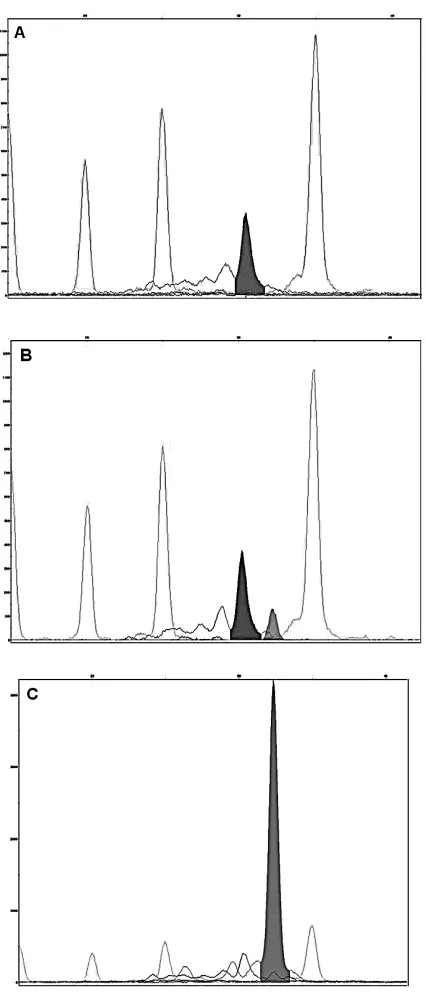
The SNapShot reaction discriminated between carriers of the wild−type, heterozygous, and vari− ant FOXA1 G559A genotypes. The detected alle− les are seen on the electrophoregram as peaks (Fig. 1). Patients were assigned as having the wild− type homozygote when there was a single peak (blue) at the 30−bp position. The heterozygous genotype was described by the occurrence of peaks for both the wild−type and variant alleles, and a variant genotype by the presence of a single peak (green) at the 32−bp position.
All genotypes were in accord with Hardy− −Weinberg equilibrium (HWE) and the genotype frequencies were GG 84%, GA 14%, and AA 2%. When compared with the frequencies available from HapMap−CEU (www.hapmap.org) obtained from residents of Utah of Northern and Western European ancestry, large discrepancies in the genotype frequencies were observed (Fig. 2), the HapMap−CEU frequencies being GG 13%, GA 48%, and AA 38%. The samples were analyzed in duplicate and the genotyping results were matched for each of them.
Discussion
Breast cancer is characterized by high rates of incidence and mortality among women worldwide [1]. In the era of personalized medicine there is a need to identify prognostic and predictive bio− markers for early detection and better management of the disease, but also for determining the most suitable analysis tools to provide efficient and reli− able molecular diagnostic methods. In this study the suitability of the SNapShot technology was tested to analyze FOXA1 G559A polymorphisms in formalin−fixed paraffin−embedded breast cancer Table 1. Primer sequences for initial PCR amplification and SNaPshot reaction
Tabela 1. Sekwencje starterów użyte do reakcji PCR i reakcji SNaPshot
Name Primer sequence
(Nazwa) (Sekwencja starterów)
FoxA1 Forward 5`− ATGACTACGAGCGGCAACAT −3` FoxA1 Reverse 5`− CCCAGGCCATTCATGGAG −3`
tissues. SNapShot is a method based on a single− base primer extension reaction [18], and so far it seems that the present authors are the first to report its assessment in the genotyping of DNA recov− ered from breast cancer archival tissues. Although it has been previously shown that FFPET samples are a valuable source for SNP genotyping using technologies such as MALDI−TOF mass spec− trometry and multiplex PCR with minisequencing [16, 17, 19], the amplification of DNA fragments longer than 200–300 bp might be problematic. The usefulness of FFPETs in PCR−based methods is limited, especially if the tissues were processed using unbuffered formalin, which was routinely practiced before 1990. Fixation time was shown to be a critical factor affecting the sensitivity of PCR amplification [20]. Due to the high impact of the fixation conditions on DNA quality and quantity, the use of amplicon sizes shorter than 180 bp is recommended [21]. Therefore, technologies such as the GeneChip Human Mapping Assay (Affymetrix) and the Infinium Genotyping Assay (Illumina), which require a relatively large amount of intact DNA, will be not as practical for typing archival DNA [16]. However, even these methods could be utilized in archival samples, especially when considering that archival tissues together with collectable clinical information are a very informative source for genetic analysis [22].
In this study a 227−bp product was amplified and 94.4% successful genotype identifications could be obtained. Due to the inclusion of DNA extracted from archival tissues prepared before 1990, an ~5.6% dropout rate in genotyping was noted. This is mostly because of poor DNA quali− ty caused by nucleic acid degradation occurring Fig. 1. Examples of electrophorograms for FOXA1
G559A polymorphisms determined by SNaPshot genotyping. Allele discrimination is based on primer size and color as a result of fluorescence−labeled ddNTPs (terminators) incorporated in the extension reaction. A – blue peak, homozygote wild−type (GG); B – blue and green peaks, heterozygote (GA); C – green peak, mutant homozygote (AA)
Ryc. 1. Elektroforogram prezentujący genotypowanie polimorfizmów FOXA1 G559A metodą SNaPshot. Al− lele określano na podstawie wielkości starterów i ko− lorów znakowanych fluorescencyjnie ddNTPs (termi− natorów) włączanych podczas reakcji wydłużania sta− retów. A – niebieski pik, homozygota typu dzikiego (GG); B – niebieski i zielony pik, heterozygota (GA); C – zielony pik, homozygota zmutowana (AA)
Fig. 2.Distribution of the FOXA1G559A polymor− phisms in the breast cancer cohort compared with the frequencies available in the HapMap−CEU database
during fixation performed in an unbuffered fixa− tion bath with an acidic pH. However, other fac− tors, such as autolysis of the sample prior to fixa− tion, tissue type, and the time and conditions of tis− sue storage, may also contribute to DNA modifications [23]. The high percentage of suc− cessful amplifications obtained in the present study is comparable with results published for other genotyping methods, i.e. an 86–100% suc− cess rate for multiplex PCR with minisequencing with trends to higher successful genotyping rates after exclusion of poor DNA material [16]. In the present study, 16 samples (22.8%) were excluded from a total of 70 because they showed too high DNA degradation on agarose gel. This helped focusing on higher quality DNA, which ensured a higher rate of successful genotyping with only a 5.6% dropout rate.
The high percentage of successful genotyping in 51 samples shows that the FFPET preparations and extraction methods were compatible with the SNapShot methodology. SNapShot is a sensitive reaction which enables the amplification and detection of very minute DNA and is ideal for car− rying out SNP analysis and thus makes it useful in analyzing FFPETs.
The distribution of FOXA1 genotypes within the cohort of breast cancer patients of the present study was different from that published by HapMap for a general population. This discrepan− cy can be the result of the difference in origin of our patients and the subjects analyzed by HapMap, although the latter group was of North and Western European ancestry and such great differ− ences in genotype frequency should not have been observed. However, this can also be explained by population bias, since our study consisted only of patients with breast cancer. As there were no
healthy controls, the frequencies obtained for the breast cancer cohort could not be compared and it was not possible to establish the genotype fre− quencies of the FOXA1 G559A polymorphisms for the general Polish population.
Furthermore, the present group of patients was relatively small; therefore all the genotype fre− quencies will be subsequently confirmed in a greater number of breast cancer patients. Also, the accessibility to archival tissues was limited and the present authors were unable to obtain any clin− ical information, including treatment type and histopathological parameters, necessary to per− form a determination of the clinical value of
FOXA1 G559A polymorphisms.
Most studies on FOXA1 in the literature focus only on the application of its expression in the prognosis and prediction of breast cancer. So far, FOXA1 expression has been associated with ERα− −positive status and favorable prognosis in patients with luminal type A breast cancer and, recently, also with a good prognosis in breast cancer patients treated by adjuvant anthracycline−based chemotherapy [24–26]. On the other hand, not much is known about the functionality and impor− tance of FOXA1 G559A and other knownFOXA1
polymorphisms. This indicates the necessity of investigating FOXA1 polymorphisms with regard to their potential contribution to either personal− ized therapy or prognosis for luminal type A breast cancer.
In summary, the SNapShot methodology is a useful and trustworthy tool for genotyping FFPETs. This technology could be used with a greater number of breast cancer patients to eval− uate the potential prognostic role of FOXA1
G559A polymorphism on a larger scale.
References
[1] Chia KS:Gene−environment interactions in breast cancer. Novartis Found Symp 2008, 293, 143–150.
[2]Adjuvant therapy for breast cancer. NIH Consensus Statement. 2000, 17, 1–35.
[3] Kaklamani VG, Gradishar WJ:Gene expression in breast cancer. Curr Treat Options Oncol 2006, 7, 123–128.
[4] Nakshatri H, Badve S:FOXA1 as a therapeutic target for breast cancer. Expert Opin Ther Targets 2007, 11, 507–514.
[5] Badve S, Turbin D, Thorat MA, Morimiya A, Nielsen TO, Perou CM, Dunn S, Huntsman DG, Nakshatri H:
FOXA1 expression in breast cancer – correlation with luminal subtype A and survival. Clin Cancer Res 2007, 13, 4415–4421.
[6] Carroll JS, Liu XS, Brodsky AS, Li W, Meyer CA, Szary AJ, Eeckhoute J, Shao W, Hestermann EV, Geistlinger TR, Fox EA, Silver PA, Brown M: Chromosome−wide mapping of estrogen receptor binding reveals long−range regulation requiring the forkhead protein FoxA1. Cell 2005, 122, 33–43.
[7] Laganičre J, Deblois G, Lefebvre C, Bataille AR, Robert F, Gigučre V:From the Cover: Location analysis of estrogen receptor alpha target promoters reveals that FOXA1 defines a domain of the estrogen response. Proc Natl Acad Sci USA 2005, 102, 11651–11656.
[8] Kouros−Mehr H, Slorach EM, Sternlicht MD, Werb Z:GATA−3 maintains the differentiation of the luminal cell fate in the mammary gland. Cell 2006, 127, 1041–1055.
[10] Collins FS, Brooks LD, Chakravarti A:A DNA polymorphism discovery resource for research on human genet− ic variation. Genome Res 1998, 8, 1229–1231.
[11] Waever TA:High−throughput SNP discovery and typing for genome−wide genetic analysis. New Technologies of Life Sciences: A Trends Guide 2000, 36–42.
[12] Taylor JG, Choi EH, Foster CB, Chanock SJ:Using genetic variation to study human disease. Trends Mol Med 2001, 7, 507–512.
[13] Ben−Ezra J, Johnson DA, Rossi J, Cook N, Wu A:Effect of fixation on the amplification of nucleic acids from paraffin−embedded material by the polymerase chain reaction. J Histochem Cytochem 1991, 39, 351–354.
[14] Srinivasan M, Sedmak D, Jewell S:Effect of fixatives and tissue processing on the content and integrity of nucleic acids. Am J Pathol 2002, 161, 1961–1971.
[15] Cawkwell L, Quirke P:Direct multiplex amplification of DNA from a formalin fixed, paraffin wax embedded tissue section. Mol Pathol 2000, 53, 51–52.
[16] Gilbert MT, Sanchez JJ, Haselkorn T, Jewell LD, Lucas SB, Van Marck E, Børsting C, Morling N, Worobey M:
Multiplex PCR with minisequencing as an effective high−throughput SNP typing method for formalin−fixed tis− sue. Electrophoresis 2007, 28, 2361–2367.
[17] Jaremko M, Justenhoven C, Abraham BK, Schroth W, Fritz P, Brod S, Vollmert C, Illig T, Brauch H:
MALDI−TOF MS and TaqMan assisted SNP genotyping of DNA isolated from formalin−fixed and paraffin− embedded tissues (FFPET). Hum Mut 2005, 25, 232–238.
[18] Pati N, Schowinsky V, Kokanovic O, Magnuson V, Ghosh S:A comparison between SNaPshot, pyrosequenc− ing, and biplex invader SNP genotyping methods: accuracy, cost, and throughput. J Biochem Biophys Methods 2004, 60, 1–12.
[19] Babol−Pokora K, Berent J:SNP−minisequencing as an excellent tool for analysing degraded DNA recovered from archival tissues. Acta Biochim Pol 2008, 55, 1–5.
[20] Inoue T, Nabeshima K, Kataoka H, Koono M:Feasibility of archival non−buffered formalin−fixed and paraffin− embedded tissues for PCR amplification: an analysis of resected gastric carcinoma. Pathol Int 1996, 46, 997–1004.
[21] Farrand K, Jovanovic L, Delahunt B, McIver B, Hay ID, Eberhardt NL, Grebe SK:Loss of heterozygosity studies revisited: prior quantification of the amplifiable DNA content of archival samples improves efficiency and reliability. J Mol Diagn 2002, 4, 150–158.
[22] Jacobs S, Thompson ER, Nannya Y, Yamamoto G, Pillai R, Ogawa S, Bailey DK, Campbell IG:Genome− wide, high−resolution detection of copy number, loss of heterozygosity, and genotypes from formalin−fixed, paraf− fin−embedded tumor tissue using microarrays. Cancer Res 2007, 67, 2544–2551.
[23] Miething F, Hering S, Hanschke B, Dressler J:Effect of fixation to the degradation of nuclear and mitochon− drial DNA in different tissues. J Histochem Cytochem 2006, 39, 351–354.
[24] Thorat MA, Marchio C, Morimiya A, Savage K, Nakshatri H, Reis−Filho JS, Badve S:Forkhead box A1 expression in breast cancer is associated with luminal subtype and good prognosis. J Clin Pathol 2008, 61, 327–332.
[25] Yamaguchi N, Ito E, Azuma S, Honma R, Yanagisawa Y, Nishikawa A, Kawamura M, Imai J, Tatsuta K, Inoue J, Semba K, Watanabe S:FoxA1 as a lineage−specific oncogene in luminal type breast cancer. Biochem Biophys Res Commun 2008, 365, 711–717.
[26] Wolf I, Bose S, Williamson EA, Miller CW, Karlan BY, Koeffler HP:FOXA1: Growth inhibitor and a favor− able prognostic factor in human breast cancer. Int J Cancer 2007, 120, 1013–1022.
Address for correspondence:
Anna Sadakierska−Chudy Department of Forensic Medicine Molecular Techniques Unit Wroclaw Medical University ul. Curie−Skłodowskiej 52 50−369 Wrocław
Poland
Tel.: +48 71−7841597
E−mail: [email protected]
Conflict of interest: None declared