R
AFAŁK
LECZYK1, A
NDRZEJM
YSIAK1, P
IOTRB
RZOSTOWICZ1,
M
AŁGORZATAK
OBUSIAK−P
ROKOPOWICZ1, M
AREKP
ELCZAR2The Dynamics of Perioperative Changes in Serum BNP
and Troponin I Concentrations in Patients Undergoing
Heart−Valve Correction
with Extracorporeal Circulation*
Dynamika zmian stężeń BNP i troponiny I w surowicy
w okresie okołooperacyjnym u chorych poddanych zabiegowi
korekty wady serca w krążeniu pozaustrojowym
1Department of Cardiology, Silesian Piasts University of Medicine in Wrocław, Poland 2Department of Cardiac Surgery, Silesian Piasts University of Medicine in Wrocław, Poland
Adv Clin Exp Med 2007, 16, 3, 383–388 ISSN 1230−025X
ORIGINAL PAPERS
© Copyright by Silesian Piasts University of Medicine in Wrocław
Abstract
Background.Measurement of BNP (brain natriuretic peptide) and troponin I in the diagnostics of heart−vessel dis− ease allows assessment of heart dysfunction (BNP) and necrosis of the heart muscle (troponin). The effectiveness of these markers in the diagnostics of myocardium function in cardiac surgery patients has not been clearly estab− lished. Heart surgery with extracorporeal circulation can lead to varied extents of damage and impairment of heart function which influence troponin and BNP concentrations.
Objectives.The aim of this study was to describe the dynamics of concentration changes in BNP and troponin I in the perioperative period in patients undergoing cardiac surgery with extracorporeal circulation to correct a valve defect.
Material and Methods.BNP and troponin I concentrations were measured in 17 patients (10 women and 7 men) aged 27–79 years who had aortic, mitral, or tricuspidal heart valve defects and therefore underwent cardiac surgery. The measurements were made before and 24 hours after surgery. The control group consisted of 18 people with− out heart−vessel diseases aged 54–75 years.
Results.Statistically significant higher concentrations of BNP in the patient group than in the control group were observed before (636.63 ± 472.13 pg/ml vs. 45.79 ± 21.92 pg/ml) and after cardiac surgery (628.22 ± 482.9 vs. 45.79 ± 21.92 pg/ml). BNP concentration did not change significantly before and after surgery. Patients in NYHA class III/IV had higher BNP concentrations before surgery than those in NYHA II (837.35 ± 580.96 pg/ml vs. 410.81 ± 116.96 pg/ml). The preoperative concentration of troponin I did not vary significantly from that of the control group. The concentration of troponin I was much higher 24 hours after the operation than the control group value. A difference in troponin I concentration in NYHA class II and class III/IV patients was not observed.
Conclusions.The increase in troponin I 24 hours after valve surgery is probably a result of damage to cardiomy− ocytes during the operation. Moreover, the chronic pathophysiology connected with the valve defect caused con− tinuation of myocardium function disturbance after cardiac surgery. The positive correlation between BNP con− centration 24 hours after surgery and aortic cross−clamping time indicates a relationship between ischemia and BNP release (Adv Clin Exp Med 2007, 16, 3, 383–388).
Key words:troponin I, BNP, extracorporeal circulation, cardiac surgery.
Streszczenie
Wprowadzenie.Jednoczesne oznaczenie troponiny oraz BNP w diagnostyce chorób układu sercowo−naczyniowe− go pozwala na ocenę dysfunkcji czynnościowej (BNP) i ewentualnej martwicy mięśnia sercowego (troponiny). Do− tychczas nie została jednoznacznie określona skuteczność wymienionych markerów w diagnostyce czynności mio−
The procedures connected with the perfor− mance of cardiac surgery with extracorporeal cir− culation can lead to a varied range of damage and impairment of heart function that influence tro− ponin and BNP (brain natriuretic peptide) concen− trations. BNP and troponin I measurements in the diagnostics of heart−vessel diseases allows assess− ment of heart dysfunction (BNP) and necrosis of heart muscle (troponin). The effectiveness of these markers in the diagnostics of myocardium action in patients after cardiac surgery has not yet been established. An incorrect BNP concentration in serum can also result from an increase in pressure in the heart’s interior due to diastolic and systolic dysfunction and hypertrophy of the heart muscle. BNP is also an indicator of myocardial function [1]. Additionally, an incorrect Tn I concentration in serum is a result of cell necrosis or an increase in the permeability of cell membrane and the release of the cytoplasmic compartment of the protein [2]. Higher troponin I and T concentrations are associated with worse prognosis in patients with acute coronary syndrome, coronary vasospasm, severe aortal stenosis, and percutaneous coronary angioplasty. It was also shown that the troponin concentration increases in patients with tachycar− dia, myocarditis, bleeding from the digestive tract, septic shock, hypertrophic muscle of the heart, sever heart failure, stroke, mechanical or electrical trauma (including electrical cardioverion), pul− monary embolism, escalated COPD, and ketone acidosis in diabetic patients. In these cases the higher troponin concentration in serum, caused by
a different mechanism from ischemia, makes the prognosis worse [3–6].
The hemodynamic dysfunction associated with valve pathology significantly influences the synthesis and release of BNP. A higher serum BNP concentration in these patients can indicate advan− cement of the defect and hemodynamic and struc− tural disturbances and can also help in choosing the optimal moment to perform surgical correc− tion. Moreover, a higher BNP concentration is related to the advancement of heart failure, which is defined by NYHA class but is not related to the ejection fraction of the left ventricular [7–9].
The aim of this study was to describe the dynamics of concentration changes of brain natri− uretic peptide and troponin I in the perioperative period in patients undergoing an operation to cor− rect a valve defect in extracorporeal circulation.
Material and Methods
Seventeen patients (10 women and 7 men) aged 27–79 years undergoing surgical valve cor− rection or replacement of an aortal, mitral, or tri− cuspidal valve were examined. Five had a mitral valve replacement (MVR), six had aortal valve replacement (AVR, one patient of whom had a patent foramen ovale closure), and one patient had both mitral and aortal valve replacements. Three other patients underwent a reconstruction of the tricuspid valve modo De Vega. One of the above had aortal valve replacement with recon− kardium u pacjentów po zabiegu kardiochirurgicznym. Procedury związane z zabiegiem kardiochirurgicznym w krążeniu pozaustrojowym mogą prowadzić do uszkodzenia mięśnia sercowego i upośledzenia jego funkcji, cze− go wyrazem jest zwiększone stężenie troponin i mózgowego peptydu natriuretycznego (BNP).
Cel pracy. Określenie okołooperacyjnej dynamiki zmian stężeń mózgowego peptydu natriuretycznego (BNP) oraz troponiny I (Tn I) w surowicy pacjentów poddanych zabiegowi korekty wady zastawkowej.
Materiał i metody.Badaniem objęto 17 pacjentów (10 kobiet i 7 mężczyzn) w wieku 27–79 lat, u których prze− prowadzono zabieg korekty wady lub wymiany zastawki serca. Oznaczanie BNP oraz troponiny I wykonano przed zabiegiem kardiochirurgicznym oraz 24 godziny po operacji. Grupę kontrolną stanowiło 18 osób bez współistnie− jących chorób układu sercowo−naczyniowego.
Wyniki.Wykazano znamiennie większe stężenia BNP w grupie badanych w porównaniu z grupą kontrolną za− równo przed (636,63 ± 472,13 pg/ml vs45,79 ± 21,92 pg/ml), jak i po zabiegu (628,22 ± 482,9 pg/ml vs45,79 ± ± 21,92 pg/ml). U badanych chorych po zabiegu operacyjnym nie wykazano znamiennych zmian stężeń BNP w po− równaniu ze stężeniem przed zabiegiem operacyjnym. U pacjentów w klasie NYHA III/IV stwierdzono większe stężenia BNP przed zabiegiem operacyjnym niż u pacjentów w klasie NYHA II (837,35 ± 580,96 pg/ml vs 410,81 ± ± 116,96 pg/ml). Stężenie troponiny I w surowicy przed zabiegiem u badanych nie różniło się istotnie statystycz− nie w porównaniu ze stężeniem w grupie kontrolnej. 24 godziny po operacji stężenie troponiny I było istotnie więk− sze niż w grupie kontrolnej. Nie wykazano różnicy w stężeniu troponiny I w grupie pacjentów w klasie NYHA II w porównaniu z pacjentami w klasie NYHA III/IV.
Wnioski. Zwiększenie stężenia troponiny I we wczesnym okresie po zabiegu chirurgicznej korekty wady zastawko− wej wskazuje na uszkodzenie kardiomiocytów podczas tej procedury. Przewlekłe zjawiska patofizjologiczne zwią− zane z wadą zastawkową sprawiają, że pomimo przeprowadzenia zabiegu chirurgicznego, czynność miokardium nadal jest zaburzona. Dodatnia korelacja między stężeniem BNP po zabiegu operacyjnym a czasem zakleszczenia aorty wskazuje na związek niedokrwienia z uwalnianiem tego peptydu (Adv Clin Exp Med 2007, 16, 3, 383–388).
struction of the mitral valve. Each patient under− went coronarography before the operation, which did not show any significant hemodynamically important narrowing in the coronary artery. Nine patients were classified as NYHA class III/IV and eight as NYHA class II. The control group con− sisted of 18 people without heart−vessel diseases aged 54–75 years.
Blood was taken twice from the patients’ cephalic vein: one day before and 24 hours after surgery. Blood for the BNP test was put into
polypropylene tubes containing 1 mg EDTA and 500 KIU aprotynin per ml of blood. The collected sam− ples were centrifuged at 1600 ×gfor 15 minutes at 4°C. The serum was stored at –70°C. The BNP con− centration was determined using the radioimmuno− logical method (RIA type, Peninsula Laboratories Inc., cat. no. RIK 9086). Blood for the troponin test was put into polypropylene tubes and centrifuged at 1600 × g for 15 minutes at 4°C. The serum was stored at –70°C. Troponin I was tested using an immunochemical method (Abbott Axsym System).
The patients were operated on using extracor− poreal circulation with antegrade cold crystalloid cardioplegia (BOSTON): 12.6 ml 15% potassium chloride, 12 ml 8.4% sodium bicarbonate, 56 ml 10% sodium chloride, 5.4 ml 40% glucose, and 914 ml 0.9% sodium chloride. Before each opera− tion, echocardiography was performed using a Hewlett Packard Sonos 5500 in accordance with the American Society of Echocardiography rules.
The statistical analysis was performed using STATISTICA 6.0 PL software. The results are given as the average ± the standard deviation. The variability of the data distribution was analyzed using the Shapiro−Wilk test. The Mann−Whitney U
and Wilcoxon tests were also used because of the nonparametric data distribution for the analysis of the results. Correlations between the parameters were analyzed using Spearman’s correlation coef− ficient. Differences were considered statistically significant with p< 0.05.
Results
The profile of the patients is presented in Table 1 and the results are shown in Tables 2 and 3. Statistically significant higher concentrations of
Table 1. Patients chracteristics before cardiac surgery (n = 17)
Tabela 1. Charakterystyka pacjentów przed operacją (n = 17)
n = 17 Average Minimum Maximum
(Średnia) (Wartość (Wartość minimalna) maksymalna)
Age – years 60.6 27 79
(Wiek – lata)
Weight 75.7 58 100
(Masa ciała) kg
Height 167.52 150 186
(Wzrost) cm
EF% 60.8 30 81
ECC (min) 94.4 50 175
Aorta stop 60.8 35 105
(min)
EF – left ventricle ejection fraction. ECC – time of extracorporeal circulation. Aorta stop – aortic cross−clamp time. EF – frakcja wyrzutowa lewej komory. ECC – czas trwania krążenia pozaustrojowego. Aorta stop – czas zakleszczenia aorty.
Table 2.Comparison of BNP and troponin I serum concentrations before surgery (BNP 0, Tn 0) and 24 hours after cardiac surgery (BNP 24, Tn I 24) and control group values
Tabela 2.Porównanie stężenia BNP i troponiny I w surowicy krwi przed operacją (BNP 0, Tn 0) i 24 godziny po operacji (BNP 24, Tn I 24) z pacjen− tami w grupie kontrolnej
Examined patients Control group p (Grupa badana) (Grupa kontrolna)
n = 17 n = 38
Tn I (0) ng/ml 0.09 ± 0.23 vs. 0.026 ± 0.01 0.503 vs.
Tn I (24) ng/ml 9.03 ± 6.72 vs. 0.026 ± 0.01 0.05
p < 0.05
BNP (0) pg/ml 636.63 ± 472.13 vs. 45.79 ± 21.92 0.05 vs.
BNP (24) pg/ml 628.22 ± 482.9 vs. 45.79 ± 21.92 0.05
BNP in the patient group before and after cardiac surgery than in the control group were observed (p< 0.05). The concentration of troponin I before operation did not differ significantly from that measured in the control group. The concentration of troponin I 24 hours after surgery was much higher than the control group value.
The patients with NYHA class III/IV had much higher serum BNP concentrations before surgery than those in the NYHA II group. After surgery the difference persisted, although the data did not reach statistical significance (p = 0.074).
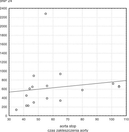
The troponin I concentration in serum increased after surgery regardless of the cardiac failure level. However, the BNP concentration did not change statistically significantly in either group (Table 3). A significant correlation between BNP con− centration before and after cardiac surgery and ejection fraction was not observed. A statistically significant correlation between aortic cross−clamp time and post−surgical serum BNP concentration was found (Fig. 1). However, a statistically signif− icant relationships between extracorporeal circula− tion time, BNP, and troponin I concentration in the short period of time after the operation was not shown. Similarly, a statistically significant correla− tion between aortic cross−clamp time and troponin I concentration in serum during the tested time after the operation was not shown.
Discussion
Many studies have shown that measuring the dynamic changes in both BNP and troponin serum concentrations can be used to specify the risk of surgical heart revascularisation and the correction of valve defects. Hutflessa et al. studied patients undergoing surgical revascularization and valve correction. They showed that intraortal contrapul− sation was more often necessary in patients with significantly higher BNP serum concentrations before cardiac surgery than those with lower con− centrations. It was also shown that patients with higher serum BNP concentrations before surgery had to stay longer than 10 days in hospital after surgery and often died within one year after surgery [10]. It was also shown that an increase in serum troponin I concentration to over 23.8 ng/ml
Table 3.Comparison of BNP and troponin I serum concentrations before cardiac surgery (BNP 0, Tn I 0) and 24 hours from cardiac surgery (BNP 24, Tn I 24) depending on NYHA class
Tabela 3.Porównanie stężenia BNP i troponiny I w surowicy przed (BNP 0, Tn I 0) i 24 godziny po operacji (BNP 24, Tn I 24) w zależności od klasy czynnościowej NYHA
Patients in NYHA II Patients in NYHA III/IV p (Pacjenci w NYHA II) (Pacjenci w NYHA III/IV)
(n = 8) (n = 9)
Tn I (0) ng/ml 0.17 ± 0.33 vs. 0.02 ± 0.02 0.42
vs. vs.
Tn I (24) ng/ml 9.72 ± 6.66 vs. 8.4 ± 7.11 0.6
p < 0.05 < 0.05
BNP (0) pg/ml 410.81 ± 116.96 vs. 837.35 ± 580.96 < 0.05 vs. vs.
BNP (24) pg/ml 431.96 ± 196.44 vs. 802.67 ± 599.91 0.074
p 0.88 0.76
p< 0.05 indicates statistical significance. p < 0,05 oznacza znamienność statystyczną.
Fig. 1.Relation between BNP concentration 24 hours after cardiac surgery and aortic cross−clamp time
Ryc. 1. Korelacja między stężeniem BNP
po 24 godzinach (BNP 24) od zakończenia operacji a czasem zakleszczenia aorty
30 40 50 60 70 80 90 100 110
0 200 400 600 800 1000 1200 1400 1600 1800 2000 2200 2400
after cardiac revascularization involves a higher risk of death caused by heart vessel disease within two years after operation [11]. It was also demon− strated that in patients with significant disturbance of heart valve function in whom surgical correc− tion of the heart valve was necessary because of structural and hemodynamic disorders, the BNP concentration before operation did not correlate with lower ejection fraction, as observed in patients with heart failure [12, 13].
In the present study the initial BNP concentra− tion corresponded with NYHA class and did not correspond with the ejection fraction of the left ventricle. BNP concentrations were much higher in the patients than in the controls. In most cases the higher troponin concentration shows necrosis of cardiomyocytes. However, the higher troponin concentration in the small group of the patients can be, as mentioned above, a consequence of higher cell membrane permeability with secon− dary release of the cytoplasmic compartment into the serum [14]. The data concerning the role of the changes in BNP concentration during a heart valve operation have not been thoroughly studied so far.
Berendea and coworkers showed that changes in serum BNP concentration during the periopera− tive period depended on the type of cardiac surgery. In the case of heart revascularization, the initially higher BNP concentration before cardiac surgery increased significantly 24 hours after the operation. It was also shown that the higher BNP concentration was dependent on the time of extra− corporeal circulation, aorta cross−clamp time, and the postoperative troponin concentration. It seems that the main cause of the higher release of BNP was ischemia of the myocardium, which led to stunning and systolic dysfunction of the heart. In patients with a defect of the mitral or aortal valve, the preoperative BNP concentration in− creased 3− and 14−times, respectively. The preop− erative BNP concentration did not change after the operation and also did not correlate with the time of extracorporeal circulation, the postoperative troponin concentration, or aorta cross−clamp time. The increase in the synthesis and release of BNP is connected with the consequences of the valve defect, such as overload of the left ventricle and higher tension of heart’s wall as a result of hyper− trophy myocardium and the increase in end dias− tolic pressure [15].
The present study showed that the BNP con− centration in serum in the patients after cardiac surgery did not change significantly from that before surgery. The BNP concentration correlated neither with the time of extracorporeal circulation nor with the postoperative troponin I concentra−
tion, but only with the aorta cross−clamp time. It can also be responsible for the influence of ischemia on higher BNP release.
On the other hand, the study did not show sig− nificant differences in BNP concentration before operation in patients with coronary artery diseases, mitral valve insufficiency, and aortal valve steno− sis who were qualified for operation. In patients with ischemic heart disease and mitral valve insuf− ficiency, significantly higher BNP concentrations 8 and 12 hours after cardiac surgery were observed [16]. However, stenosis of the aortal valve did not cause the BNP concentration to increase. The high diversity of the obtained BNP concentrations in the groups of patients in that study, as in the pre− sent study, decrease the credibility of the obtained results. It was also shown that the troponin I serum concentration after cardiac surgery increased sig− nificantly from the concentration before operation. However, aorta cross−clamp time, extracorporeal circulation time, NYHA class, BNP concentration, and the type of cardiac surgery did not show any statistically significant correlations. These studies just proved the result obtained by Opfermann and coworkers, who did not observe any relationship between the increase in troponin concentration, time of operation, extracorporeal circulation time, aorta cross−clamp time, and the dose of cardiople− gin [17]. It was shown that OPCAB (off−pump coronary artery bypass graft) operation is connect− ed with lower cardiac damage described by lower troponin I and T concentrations after cardiac surgery compared with patients operated with extracorporeal circulation [18].
According to some other researchers, an in− crease in serum BNP concentration over 450 pg/ml and an increase in serum troponin I concentration over 5.4 ng/ml result in a 12−fold increase in the risk of heart failure. However, the determination of BNP and troponin concentrations in serum in the early perioperative period indicated heart failure more correctly than each of these markers sepa− rately. A BNP concentration over 352 pg/ml influ− enced higher mortality within the first year of observation [19]. It also seems that determination of the BNP and troponin serum concentrations before operation and a short time after operation can be useful in defining the overload of the left ventricle, its dysfunction, ischemia, and also necrosis of the heart muscle.
physiological phenomenon during disease, the consequences of which did not recede directly after surgery. The BNP concentration in serum, regardless of initial the BNP concentration, did not change within the short time after cardiac surgery.
The positive correlation between aortic cross− clamp time and serum BNP concentration 24 hours after surgical correction of a valve defect showed that perioperative ischemia of the heart muscle influenced BNP release.
References
[1] Bursi F, Weston SA, Redfield MM, Jacobsen SJ, Pakhomov S, Nkomo VT, Meverden RA, Roger VL:
Systolic and diastolic heart failure in the community. JAMA 2006, 8, 2209–2216.
[2] Bleier J, Vorderwinkler KP, Falkensammer J, Mair P, Dapunt O, Puschendorf B, Mair J:Different intracel− lular compartmentations of cardiac troponins and myosin heavy chains: a causal connection to their different early release after myocardial damage. Clin Chem 1998, 44, 1912–1918.
[3] Mahajan N, Mehta Y, Rose M, Shani J, Lichstein E:Elevated troponin level is not synonymous with myocar− dial infarction. Int J Cardiol 2006, 111, 442–449.
[4] Nienhuis MB, Ottervanger JP, Dikkeschei B, Suryapranata H, de Boer MJ, Dambrink JH, Hoorntje JC, van ‘t Hof AW, Gosselink M, Zijlstra F:Prognostic importance of troponin T and creatine kinase after elective angioplasty. Int J Cardiol 2006, 18, Abstract.
[5] Mehta NJ, Khan IA, Gupta V, Jani K, Gowda RM, Smith PR:Cardiac troponin I predicts myocardial dys− function and adverse outcome in septic shock. Int J Cardiol 2004, 95, 13–17.
[6] Janata K, Holzer M, Laggner AN, Mullner M:Cardiac troponin T in the severity assessment of patients with pulmonary embolism: cohort study. BMJ 2003, 326, 312–313.
[7] Iltumur K, Karabulut A, Yokus B, Yavuzkir M, Taskesen T, Toprak N:N−terminal proBNP plasma levels cor− relate with severity of mitral stenosis. J Heart Valve Dis 2005, 14, 735–741.
[8] Arat−Ozkan A, Kaya A, Yigit Z, Balci H, Okcun B, Yazicioglu N, Kucukoglu S:Serum N−terminal pro−BNP levels correlate with symptoms and echocardiographic findings in patients with mitral stenosis. Echocardiography 2005, 22, 473–478.
[9] Weber M, Arnold R, Rau M, Brandt R, Berkovitsch A, Mitrovic V, Hamm C:Relation of N−terminal pro−B−type natriuretic peptide to severity of valvular aortic stenosis. Am J Cardiol 2004, 15, 740–745.
[10] Hutfless R, Kazanegra R, Madani M, Bhalla MA, Tulua−Tata A, Chen A, Clopton P, James C, Chiu A, Maisel AS: Utility of B−type natriuretic peptide in predicting postoperative complications and outcomes in patients undergoing heart surgery. J Am Coll Cardiol 2004, 19, 1873–1879.
[11] Fellahi JL, Gue X, Richomme X, Monier E, Guillou L, Riou B:Short− and long−term prognostic value of post− operative cardiac troponin I concentration in patients undergoing coronary artery bypass grafting. Anesthesiology 2003, 99, 270–274.
[12] Bursi F, Weston SA, Redfield MM, Jacobsen SJ, Pakhomov S, Nkomo VT, Meverden RA, Roger VL:
Systolic and diastolic heart failure in the community. JAMA 2006, 8, 2209–2216.
[13] Sutton TM, Stewart RA, Gerber IL, West TM, Richards AM, Yandle TG, Kerr AJ:Plasma natriuretic pep− tide levels increase with symptoms and severity of mitral regurgitation. J Am Coll Cardiol 2003, 41(12), 2280–2287.
[14] Bleier J, Vorderwinkler KP, Falkensammer J, Mair P, Dapunt O, Puschendorf B, Mair J:Different intracel− lular compartmentations of cardiac troponins and myosin heavy chains: a causal connection to their different early release after myocardial damage. Clin Chem 1998, 44, 1912–1918.
[15] Berendes E, Schmidt C, Van Aken H, Hartlage MG, Rothenburger M, Wirtz S, Scheld HH, Brodner G, Walter M:A−type and B−type natriuretic peptides in cardiac surgical procedures. Anesth Analg 2004, 98, 11–19.
[16] Georges A, Forestier F, Valli N, Plogin A, Janvier G, Bordenave L:Changes in type B natriuretic peptide (BNP) concentrations during cardiac valve replacement. Eur J Cardiothorac Surg 2004, 25, 941–945.
[17] Opfermann UT, Peivandi AA, Dahm M, Hilgenstock H, Hafner G, Loos A, Oelert H:Postoperative patterns and kinetics of cTnI, cTnT, CK−MB−activity and CK−activity after elective aortic valve replacement. Swiss Med Wkly 2001, 22, 550–555.
[18] Guerin V, Ayed SB, Varnous S, Golmard JL, Leprince P, Beaudeux JL, Gandjbakhch I, Bernard M:Release of brain natriuretic−related peptides (BNP, NT−proBNP) and cardiac troponins (cTnT, cTnI) in on−pump and off− pump coronary artery bypass surgery. Surg Today 2006, 36, 783–789.
[19] Provenchere S, Berroeta C, Reynaud C, Baron G, Poirier I, Desmonts JM, Iung B, Dehoux M, Philip I, Benessiano J:Plasma brain natriuretic peptide and cardiac troponin I concentrations after adult cardiac surgery: association with postoperative cardiac dysfunction and 1−year mortality. Crit Care Med 2006, 34, 995–1000.
Address for correspondence:
Rafał Kleczyk Szybka 3/50 50−421 Wrocław Poland
Tel.: +48 600 92 88 59
e−mail: [email protected]
Conflict of interest: None declared