Cite as
Lee H, Lee H, Na C-B, Park J-B. The effects of simvastatin on cellular viability, stemness and osteogenic differentiation using 3-dimensional cultures of stem cells and osteoblast-like cells. Adv Clin Exp Med. 2019;28(5):699–706. doi:10.17219/acem/94162
DOI
10.17219/acem/94162
Copyright
© 2019 by Wroclaw Medical University This is an article distributed under the terms of the Creative Commons Attribution Non-Commercial License (http://creativecommons.org/licenses/by-nc-nd/4.0/)
Address for correspondence
Jun-Beom Park
E-mail: [email protected]
Funding sources
This research was supported by the Basic Science Research Program through the National Research Foundation of Korea (NRF), funded by the Ministry of Science, Information and Communication Technology & Future Planning of South Korea (NRF-2017R1A1A1A05001307).
Conflict of interest
None declared
Acknowledgements
The first 2 authors contributed equally to the study. The authors gratefully acknowledge the support provided by the Basic Science Research Program through the NRF, funded by the Ministry of Science, Information and Communication Technology & Future Planning.
Received on May 18, 2017 Reviewed on June 30, 2017 Accepted on August 9, 2018
Published online on February 5, 2019
Abstract
Background. Simvastatin has been reported to increase the therapeutic effects of many kinds of stem cells by increasing the number of those cells. However, the effects of simvastatin on the differentiation potential of stem cells have not been clearly determined.
Objectives. The aim of the study was to evaluate the effects simvastatin has on cellular viability, stemness and osteogenic differentiation using 3-dimensional cell spheroids of stem cells and osteoblast-like cells.
Material and methods. Three-dimensional cell spheroids were fabricated using concave silicon elastomer-based microwells in the presence of simvastatin at concentrations of 1 μM and 10 μM. Qualitative cellular viability was determined with a confocal microscope, and quantitative cellular viability was evaluated using a cell-counting assay kit. The expression of stem cell surface markers was tested. A quantitative real-time polymerase chain reaction (qRT-PCR) was performed to evaluate the expression of collagen I and RUNx2. Alkaline phosphatase activity and alizarin red S staining were used to assess osteogenic differentiation.
Results. The spheroids formed well in the concave silicon elastomer-based microwells, and the application of simvastatin caused no significant morphological changes. No significant changes in cellular viability were noted with the addition of simvastatin on days 1, 3 and 5. Secretion of the vascular endothelial growth factor (VEGF) was observed on day 1 and remained stable throughout the culture period. Expression of the CD90 surface marker was seen on day 7. The addition of simvastatin caused a statistically significant increase in the expression of collagen I and RUNX2. It also caused decreases in alkaline phosphatase activity and alizarin red S staining.
Conclusions. The study clearly showed that the application of simvastatin enhanced collagen I and RUNX2 expression; however, this did not lead to increases in alkaline phosphatase activity or alizarin red S staining.
Key words: gingiva, simvastatin, stem cells, cell differentiation, cellular spheroids
The effects of simvastatin on cellular viability, stemness
and osteogenic differentiation using 3-dimensional
cultures of stem cells and osteoblast-like cells
Hyunjin Lee
A–F, Hyuna Lee
A–F, Chae-Bin Na
A–F, Jun-Beom Park
A–FDepartment of Periodontics, College of Medicine, The Catholic University of Korea, Seoul, South Korea
A – research concept and design; B – collection and/or assembly of data; C – data analysis and interpretation; D – writing the article; E – critical revision of the article; F – final approval of the article
Simvastatin is a chemical modification of lovastatin, a rate-limiting enzyme of the cholesterol synthesis path-way.1,2 Simvastatin has been reported to promote osteoblas-tic activity and inhibit osteoclasosteoblas-tic activity.3 Previous reports have shown that simvastatin positively affects the differen-tiation and mineralization of osteoprecursor cells through the estrogen receptor pathway by increasing estrogen receptor-α expression.4 A combination of simvastatin and bone morphogenetic protein-2 enhanced the differentiation of osteoprecursor cells through the bone morphogenetic protein pathway by enhancing pSmad1/5/8 expression.5
The effects of simvastatin on mesenchymal stem cells (MSCs) have been observed in previous studies.6–9 Simvas-tatin has been reported to increase the therapeutic effects of nucleus pulposus stem cells by increasing the number of those cells.8 It has also been shown to induce enhanced proliferation of cultured neural stem cells.7 Conversely, simvastatin has been reported to negatively modulate bone marrow MSC proliferation in a dose-dependent manner and to regulate the expression of proliferation-related genes.6 One report showed that simvastatin both directly interfered with the proliferation of stem cells and promoted cell death.9 Moreover, the effects of simvastatin on the dif-ferentiation potential of stem cells have not been clearly determined. Recently, 3-dimensional culture systems us-ing stem cells have been applied to cell therapy.10 These cell spheroids maintained the viability and stem-cell characteristics of stem-stem-cell surface-marker expression and differentiation potential.11 In a previous study, cell spheroids from gingival cells and osteoprecursor cells were shown to be a promising strategy for stem-cell therapy.12 The current study was therefore performed to evaluate the effects of simvastatin on cellular viability, stemness and osteogenic differentiation using 3-dimensional cell spheroids of stem cells and osteoblast-like cells.
Material and methods
Formation of cell spheroids
with human gingiva-derived stem cells
and osteoblast-like cells
Stem cell spheroids were formed in concave silicon elastomer-based microwells (StemFit 3D; Micro Fit Co. Ltd., Seongnam, South Korea) with a diameter of 600 μM. Gingiva-derived stem cells (GMSCs) and osteoblast-like cells (MC3T3-E1 cells; American Type Culture Collection, Manassas, USA) were plated in a 1:1 ratio (5 × 10⁵ cells/well), cultured in an alpha-minimal essential medium (α-MEM; Gibco, Grand Island, USA) with a total volume of 1.5 mL, and then supplemented with 15% fetal bovine serum (Gibco), 100 U/mL of penicillin, 100 μg/mL of streptomycin (Sigma-Aldrich, St. Louis, USA), 200 mM of L-glutamine (Sigma-Al-drich), 10 mM of ascorbic acid 2-phosphate (Sigma-Al(Sigma-Al-drich), 38 μg/mL of dexamethasone (Fujifilm Wako Pure Chemical
Corp., Osaka, Japan), and 2 mg/mL of glycerophosphate disodium salt hydrate (Sigma-Aldrich). The culturing was performed in the presence of simvastatin (Sigma-Aldrich) at concentrations of 0 μM (the untreated control group), 1 μM and 10 μM with dimethyl sulfoxide (DMSO) used as a dissolving agent. The media were changed every second day. The GMSCs were obtained from the mandibular area of a healthy female participant undergoing second-stage implant surgery, using a previously reported method.13 The study design was reviewed and approved by the Insti-tutional Review Board of Seoul St. Mary’s Hospital (College of Medicine, Catholic University of Korea, Seoul, South Korea; approval No. KC11SISI0348), and informed consent was obtained from the patient. On days 1, 3 and 5, inverted microscopy (Olympus Corporation, Tokyo, Japan) was used to evaluate the morphology of the stem cells being tested.
Determination of cell viability
The viability of the spheroids was qualitatively ana-lyzed by an assay using the Live/Dead Kit (Molecular Probes, Eugene, USA). The spheroids were washed twice with the growth media, followed by suspension in 2 mL of α-MEM (containing 1 μL of 4 mM calcein acetoxymethyl ester working solution and 4 μL of 2 mM ethidium ho-modimer-1) for 30 min at room temperature. The spher-oids stained with calcein acetoxymethyl ester and ethid-ium homodimer-1 were observed under a confocal laser microscope (LSM800 w/Airyscan; Carl Zeiss, Oberkochen, Germany) on days 3 and 5 (n = 3).
Quantitative cell-viability analyses were performed on days 1, 3 and 5. Using a Dojindo Cell Counting Kit-8 (CCK-8; Dojindo Molecular Technologies, Tokyo, Japan), we added 2-(2-methoxy-4-nitrophenyl)-3-(4-nitrophenyl)-5-(2,4-disulfophenyl)-2H tetrazolium monosodium salt (WST-8) to the cultures, and the spheres were incubat-ed for 1 h at 37°C. Viable cells were identifiincubat-ed through the assay, which relies on the ability of mitochondrial de-hydrogenases to oxidize WST-8 into a formazan product. The spectrophotometric absorbance of the samples was measured at 450 nm using a microplate reader (BioTek Instruments Inc., Winooski, USA) (n = 3).
Evaluation of the secretion
of human VEGF for paracrine effects
Evaluation of maintenance of stemness
Approximately 1.5 × 10⁵ GMSCs and 1.5 × 10⁵ osteoblast-like cells were incubated with specific fluorescein isothio-cyanate-conjugated mouse monoclonal antibodies for hu-man CD90, CD 73 and CD 34 (BD Biosciences, San Jose, USA) (n = 2). A flow cytometric analysis was performed using a FACSCanto II flow cytometer (BD Biosciences) and FACSDiva software (BD Biosciences).
Total RNA extraction and quantification
using RT-PCR
The cell spheroids were harvested on day 7. Total RNA was isolated using a GeneJET RNA Purification Kit (Thermo-Fisher Scientific Inc., Waltham, USA), and the quantities were determined with an ND-2000 spectrophotometer (Thermo-Fisher Scientific Inc.) using ratios of absorbance at 260 nm and 280 nm. The sense and antisense primers were based on GenBank. The primer sequences were as follows: RUNX2 Forward 5’ – AAT GAT GGT GTT GAC GCT GA – 3’; Re-verse 5’ – TTG ATA CGT GTG GGA TGT GG – 3’; Colla-gen I Forward 5’ – TCA TGG CCC TCC AGC CCC CAT3’; and Reverse 5’ – ATG CCT CTT GTC CTT GGG GTT C – 3’. β-actin served as a housekeeping gene for normaliza-tion. The mRNA expression was detected with real-time polymerase chain reaction (RT-PCR) using SYBR Green Real-Time PCR Master Mixes (Enzynomics Inc., Daejeon, South Korea) according to the manufacturer’s protocol. The quan-titative RT-PCR experiments were conducted 3 times.
Alkaline phosphatase activity assays
and alizarin red S staining
Cell spheroids that had been grown on culture plates with an osteogenic medium were harvested on day 14. Alkaline phosphatase activity assays were performed with a commercially available kit (BioVision Inc., Milpitas, USA). The cells were suspended again with an assay buffer, sonicated and then centrifuged to remove insoluble mate-rial. The supernatant was mixed with a p-nitrophenylphos-phate substrate and incubated at 25°C for 60 min. The opti-cal density of the resultant p-nitrophenol at 405 nm was determined spectrophotometrically (n = 3).
On days 7 and 14, the spheroids were washed twice with phosphate-buffered saline (PBS) (Welgene, Gyeong-san, South Korea), fixed with 4% paraformaldehyde and rinsed twice with deionized water. The cultures were then stained with alizarin red S for 30 min at room temperature. To remove nonspecifically-bound stains, the cultures were washed 3 times with deionized water. A morphological eval-uation was then performed using a Leica DM IRM inverted microscope (Leica Camera AG, Wetzlar, Germany) (n = 3).
Statistical analysis
The data is presented as means ± standard devia-tions (SD). A test of normality was conducted and a one-way analysis of variance (ANOVA) with a post hoc test were performed to determine the differences between the groups. The analysis was done using SPSS v. 12 soft-ware for Windows (SPSS Inc., Chicago, USA), and the level of significance was p < 0.05.
Results
Evaluation of cell morphology and viability
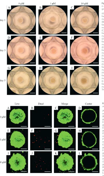
On day 1, the spheroids were properly formed in concave silicon elastomer-based microwells (Fig. 1A). The addition of simvastatin caused no significant changes in the mor-phology on day 1 (Fig. 1B,C). The morphological results for days 3 and 5 are shown in Fig. 1D–I. In general, the shapes of the cells in the experimental groups were similar to those in the control group (0 μM).
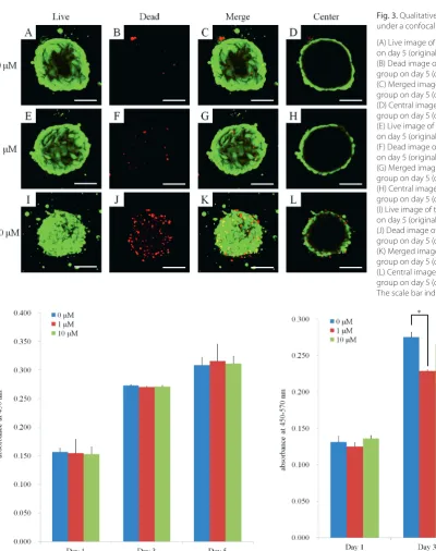
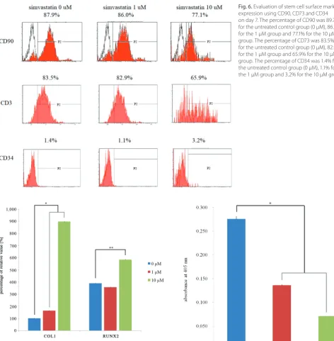
Using a confocal microscope, the cellular viability was determined using live/dead assays on days 3 and 5, as shown in Fig. 2 and 3, respectively. For day 3, most of the cells in the spheroids emitted green fluorescence and had a round morphology (Fig. 2). Similarly, most of the cells in the spheroids emitted green fluorescence, but an increase in red fluorescence was noted at the higher dose of simvastatin (10 μM). The CCK-8 results for days 1, 3 and 5 are shown in Fig. 4. In terms of quantitative results, no significant changes in cellular viability were noted with the addition of simvastatin on days 1, 3 and 5.
Secretion of human VEGF from spheroids
and expression of stem cell markers
The results clearly showed that VEGF secretion was observable on the 1st day, and that stable VEGF secretion occurred throughout the culture period (Fig. 5). A decrease in VEGF secretion was noted with the addition of simvas-tatin (1 μM) on day 3. A statistically significant decrease in secretion was noted with the addition of simvastatin (1 μM and 10 μM) on day 5 (p < 0.05).
Validation of mRNA expression
using RT-PCR
The quantitative RT-PCR results for the mRNA levels of collagen I and RUNX2 are shown in Fig. 7. The addition of simvastatin produced statistically significant increases
in the expression of collagen I and RUNX2. For the un-treated control group, the 1 μM group and the 10 μM group, the relative expressions of collagen I were 100.0 ±1.7%, 164.1 ±2.4% and 898.4 ±1.2%, respectively (p < 0.05), and the relative expressions of RUNX2 were 100.0 ±1.2%, 91.5 ±0.6% and 150.1 ±0.9%, respectively (p < 0.05).
Fig. 2. Qualitative results of cellular viability under a confocal microscope on day 3 (A) Live image of the untreated control group on day 3 (original magnification ×100) (B) Dead image of the untreated control group on day 3 (original magnification ×100) (C) Merged image of the untreated control group on day 3 (original magnification ×100) (D) Central image of the untreated control group on day 3 (original magnification ×100) (E) Live image of the simvastatin 1 μM group on day 3 (original magnification ×100) (F) Dead image of the simvastatin 1 μM group on day 3 (original magnification ×100) (G) Merged image of the simvastatin 1 μM group on day 3 (original magnification ×100) (H) Central image of the simvastatin 1 μM group on day 3 (original magnification ×100) (I) Live image of the simvastatin 10 μM group on day 3 (original magnification ×100) (J) Dead image of the simvastatin 10 μM group on day 3 (original magnification ×100) (K) Merged image of the simvastatin 10 μM group on day 3 (original magnification ×100) (L) Central image of the simvastatin 10 μM group on day 3 (original magnification ×100) The scale bar indicates 100 μM.
Fig. 1. Morphology of cell spheroids cultured in osteogenic media, the simvastatin 1 μM group and the simvastatin 10 μM group (A) Morphology of the cell spheroids on day 1 for the untreated control group (original magnification ×200)
(B) Morphology of the cell spheroids on day 1 for the simvastatin 1 μM group (original magnification ×200)
(C) Morphology of the cell spheroids on day 1 for the simvastatin 10 μM group (original magnification ×200)
(D) Morphology of the cell spheroids on day 3 for the untreated control group (original magnification ×200)
(E) Morphology of the cell spheroids on day 3 for the simvastatin 1 μM group (original magnification ×200)
(F) Morphology of the cell spheroids on day 3 for the simvastatin 10 μM group (original magnification ×200)
(G) Morphology of the cell spheroids on day 5 for the untreated control group (original magnification ×200)
(H) Morphology of the cell spheroids on day 5 for the simvastatin 1 μM group (original magnification ×200)
(I) Morphology of the cell spheroids on day 5 for the simvastatin 10 μM group (original magnification ×200)
The scale bar indicates 200 μM.
day 1
day 3
Alkaline phosphatase activity assays
and alizarin red S staining
Alkaline phosphatase activity decreased on day 14 after treatment with simvastatin, as shown in Fig. 8. The relative values on day 14 for the untreated control group, the 1 μM group and the 10 μM group were 100.0 ±1.6%, 49.3 ±0.4% and 25.8 ±0.0%, respectively.
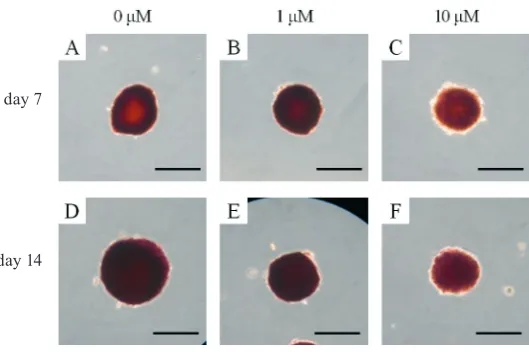
Mineralized extracellular deposits were evaluated us-ing alizarin red S stainus-ing on days 7 and 14 (Fig. 9) and were found to be equivalent in all 3 groups. An increase
in mineralized deposits was noted between day 7 and day 14. Alizarin red S staining decreased with the addi-tion of simvastatin.
Discussion
This report discusses the effects of simvastatin on cellu-lar viability, stemness and osteogenic differentiation using 3-dimensional cell spheroids of stem cells and osteoblast-like cells. The addition of simvastatin clearly enhanced
Fig. 3. Qualitative results of cellular viability under a confocal microscope on day 5 (A) Live image of the untreated control group on day 5 (original magnification ×100) (B) Dead image of the untreated control group on day 5 (original magnification ×100) (C) Merged image of the untreated control group on day 5 (original magnification ×100) (D) Central image of the untreated control group on day 5 (original magnification ×100) (E) Live image of the simvastatin 1 μM group on day 5 (original magnification ×100) (F) Dead image of the simvastatin 1 μM group on day 5 (original magnification ×100) (G) Merged image of the simvastatin 1 μM group on day 5 (original magnification ×100) (H) Central image of the simvastatin 1 μM group on day 5 (original magnification ×100) (I) Live image of the simvastatin 10 μM group on day 5 (original magnification ×100) (J) Dead image of the simvastatin 10 μM group on day 5 (original magnification ×100) (K) Merged image of the simvastatin 10 μM group on day 5 (original magnification ×100) (L) Central image of the simvastatin 10 μM group on day 5 (original magnification ×100) The scale bar indicates 100 μM.
Fig. 4. Cellular viability on days 1, 3 and 5 using CCK-8. No significant changes in cellular viability were noted with the addition of simvastatin on days 1, 3 and 5
collagen I and RUNX2 expression, but it did not lead to in-creased alkaline phosphatase activity or into in-creased alizarin red S staining.
Simvastatin has been shown to increase therapeutic ef-fects by enhancing stem cell function.8 Simvastatin induces increased pluripotency and protects against cellular senes-cence in MSCs.6 It also improves the migration of bone marrow-derived MSCs via the PI3K/AKT pathway,14 and enhances the neurogenesis of cultured neural stem cells and elevates Notch-1 protein expression.7 Simvastatin
exhibits increased dentin sialoprotein and calcium de-position, and it has been suggested that it can promote odontoblastic differentiation in dental pulp stem cells.15 Moreover, previous reports have shown that simvastatin stimulates osteogenic effects in bone marrow-derived MSCs.16 It has also been shown that it enhances the osteo-genic differentiation of stem cells without affecting their immunosuppressive properties.17 Additionally, simvastatin increases bone morphogenetic protein-2 gene expression on titanium surfaces that have been sandblasted with large Fig. 7. Quantitative real-time polymerase chain reaction results for
collagen I and RUNX2 expression on day 7. The addition of simvastatin produced statistically significant increases in the expression of collagen I and RUNX2. For the untreated control group, the 1 μM group, and the 10 μM group, the relative expressions of collagen I are 100.0 ±1.7%, 164.1 ±2.4% and 898.4 ±1.2%, respectively, and the relative expressions of RUNX2 are 100.0 ±1.2%, 91.5 ±0.6% and 150.1 ±0.9%, respectively
Fig. 6. Evaluation of stem cell surface marker expression using CD90, CD73 and CD34 on day 7. The percentage of CD90 was 89.7% for the untreated control group (0 μM), 86.0% for the 1 μM group and 77.1% for the 10 μM group. The percentage of CD73 was 83.5% for the untreated control group (0 μM), 82.9% for the 1 μM group and 65.9% for the 10 μM group. The percentage of CD34 was 1.4% for the untreated control group (0 μM), 1.1% for the 1 μM group and 3.2% for the 10 μM group
grit and etched with acid.18 To add to these observations, the present study showed that simvastatin enhanced col-lagen I and RUNX2 expression.
This report discusses the effects that simvastatin has on the viability and differentiation of cells at predeter-mined concentrations (1 μM and 10 μM). Previous studies have shown that simvastatin at concentrations of 0.1 μM and 1 μM can be used for the differentiation of osteoblast-like cells4. In another report, 0.01 μM and 0.1 μM con-centrations of simvastatin were used for the osteoblas-tic differentiation of osteoblast-like cells.19 In a previous report, 1 μM concentration of simvastatin was applied to bone marrow-derived MSCs17; simvastatin significantly decreased the proliferation of the MSCs and notably en-hanced osteogenic differentiation.20 The present study clearly proved that at 1 μM and 10 μM concentration, simvastatin had significant effects on a 3-dimensional cul-ture model. Variations in doses could be used to achieve optimal effects based on the culture system, the duration of the culture period and the type of cells.4,19
Various scaffolds have been utilized for simvastatin use.15,21–24 A hydrogel composed of gelatin-poly(ethylene glycol)-tyramine has been used as an efficient simv-astatin delivery vehicle to trigger osteogenic differen-tiation.21 Calcium phosphate composite scaffolds con-taining simvastatin-loaded poly(lactic-co-glycolic acid) microspheres have been fabricated, and MSCs have been seeded onto the scaffolds.22 Simvastatin has been incor-porated into the micelles of gelatin grafted with L-lactic acid oligomers, and enhanced alkaline phosphatase ac-tivity was achieved.15 Simvastatin has been incorporated into mesoporous hydroxyapatite microspheres; this sys-tem led to superior bone regeneration from sustained re-lease.23 It has also been incorporated into hydroxyapatite in poly(ε-caprolactone)/polylactic acid polymeric scaffolds for osteogenic bioengineering.24
Simvastatin-stimulated osteogenesis is mediated by es-trogen receptor α.16 It has also been noted that simvastatin enhances the Rho/actin/cell rigidity pathway, contribut-ing to osteogenic differentiation of MSCs; both keepcontribut-ing
the actin cytoskeletons intact and enhancing cell rigidity is crucial in simvastatin-induced osteogenesis.25 The cur-rent study added the observation that simvastatin was involved with the RUNX2 pathway.
In this study, cell viability was evaluated using a CCK-8 assay. CCK-8 is based on mitochondrial enzyme activ-ity and quantification of the formazan generated.12,26 Alkaline phosphatase activity is used as an early marker of osteogenesis.27 Alizarin red S staining is used to evalu-ate the quantity of calcium deposits after matrix matura-tion.28 There may be limitations for applying these assays to spheroid cultures because they were initially dedicated for 2-dimensional cultures.
This study clearly showed that the application of sim-vastatin enhances collagen I and RUNX2 expression but does not lead to increased alkaline phosphatase activity or increased alizarin red S staining.
References
1. Park JB. The use of simvastatin in bone regeneration. Med Oral Patol Oral Cir Bucal. 2009;14(9):e485–488.
2. Zhang H, Wang L, Park JB, et al. Intradiscal injection of simvastatin retards progression of intervertebral disc degeneration induced by stab injury. Arthritis Res Ther. 2009;11(6):R172.
3. Park JB. Combined effects of simvastatin and fibroblast growth factor-2 on the proliferation and differentiation of preosteoblasts. Biomed Rep. 2013;1(5):812–814.
4. Park JB, Zhang H, Lin CY, et al. Simvastatin maintains osteoblas-tic viability while promoting differentiation by partially regulating the expressions of estrogen receptors alpha. J Surg Res. 2012;174(2): 278–283.
5. Park JB. Combination of simvastatin and bone morphogenetic protein-2 enhances the differentiation of osteoblasts by regulat-ing the expression of phospho-Smad1/5/8. Exp Ther Med. 2012;4(2): 303–306.
6. Zanette DL, Lorenzi JC, Panepucci RA, et al. Simvastatin modulates mesenchymal stromal cell proliferation and gene expression. PLoS One. 2015;10(4):e0120137.
7. Xie C, Cong D, Wang X, et al. The effect of simvastatin treatment on proliferation and differentiation of neural stem cells after trau-matic brain injury. Brain Res. 2015;1602:1–8.
8. Huang Z, Zhang L, Feng X, Chen T, Bi S. A new in vivo method to retard progression of intervertebral disc degeneration through stimulation of endogenous stem cells with simvastatin. Med Hypotheses. 2017;101: 65–66.
Fig. 9. Mineralization assay using alizarin red S staining
(A) Alizarin red S staining results on day 7 for the untreated control group (original magnification ×200)
(B) Alizarin red S staining results on day 7 for the simvastatin 1 μM group (original magnification ×200)
(C) Alizarin red S staining results on day 7 for the simvastatin 10 μM group (original magnification ×200)
(D) Alizarin red S staining results on day 14 for the untreated control group (original magnification ×200)
(E) Alizarin red S staining results on day 14 for the simvastatin 1 μM group (original magnification ×200)
(F) Alizarin red S staining results on day 14 for the simvastatin 10 μM group (original magnification ×200)
The scale bar indicates 100 μM.
day 7
9. Mendes Jr D, Domingues JA, Hausen MA, et al. Study of mesenchy-mal stem cells cultured on a poly(lactic-co-glycolic acid) scaffold containing simvastatin for bone healing. J Appl Biomater Funct Mater. 2017;15(2):e133–e141.
10. Lee SI, Yeo SI, Kim BB, Ko Y, Park JB. Formation of size-controllable spheroids using gingiva-derived stem cells and concave microwells: Morphology and viability tests. Biomed Rep. 2016;4(1):97–101. 11. Lee SI, Ko Y, Park JB. Evaluation of the maintenance of stemness,
viability, and differentiation potential of gingiva-derived stem-cell spheroids. Exp Ther Med. 2017;13(5):1757–1764.
12. Lee SI, Ko Y, Park JB. Evaluation of the shape, viability, stemness and osteogenic differentiation of cell spheroids formed from human gin-giva-derived stem cells and osteoprecursor cells. Exp Ther Med. 2017; 13(6):3467–3473.
13. Jin SH, Lee JE, Yun JH, Kim I, Ko Y, Park JB. Isolation and character-ization of human mesenchymal stem cells from gingival connective tissue. J Periodontal Res. 2015;50(4):461–467.
14. Bing W, Pang X, Qu Q, et al. Simvastatin improves the homing of BMSCs via the PI3K/AKT/miR-9 pathway. J Cell Mol Med. 2016;20(5):949–961. 15. Miyazawa A, Matsuno T, Asano K, Tabata Y, Satoh T. Controlled release
of simvastatin from biodegradable hydrogels promotes odontoblas-tic differentiation. Dent Mater J. 2015;34(4):466–474.
16. Chuang SC, Chen CH, Fu YC, et al. Estrogen receptor mediates sim-vastatin-stimulated osteogenic effects in bone marrow mesenchy-mal stem cells. Biochem Pharmacol. 2015;98(3):453–464.
17. Niu J, Ding G, Zhang L. Effects of simvastatin on the osteogenic dif-ferentiation and immunomodulation of bone marrow mesenchymal stem cells. Mol Med Rep. 2015;12(6):8237–8240.
18. Arpornmaeklong P, Pripatnanont P, Chookiatsiri C, Tangtrakul-wanich B. Effects of titanium surface microtopography and simv-astatin on growth and osteogenic differentiation of human mesen-chymal stem cells in estrogen-deprived cell culture. Int J Oral Maxil-lofac Implants. 2017;32(1):e35–e46.
19. Maeda T, Matsunuma A, Kawane T, Horiuchi N. Simvastatin promotes osteoblast differentiation and mineralization in MC3T3-E1 cells. Bio-chem Biophys Res Commun. 2001;280(3):874–877.
20. Baek KH, Lee WY, Oh KW, et al. The effect of simvastatin on the pro-liferation and differentiation of human bone marrow stromal cells. J Korean Med Sci. 2005;20(3):438–444.
21. Park YS, David AE, Park KM, et al. Controlled release of simvastatin from in situ forming hydrogel triggers bone formation in MC3T3-E1 cells. AAPS J. 2013;15(2):367–376.
22. Zhang HX, Xiao GY, Wang X, et al. Biocompatibility and osteogenesis of calcium phosphate composite scaffolds containing simvastatin-loaded PLGA microspheres for bone tissue engineering. J Biomed Mater Res A. 2015;103(10):3250–3258.
23. Yu WL, Sun TW, Qi C, et al. Enhanced osteogenesis and angiogenesis by mesoporous hydroxyapatite microspheres-derived simvastatin sustained release system for superior bone regeneration. Sci Rep. 2017;7:44129.
24. Samiei M, Aghazadeh M, Alizadeh E, et al. Osteogenic/odontogenic bioengineering with co-administration of simvastatin and hydroxy-apatite on poly caprolactone based nanofibrous scaffold. Adv Pharm Bull. 2016;6(3):353–365.
25. Tai IC, Wang YH, Chen CH, Chuang SC, Chang JK, Ho ML. Simvastatin enhances Rho/actin/cell rigidity pathway contributing to mesen-chymal stem cells’ osteogenic differentiation. Int J Nanomedicine. 2015;10:5881–5894.
26. Jeong SH, Kim BB, Lee JE, Ko Y, Park JB. Evaluation of the effects of Angelicae dahuricae radix on the morphology and viability of mes-enchymal stem cells. Mol Med Rep. 2015;12(1):1556–1560.
27. Park JB. Low dose of doxycycline promotes early differentiation of preosteoblasts by partially regulating the expression of estrogen receptors. J Surg Res. 2012;178(2):737–742.