R E S E A R C H
Open Access
Acceleration rate of mitral inflow E wave:
a novel transmitral doppler index for
assessing diastolic function
Roya Sattarzadeh
1, Anahita Tavoosi
1*, Mostafa Jabbari
1, Amir Farhang Zand Parsa
1, Babak Geraiely
1,
Mohammad Saadat
1, Farnoosh Larti
1, Ali Pasha Meysamie
2and Mehrdad Salehi
1Abstract
Background:We performed comprehensive transmitral and pulmonary venous Doppler echocardiographic studies to devise a novel index of diastolic function. This is the first study to assess the utility of the acceleration rate (AR) of the E wave of mitral inflow as a primary diagnostic modality for assessing diastolic function.
Methods:Study group consisted of 84 patients (53 + 11 years) with left ventricle (LV) diastolic dysfunction and 34 healthy people (35 ± 9 years) as control group, who were referred for clinically indicated two-dimensional transthoracic echocardiogram (TTE) during 2012 and 2013 to Imam Hospital. Normal controls were defined as patients without clinical evidence of cardiac disease and had normal TTE. LV diastolic function was determined according to standardized protocol of American Society of Echocardiography (ASE). As our new parameter, AR of E wave of mitral inflow was also measured in all patients. It was represented by the slope of the line between onset of E wave and peak of it. Correlation between AR of E wave and LV diastolic function grade was measured using the Spearman correlation coefficient. Receiver operating characteristic (ROC) curve was used to determine the sensitivity and specificity of AR of E wave in diagnosing LV diastolic dysfunction in randomly selected two-thirds of population then its derived cutoff was evaluated in rest of the population. The institutional review board of the hospital approved the study protocol. All participants gave written informed consent. This investigation was in accordance with the Declaration of Helsinki.
Results: The mean value of AR was 1010 ± 420 cm/s2 in patients whereas the mean value for the normal controls was 701 ± 210 cm/s2. There was a strong and graded relation between AR of E wave of mitral inflow and LV diastolic function grade (SpearmanP≤0.0001, rs=0.69). ROC curve analysis revealed that AR of E wave of mitral inflow =750 cm/s2predicted moderate or severe LV diastolic dysfunction with 89 % sensitivity and 89 % specificity (area under curve [AUC] = 0.903,P<0.0001). Application of this cutoff on test group showed 96 % sensitivity and 77 % specificity with AUC = 0.932 andP<0.0001.
Conclusion:The AR of E wave of mitral inflow could be used for assessment of diastolic function, especially moderate or severe diastolic dysfunction. However, before its clinical application, external validation should be considered.
* Correspondence:[email protected]
1Cardiology Department of Imam Khomeini Hospital, Tehran University of Medical Sciences, Tehran, Iran
Full list of author information is available at the end of the article
Background
Over the past two decades, the prevalence of heart failure due to diastolic dysfunction has been gradually rising. Des-pite the growing incidence of this disorder, no effective therapies exist to treat the disease, halt its progression or reduce the associated mortality [1]. The assessment of left ventricular (LV) diastolic function and filling pressures is of paramount clinical importance to distinguish this syndrome from other diseases such as pulmonary disease resulting in dyspnea, to assess prognosis, and to identify underlying car-diac disease and its best treatment. LV filling pressures as measured invasively include mean pulmonary wedge pres-sure or mean left atrial (LA) prespres-sure, and LV end-diastolic pressure [2, 3]. Echocardiography has played a central role in the evaluation of LV diastolic function over the past two decades. Transmitral Doppler echocardiography has been routinely used to identify left ventricular diastolic dysfunc-tion in patients [4, 5]. However, problems related to the complexity of interpreting the transmitral flow profile still exist, and some of the better established clinical indices may need to be re-evaluated for their relevance.
The estimation of LV filling pressures in patients with normal ejection fractions (EF)s is more challenging than in patients with depressed EFs. In this patient group, the ratio of mitral peak velocity of early filling (E) to early diastolic mitral annular velocity (e’), the E/e’ ratio, should be calculated. An average ratio ≤8 identifies pa-tients with normal LV filling pressures, whereas a ratio
≥13 indicates an increase in LV filling pressures. When the ratio is between 9 and 13, other measurements are essential. A pulmonary venous (PV) atrial reversal wave (Ar) duration longer than 30 ms of that of the transmi-tral A wave, a change in E/A ratio with the Valsalva maneuver of≥0.5, ratio between isovolumetric relaxation time (IVRT) and the time delay (TE- e’) between onset of mitral E and annular e,less than 2 (IVRT/TE-e’ <2), pul-monary artery systolic pressure ≥35 mm Hg (in the ab-sence of pulmonary disease), and maximal LA volume
≥34 mL/m2are all indicative of increased LV filling pres-sures. The presence of ≥2 abnormal measurements in-creases the confidence in the conclusions [6]. By the way according to literature each of the mentioned criteria has some limitations which cause some difficulties in explanation of echocardiography results.
Therefore, we performed comprehensive transmitral and pulmonary venous Doppler echocardiographic stud-ies to devise a novel index of diastolic function. This is the first study to assess the utility of the acceleration rate and time of the E wave of mitral inflow as a primary diagnostic modality for assessing diastolic function.
Methods
Study group consisted of 84 patients with LV diastolic dysfunction and 34 healthy people as control group,
who were referred for clinically indicated two-dimensional transthoracic echocardiogram (TTE) between 2012 January and 2013 May. The inclusion criterion was pres-ence of LV diastolic dysfunction in TTE. Patients with unstable hemodynamic state, arrhythmia, valvular heart disease, congenital heart disease, constrictive pericarditis or permanent pacemaker implantation were excluded. Normal controls were defined as patients without clinical evidence of cardiac disease and had normal TTE. Clinical data were obtained through a comprehensive review of patient’s medical records.
The institutional review board of Imam Khomeini Hospital which is a tertiary hospital approved the study protocol. All participants gave written informed consent. This investigation was in accordance with the Declaration of Helsinki.
Standard transthoracic echocardiography
Complete M-mode, two-dimensional and Doppler echocar-diogram was performed by two experienced cardiologists according to standardized protocol of American Society of Echocardiography [7, 8] using a commercially available instrument (VIVID 7, GE-Ving Med, Horten, Norway) equipped with a 3.5 MHz transducer. Making use of the modified Simpson method, LV ejection fraction was mea-sured at the apical four-chamber view.
Assessment of diastolic function
Mitral inflow was assessed from the apical 4-chamber view with pulsed wave Doppler by placing a 1–2 mm sample volume between the tips of the mitral leaflets during diastole. From the mitral inflow profile, the E-and A-wave velocity, E-deceleration time (DT), A-wave duration, andE/Avelocity ratio were measured. Pulmon-ary venous velocities were obtained from the same window with the sample volume placed 1 cm into the right upper pulmonary vein. The flow velocities were recorded, the ratio of systolic to diastolic flow (S/Dratio) was calculated and duration of atrial reversal flow was measured. Doppler tissue imaging was used to measure
E' andA'velocities by placing a 1–2 mm sample volume in the septal and lateral mitral annulus.
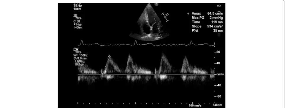
Acceleration rate of E wave of mitral inflow (AR)
As our new parameter acceleration rate and time of E was also measured in all patients. Acceleration rate of E (cm/sec2) was represented by the slope of the line be-tween an anchored point and a crosshair (Fig. 1). This linear measurement was made on the velocity spectrum. Acceleration time (AT) of E was measured from onset to peak of E These recordings were shown on a strip chart with a sweep speed of 100 mm/s to determine correct temporal observations. Measurements were performed off line by an independent observer who had no know-ledge of the Doppler or Tissue Doppler findings. At least three measurements were taken of each parameter and these were averaged.
Statistical analysis
SPSS release 21.0 statistical package was used for data analysis. All values were expressed as mean ± SD. Correl-ation between AR and AT of E wave and LV diastolic function grade was measured using the Spearman cor-relation coefficient. Multivariate Logistic Regression ana-lysis was also done to adjust the age and gender effect. Receiver operating characteristic (ROC) curve was used to determine the sensitivity and specificity of AR of E wave in diagnosing LV diastolic dysfunction and elevated left ventricle diastolic pressure (LVDP). Study population randomly assigned into two groups with 2:1 ratio. ROC curve was performed in randomly selected two-thirds of population (derivation group), then the derived cut-off was evaluated in the rest of the population (Test group). Therefore, ROC curve analysis was performed two times. First, we recoded the “diastolic function” into a dichotic variable just based on presence or ab-sence of any degree of diastolic dysfunction (mild, mod-erate, and severe). Second, we recoded the “diastolic function” into another dichotic variable, this time based
on presence or absence of elevated LVDP (moderate and severe diastolic dysfunction).
Result
The study population consisted of 84 adult patients (64 % men), mean age 53 ± 11(range, 20–70 years) and 34 nor-mal controls, who were referred for clinically indicated two-dimensional echocardiogram. They were referred be-cause of the following reasons: dyspnea/peripheral edema/ congestive heart failure (48 %), cerebrovascular accident (12 %), preoperative assessment (10 %), coronary artery disease (9 %), and others (21 %).
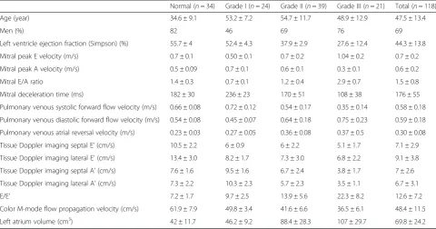
The baseline clinical and echocardiographic characteris-tics are listed in Table 1.
The mean value of E acceleration time and E acceler-ation rate were 85.6 ± 19.1 ms, and 1010 ± 420 cm/s2in case group respectively, whereas these mean values for the normal controls were 96.7 ± 15.1 ms, and 701 ± 210 cm/s2respectively. The mean values of AT and AR according to grade of LV diastolic dysfunction is showed in Table 2. There was a strong and graded relation be-tween AR of E wave of mitral inflow and LV diastolic function grade (Spearman P ≤0.0001, rs =0.69) (Fig. 2). The Logistic Regression Analysis analysis showed that AR of E could predict the diastolic dysfunction after adjust-ment for age and gender (P= 0.001). Receiver operating characteristic (ROC) curve analysis in “derivation group” revealed that AR of E wave of mitral inflow =655 cm/s2 predicted presence of LV diastolic dysfunction with 71 % sensitivity and 65 % specificity (area under curve [AUC] = 0. 0.727, P= 0.003). Application of this cutoff on test group showed 84 % sensitivity and 64 % specificity with AUC = 0.800 andP= 0.001. When considering only Grade II and III (moderate and severe) diastolic dysfunction, an AR of 750 cm/s2 predicted at least moderate diastolic dysfunction with 89 % sensitivity and 89 % specificity (area
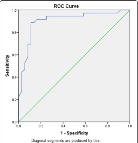
under curve 0.903,P<0.0001) (Fig. 3, Table 3). Application of this cutoff on test group showed 96 % sensitivity and 77 % specificity with AUC = 0.932 andP<0.0001. AR of E = 1250 cm/s2 was 100 % specific for the detection of elevated LVDP (moderate or severe diastolic dysfunc-tion) but with a low sensitivity of 22 %.
Discussion
The present study showed that AR of E wave of mitral inflow has a strong and graded relation to the LV dia-stolic function grade, and could be used especially to identify moderate or severe LV diastolic dysfunction. This result was predictable because according to Newton’s second law there is a direct relationship between acceler-ation and pressure. E wave acceleracceler-ation is directly deter-mined by LA pressure and inversely related to myocardial relaxation [9]. Thomas and weyman [10] demonstrated in a mathematical model of LV filling that AR is greatly af-fected by changes in left atrial pressure compared with peak velocity, peak deceleration, and the total integral of the inflow velocity.
Despite the theoretical observations by Thomas and Weyman [10], AR has not been used clinically for esti-mation of LA pressure. There are two studies which re-ported AT and AR as parameters which can be useful
for assessing left ventricular diastolic function in patients with diabetes or coronary artery diseases [11, 12]. There is also an experimental study on mice which reported AT of E wave was sensitive for detecting early stages of diastolic function, and appeared to add incremental value over that provided by the E/A ratio and IVRT for detecting later stages of diastolic dysfunction in murine models [13]. To our knowledge none of these studies reported a cut of value for AR of E wave to predict LV diastolic dysfunction. However in 1996 Nagueh et al. showed that peak AR≥1900 cm/s2had a 77 % sensitivity and 94 % specificity for left ventricle diastolic pressure (LVDP) >15 mmHg in patients with atrial fibrillatio-n(AF) [14]. In our study we showed that by simple measurement of AR of E of mitral valve we might be able to identify patients with elevated LVDP. This applica-tion could be clinically important and valuable because it might explain patients’symptoms in some conditions. The cut off value of 750 cm/s2 had a suitable sensitivity and specificity for elevated LVDP according to diastolic func-tion grade II or more. And the cut off value of 1250 cm/s2 had 100 % specificity for diastolic dysfunction of grade II or more. The difference in our cut off value and the previ-ous one could have three reasons. First, in our study we used AR which conventionally could be measured by Table 1Echocardiographic parameters of the study population
Normal (n= 34) Grade I (n= 24) Grade II (n= 39) Grade III (n= 21) Total (n= 118)
Age (year) 34.6 ± 9.1 53.2 ± 7.2 54.7 ± 11.7 48.9 ± 12.9 47.5 ± 13.4
Men (%) 82 46 69 76 69
Left ventricle ejection fraction (Simpson) (%) 55.7 ± 4 52.4 ± 4.3 37.9 ± 2.9 27.6 ± 12.4 44.3 ± 13.8
Mitral peak E velocity (m/s) 0.7 ± 0.1 0.50 ± 0.1 0.7 ± 0.2 1.04 ± 0.2 0.7 ± 0.2
Mitral peak A velocity (m/s) 0.5 ± 0.09 0.7 ± 0.1 0.6 ± 0.1 0.3 ± 0.1 0.6 ± 0.2
Mitral E/A ratio 1.4 ± 0.3 0.7 ± 0.1 1.2 ± 0.4 2.9 ± 0.7 1.5 ± 0.8
Mitral deceleration time (ms) 182 ± 30 236 ± 23 170 ± 51 108 ± 38 176 ± 55
Pulmonary venous systolic forward flow velocity (m/s) 0.66 ± 0.08 0.72 ± 0.12 0.54 ± 0.17 0.35 ± 0.14 0.58 ± 0.18
Pulmonary venous diastolic forward flow velocity (m/s) 0.54 ± 0.08 0.45 ± 0.07 0.64 ± 0.18 0.75 ± 0.23 0.59 ± 0.18
Pulmonary venous atrial reversal velocity (m/s) 0.23 ± 0.03 0.27 ± 0.05 0.36 ± 0.08 0.37 ± 0.5 0.30 ± 0.08
Tissue Doppler imaging septal E' (cm/s) 10.5 ± 2.2 6 ± 0.9 6 ± 2.2 5.1 ± 1.7 7.1 ± 2.9
Tissue Doppler imaging lateral E' (cm/s) 13.4 ± 3.0 8.2 ± 1.7 7.3 ± 3.0 6.8 ± 2.2 9.1 ± 3.8
Tissue Doppler imaging septal A' (cm/s) 7.6 ± 1.6 9.5 ± 1.6 6.7 ± 2.4 3.8 ± 1.7 7 ± 2.6
Tissue Doppler imaging lateral A' (cm/s) 7.3 ± 2.2 10.3 ± 2.3 5.7 ± 2.3 3.5 ± 1.1 6.7 ± 3.1
E/E' 7.2 ± 1.7 9.7 ± 2.5 13.9 ± 5.6 22.3 ± 8.2 12.6 ± 7.2
Color M-mode flow propagation velocity (cm/s) 61.9 ± 7.9 49.8 ± 3.4 41.6 ± 6.6 36.5 ± 6.1 48.4 ± 11.5
Left atrium volume (cm3
) 42 ± 11.7 46.2 ± 9.2 88.4 ± 28.3 107 ± 29.7 69.8 ± 24.2
Table 2Acceleration rate and time of E wave by diastolic function grade
Normal (n= 34) Grade I (n= 24) Grade II (n= 39) Grade III (n= 21) Total (n= 118)
Acceleration time (ms) 96.7 ± 15.1 95.7 ± 16.7 83 ± 18.9 79.1 ± 18.5 88.8 ± 18.7
every machine of echocardiography, but Naughueh et.al used a computer software to measure peak of AR. Second the method of estimation of elevated LVDP was different in two studies. In our study we postulated that presence of diastolic dysfunction of grade II or more would be associ-ated with elevassoci-ated LVDP, however Naughueh et al. directly measured LVDP or LA pressure in their patients. Third,
all of our patients had sinus rhythm, but the rhythm of patients in that study was AF.
Limitation
The limitations of our study include its relatively small size. The‘normal controls’are included on the basis of ab-sence of a history of cardiovascular disease and a normal resting two-dimensional echocardiogram. Stress tests were not performed to rule out occult coronary artery disease. We used previously published Doppler echocardiographic referenced standards to define the different grades of LV diastolic function. Cardiac catheterization was not per-formed to evaluate LV diastolic function. However, these reference standards were previously validated with cardiac catheterization and are widely accepted as standards for classification of LV diastolic function grade. Although the results of our study about AR are promising, before its clinical application external validation should be consid-ered. Further studies with invasive haemodynamic measure-ments are needed to show if AR measurement increase the
Fig. 2Relationship between acceleration rate of E wave of mitral inflow and LV diastolic function grade
Fig. 3Receiver operating characteristic curve for the detection of Moderate or Severe diastolic dysfunction using acceleration rate of E wave of mitral inflow (area under curve 0.903,P<0.0001)
Table 3Positive Predictive Value (PPV), Negative Predictive Value (NPV), sensitivity and specificity of acceleration rate of E to detect moderate or severe diastolic dysfunction
Moderate or severe diastolic dysfunction
No Yes
Acceleration rate of E <750 32 4 NPV = 89 %
≥750 4 32 PPV = 89 %
diagnostic accuracy of LV diastolic dysfunction and elevated LA pressure with respect to validated echo-Doppler parameters.
Conclusion
AR of EARE wave of mitral inflow could be used for as-sessment of diastolic function, especially moderate to se-vere diastolic dysfunction. The cutoff value of 750 cm/s2 could deserve as suitable cutoff point with 89 % sensitiv-ity and 89 % specificsensitiv-ity in detection of moderate to se-vere diastolic dysfunction. Before its clinical application external validation should be considered.
Acknowledgements
Authors’contributions
Roya Sattarzadeh was responsible for idea formation, study designing, performing echocardiography and reviewing the article. Anahita Tavoosi contributed in idea formation, study designing, performig
echocardiography,drafting and reviewing the article. Mostafa Jabbari was responsible for data gathering and helped in analysis and drafting the article. Amir Farhang Zand Parsa, Babak Geraiely and Mehrdad Salehi were responsible for critical review and editing the manuscript. Mohammad Saadat and Farnoosh Larti had significant contribution in revision of the article by performing requested analysis and rewriting some parts of the manuscript. Alipasha Meysami contributed by consultation about study designing, and analysis of the data. All authors read and approved the final manuscript.
Competing interests
We also confirm that this study complied with the principles of the declaration of Helsinki. Study protocol was approved by Local Ethics Committee of Tehran University of Medical Sciences. Informed consent was obtained from all physicians participated in this study. No conflict of interest existed for this study. No fund or grant was received to support this study.
Author details
1Cardiology Department of Imam Khomeini Hospital, Tehran University of Medical Sciences, Tehran, Iran.2Department of community medicine, Tehran University of Medical Sciences, Tehran, Iran.
Received: 21 January 2016 Accepted: 4 June 2016
References
1. Nishimura RA, Jaber W. Understanding“diastolic heart failure”: the tip of the iceberg. J Am Coll Cardiol. 2007;49(6):695–7.
2. Dokainish H, Nguyen JS, Bobek J, Goswami R, Lakkis NM. Assessment of the American Society of Echocardiography-European Association of Echocardiography guidelines for diastolic function in patients with depressed ejection fraction: an echocardiographic and invasive haemodynamic study. Eur J Echocardiogr. 2011;12(11):857–64. 3. Little WC, Oh JK. Echocardiographic evaluation of diastolic function can be
used to guide clinical care. Circulation. 2009;120(9):802–9.
4. Appleton CP, Hatle LK, Popp RL. Relation of transmitral flow velocity patterns to left ventricular diastolic function: new insights from a combined hemodynamic and Doppler echocardiographic study. J Am Coll Cardiol. 1988;12(2):426–40. 5. Tabata T, Thomas JD, Klein AL. Pulmonary venous flow by doppler
echocardiography: revisited 12 years later. J Am Coll Cardiol. 2003;41(8):1243–50. 6. Nagueh SF, Appleton CP, Gillebert TC, Marino PN, Oh JK, Smiseth OA, et al.
Recommendations for the evaluation of left ventricular diastolic function by echocardiography. J Am Soc Echocardiogr. 2009;22(2):107–33.
7. Lang RM, Bierig M, Devereux RB, Flachskampf FA, Foster E, Pellikka PA, et al. Recommendations for chamber quantification: a report from the American Society of Echocardiography’s Guidelines and Standards Committee and the Chamber Quantification Writing Group, developed in conjunction with the European Association of Echocardiography, a branch of the European Society of Cardiology. J Am Soc Echocardiogr. 2005;18(12):1440–63.
8. Lang RM, Badano LP, Mor-Avi V, Afilalo J, Armstrong A, Ernande L, et al. Recommendations for cardiac chamber quantification by echocardiography in adults: an update from the American Society of Echocardiography and the European Association of Cardiovascular Imaging. J Am Soc Echocardiogr. 2015;28(1):1–39. e14.
9. Ohno M, Cheng CP, Little WC. Mechanism of altered patterns of left ventricular filling during the development of congestive heart failure. Circulation. 1994; 89(5):2241–50.
10. Thomas JD, Weyman AE. Echocardiographic Doppler evaluation of left ventricular diastolic function. Physics and physiology. Circulation. 1991;84(3):977–90. 11. Bajraktari G, Qirko S, Fusco R, Milazzo A, Xhaxho B, Pezzano A. Transmitral
pulsed-Doppler echocardiography is a more accurate technique compared with two-dimensional echocardiography using dobutamine, in patients with one vessel coronary artery disease. Eur J Heart Fail. 2003;5(1):63–72. 12. Riordan MM, Chung CS, Kovacs SJ. Diabetes and diastolic function: stiffness
and relaxation from transmitral flow. Ultrasound Med Biol. 2005;31(12):1589–96. 13. Yuan L, Wang T, Liu F, Cohen ED, Patel VV. An evaluation of transmitral and
pulmonary venous Doppler indices for assessing murine left ventricular diastolic function. J Am Soc Echocardiogr. 2010;23(8):887–97. Pubmed Central PMCID: 2910830.
14. Nagueh SF, Kopelen HA, Quinones MA. Assessment of left ventricular filling pressures by Doppler in the presence of atrial fibrillation. Circulation. 1996; 94(9):2138–45.
• We accept pre-submission inquiries
• Our selector tool helps you to find the most relevant journal • We provide round the clock customer support
• Convenient online submission • Thorough peer review
• Inclusion in PubMed and all major indexing services • Maximum visibility for your research
Submit your manuscript at www.biomedcentral.com/submit