Cite this article Nadaroğlu H, Dikbaş N. Purification and Characterization of A -Mannanase from Lactobacillus plantarum (ATCC® 14917TM). International Journal of Innovative Research and Reviews (INJIRR) (2018) 2(1):1-6
Link to this article: http://www.injirr.com/article/view/14 Copyright © 2017 Authors.
International Journal of Innovative Research and Reviews
ISSN: 2636-8919Website: www.injirr.com doi:
Research paper, Short communication, Review, Technical paper
RE S E A R C H AR T I C L E
Purification and Characterization of A
-Mannanase from
Lactobacillus
plantarum
(ATCC® 14917TM)
Hayrunnisa NADAROĞLU
1,2*, Neslihan DİKBAŞ
31 Ataturk University, Erzurum Vocational Training School, Department of Food Technology, 25240 Erzurum, Turkey 2 Ataturk University, Faculty of Engineering, Department of Nano-Science and Nano-Engineering, 25240 Erzurum, Turkey
3 Ataturk University, Faculty of Agriculture, Department of Biotechnology, Erzurum, Turkey
*Corresponding author E-mail: [email protected] HI G H L I G H T S
Mannanase was purified alkaline thermo Lactobacillus plantarum (ATCC® 14917TM).
Mannanase enzyme was purified using ammonium sulfate precipitation and DEAE-Cellulose ion exchange chromatog-raphy and Sephacryl S200.
All the metal ions were increased the enzyme activity from 100 to 344 %.
AR T I C L E IN F O
Received : 06.20.2018 Accepted : 07.01.2018 Published : 07.15.2018
Keywords: Mannanase,
Lactobacillus plantarum (ATCC® 14917TM) Purification Clarification
AB S T R A C T
Mannanase alkaline was purified from thermo Lactobacillus plantarum (ATCC® 14917TM) using ammonium sulfate precipitation and DEAE- cellulose ion exchange chromatography and Sephacryl S200 Gel filtration chromatography techniques, in 36% yield and 111 -fold. Gel filtration chromatography and SDS-PAGE electrophoresis were executed pure enzyme systems 35 kDa and 55 kDa. Enzyme subunits were determined as two subunits. Optimum pH 10 and 40 °C was determeined at the optimum temperature. Enzyme activities were retained fairly between 30-80 oC. It was observed that purified mannanase in a wide range was mostly stable at elevated temperatures. In addition, the effects of some metal ions such as Ca2+, Mn2+, Co2+, Zn2+, Cu2+, Fe2+ and Ni2+ on mannanase enzymes activity was assayed and all the metal ions were increased the enzyme activity from 100 to 344 %.
Contents
1. Introduction ... 2
2. Materials and methods ... 2
2.1. Characterization of Lactic acid Bacteria (LAB) ... 2
2.2. Purification of mannanase from L. plantarum ... 2
2.2.1. Anion exchange chromatography ... 2
2.2.2. Gel filtration ... 2
2.3. Protein concentration ... 2
2.4. Determination of mannanase activity ... 2
2.5. SDS polyacrylamide gel electrophoresis ... 3
2.6. Molecular weight determination by gel filtration ... 3
2.7. Studies on certain in vitro chemicals and metal ions ... 3
2.8. Statistical analysis ... 3
3. Results and Discussion ... 3
3.1. Purification and characterization of mannanase from L. plantarum ... 3
3.2. Optimum temperature ... 4
3.3. The results of Vmax and Km values ... 5
3.4. Effects of some metal ions on the activity of the mannanase enzyme purified from L. plantarum ... 5
4. Conclusion ... 5
1.
Introduction
β-Mannanases are extracellular enzymes hydrolyzing the 1,4-n beta-D-mannosidic linkages in mannans, galactoman-nans, glucomannans and galactoglucomannans. Mannans and widely heteromannans hard and soft woods, legumes and beans as part of the hemicellulose fraction is distributed in nature. Hemicelluloses are copolymers of both hexose and pentose sugars. The branched structure allows hemicel-lulose to exist in an amorphous form that is more suscepti-ble to hydrolysis.
The demands for microbial enzymes for industrial applica-tion have increased in the search of Microorganisms with potential and adoption of biotechnological methods of en-hancing enzyme production [1]. Biotechnology research into microbial enzymes has been driven by the need to iso-late and identify organisms which are either hyper-producer and/or sufficiently robust to with stand conditions of the intended application and/or are producers of novel extra cellular enzymes. Therefore, importance is placed on indus-trial enzymes of microbial origin with catalytic efficiency that is relatively stable under wide pH range, high tempera-ture, and salts concentration with a view of producing in-dustrial enzymes of desirable physicochemical properties [2].
In recent years, mannanases (1,4-β-D-mannanmanno hydro-lase, EC 3.2.1.78) have gained increasing attention because of their various biotechnological applications in the food, feed, coffee extraction, oil drilling, detergent, as well as pulp and paper industries [3, 4]. They can also be used in the production of mannooligosaccharides, which were re-ported to be excellent prebiotics stimulating growth of ben-eficial intestinal microorganisms [5, 6]. Many microorgan-isms have been reported as mannanase producers. Microbi-al species known to actively produce mannanases include
Bacillus subtilis [7, 8],Aeromonas hydrophila [9], Entero-coccus casseliflavus [10], Pseudomonas sp. [11], Vibrio sp. [12], Streptomyces sp. [13], fungi [14], and yeast [15]. Mannanases are also distributed in higher plants [16] and animals [11, 17]. Although it is presumed that the hemicel-luloses are degraded both aerobically and anaerobically in natural environments, there are no reports available con-cerning mannanase production or -1,4- mannan-degradation by anaerobic bacteria in natural environments.
In this experiment; under optimum conditions of tempera-ture, pH and metal resistance, L. plantarum which produce mannanase enzyme was obtained purified and characterized using ion exchange and gel filtration chromatographic tech-niques.
2.
Materials and methods
2.1. Characterization of Lactic acid Bacteria (LAB)
Lactic acid bacteria used in the study, strain L. plantarum
(ATCC® 14917TM), has been obtained from Sigma-Aldrich. Our bacteria have been transferred to MRS broth. L. planta-rum bacteria wereincubated in liquid medium at pH 5-7, at 30-35 °C range for 48 hours. After 7 days of incubation, mannanase enzyme activity was determined.
Subsequent to the filtering process, the crude enzyme ex-tract was centrifuged at 5000 rpm for 15 min and the en-zyme homogenate ammonium sulfate was precipitated at 0-20% 20-40% 40-60% 60-80% and 80-100% intervals, re-spectively. Following the filtration, raw enzyme extract was centrifuged at 5000 rpm for 15 min.
The obtained supernatant was saturated up to 40-80%, us-ing ammonium sulphate. Thereafter, the precipitate was dissolved in 20 mM sodium citrate (pH: 6.0), and it was dialyzed against the same buffer.
2.2.1. Anion exchange chromatography
Suspension dialyzed ammonium sulfate, DEAE-cellulose ion exchange column (2.5 x 30 cm) was performed after it was previously equilibrated with 20 mM sodium citrate (pH: 6.0). Column was washed with the same buffer. Af-terwards, the proteins attached to the column were eluted by applying NaCl gradient from 0 up to 1 M. The fractions were collected as 3 mL, with a 3 mL/min flow rate. Protein elution absorbance was measured at 280 nm as spectropho-tometrically. Hyrolysis activities of locust bean gum were determined for all enzyme fractions. Active fractions were combined and it was allowed to stand at 4 °C.
2.2.2. Gel filtration
By using an Amicon membrane concentrator with a 10 kDa cut off, the active fractions which were obtained from anion exchange column were combined, dialyzed, desalted and concentrated, respectively. The aforementioned method, obtained by enzyme solution, previously equilibrated with 20 mM phosphate buffer (pH 5.0) to the Sephacryl S-200 column (120 x 1 cm), then the enzyme was eluted using 0.5 M of same NaCl-containing buffer. After all the tions had been analyzed as described above the active frac-tions were combined, concentrated, they were allowed to stand at 4 °C for later use.
2.3. Protein concentration
By the method of Bradford [18], the protein concentration, as mentioned previously, are determined spectrophotomet-rically. In this method, bovine serum albumin (BSA) was used as a standard.
2.4. Determination of mannanase activity
The mannanase activity was principally determined by us-ing a locust bean gum as a substrate [19]. The mixture of the reaction was formed by adding a 0.1 mL enzyme solu-tion onto the locust bean gum substrate dissolved with 0.9 ml of 20 mM sodium citrate buffer (pH: 6.0).
After this process, it was incubated at 50 C, in which the mannanase activity was determined through monitoring the absorbance at 540 nm by adding in the increased an average of DNSA (3,5-dinitrosalisilic acid) [20].
plantarum (ATCC® 14917TM)
2.5. SDS polyacrylamide gel electrophoresis
Sodium dodecyl sulfate-polyacrylamide gel electrophoresis (SDS-PAGE) method was identified by Laemmli [21], 10-30% discontinued, was realized.
20 g protein sample, each being disposed into a gel cast-ing apparatus (Bio-Rad) was applied to buffer (0.25 M Tris, 1.92 M glycine, 1% SDS, pH the electrophoresis process: 8.3) at 4 °C. Gel were stained in a 50% methanol, 10% ace-tic acid and 40% distilled water mixture for 1.5 hours, until it was clear. Afterwards, electrophoretic pattern was photo-graphed.
2.6. Molecular weight determination by gel filtration
Sephadex G100 column (3.0 x 70 cm) was used for deter-mination of the molecular weight of mannanase enzyme. Column was balanced using 0.05 M Na2HPO4, 1 mM dithi-oerythretol pH 7.0 buffer.
Standard protein solution (bovine serum albumin, 66 kDa; egg ovalbumin, 45 kDa; pepsin, 34 kDa; trypsinogen, 24 kDa; ß-lactoglobulin and lysozyme, 14 kDa) was applied to the column and eluted from the column with the same buff-er. Afterwards, the pure mannanase enzyme was applied to the column and eluted under same conditions. The flow-rate through the column was 20 mL/h. The elution volume was compared with those of standard proteins [22].
2.7. Studies on certain in vitro chemicals and metal ions
Effect of different concentrations of inhibitors (Mn2+, Co2+, Cu2+, Zn2+, Fe2+, Ca2+ and Ni 2+) on mannanase enzyme ac-tivity was investigated. All the metal ion concentrations were tested for each of the three runs. Mannanase inhibi-tor/activator activity was measured at different concentra-tions.
2.8. Statistical analysis
For all testing samples, mannanase activity was determined at three different runs. Data were expressed as mean ± standard error. Using SPSS 20.0 software (SPSS Inc, Chi-cago, IL., USA), statistical analysis had been performed. Significant differences were determined as 95% confidence intervals (p <0.05) using Tukey’s test.
3.
Results and Discussion
3.1. Purification and characterization of mannanase from L. plantarum
The mannanase enzyme manufactured extracellularly from the bacteria called L. plantarum, was refined in 3 steps as ammonium sulfate precipitation, DEAE-cellulose and Se-phacryl S200 gel filtration chromatography. All the purifi-cation steps of the mannanase enzyme are shown in Table 1.
In the first stage, mannanase enzyme was collapsed the ammonium sulfate saturation between the range 0-90%.
74.5% yield was achieved in this first purification step (Table 1).
In the second step, sample was applied to the DEAE-cellulose ion exchange column, enzyme rose to 31.5% and a yield 69 times greater purified-fold.
In the last step, obtained fractions from ion exchange col-umn were concentrated, then it was applied to a Sephacryl S 200 column. A single peak was obtained from this col-umn, and 110.6 purification-fold and 36% yield. 22 EU / g protein, specific activity (Table 1) was calculated from re-sults.
Table 1 The purification process of purified β -mannanase enzyme from L. plantarum Enzyme Fraction Crude extract (NH4)2SO4 (40-80%) DEAE-Cellulose Sephacryl S200 Volume
(mL) 50 48 20 20
Activity
(EU/mL) 41.6±1.3 31.5±1.3 15.2±0.32 7.78±2.5 Total
Activity
EU 2.08x10³ 1.51 x10³ 3.04 x10² 1.56 x10²
% 100 72.7 20.1 51.2
Protein
g protein/mL) 152.8±1.8 3.88±0.38 0.92±0.11 0. 18±0.4 Specific
Activity
(EU/g protein) 0.272 8.12 16.52 43.2
Purification
Fold - 29.85 60.74 158.8
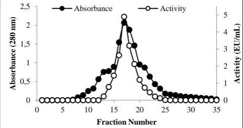
Mannanases from bacteria elution profiles of anion ex-change chromatography and gel filtration was purified by chromatography, L. plantarum, is presented in Figure 1 and Figure 2.
Figure 1 Purification of β-mannanase enzyme from L. plantarum by ion exchange chromatography using DEAE-Cellulose.
0 2 4 6 0 0,2 0,4 0,6 0,8 1
0 4 8 12 16 20 24 28 32
A ct iv it y ( EU /m L) A b so rb a n ce ( 2 8 0 n m ) Fraction Number Absorbance Activity 0 1 2 3 4 5 0 0,5 1 1,5 2 2,5
0 5 10 15 20 25 30 35
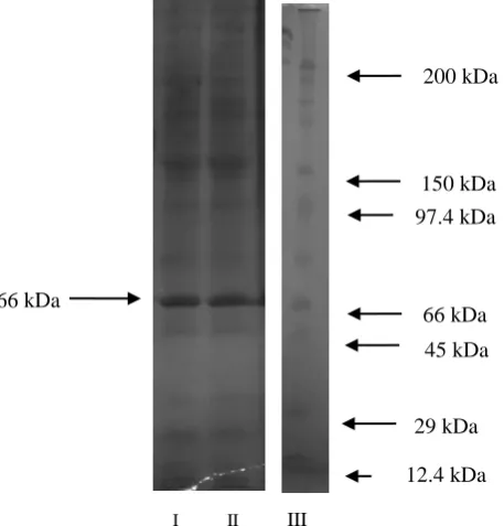
(SDS-PAGE) results of the purified mannanase enzyme were observed to have consisted of two subunits as 35 kDa and 55 kDa (Figure 3). To find the molecular weights of the active form of the enzyme, the Sephadex G-100 gel filtra-tion chromatography was applied and the graph was drawn as log MA-Kav.
Figure 3 SDS-PAGE electrophoretic pattern of peroxidase [standart pro-tein (β-Amylase, sweet potato, 200 kDa; alcohol dehydrogenase, yeast, 150 kDa bovine serum albumin, 97.4 kDa; rabbit muscle phosphorylase A, 66 kDa; egg ovalbumin, 45 kDa; pepsin, 29 kDa; carbonic anhydrase); cytochrome c, horse heart 12.4 kDa (I); purified laccase enzyme from rocket peroxidase (II)].
In this study, the amplified beta-mannanase gene was placed from Aspergillus aculeatus to Aspergillus oryzae
and the molecular weight of the resultant recombinant en-zyme was found to be 45 kDa [23]. Also, molecular weight of the mannanase enzyme of Aspergillus niger was ob-tained as 40 kDa [24].
Activity measurements were carried out using locust bean gum as a substrate, and the results were also calculated. Results were displayed using Activity - pH graph in Figure 4. The optimal pH values of the purified mannanase en-zyme from L. plantarum (ATCC® 14917TM), was deter-mined to be 5.0 for the substrate locust bean gum.
The mannanase enzyme from L. plantarum was produced, purified and the pH optimum was found in range of pH 2 to 9. From pH 2 to pH 5, acetate buffer, from pH 6 to pH 8 phosphate buffer, and from pH 8 to pH 9 Tris / HCl buffer was used. Optimum pH was determined as pH 7.
Activity measurements, locust bean gum were used as a substrate, were calculated from the results obtained and were also plotted Activity - pH and the results were given in Figure 4. P. plantarum produced a purified mannanase enzyme and the optimal pH value was determined to be 5.0.
Figure 4 Effect of pH on the activity of the purified β -mannanase of L. acidophilus.
Enzymes and substrate were dissolved either in 10 mM buffers of various pH. Other conditions were as given for the standard assay method. Also, the pH stability was de-termined and related results were given in Figure 5.
Figure 5 Effect of pH on the activity and stability of the purified purified β-mannanase of L. plantarum
3.2. Optimum temperature
To determine the optimum temperature of the purified mannanase enzyme in the range of 20-90 °C, an activity measurement was applied by keeping for 5 minutes at each temperature using the locust bean gum as a substrate. As seen in Figure 5, the mannanase enzyme was found to be quite active between 30-80 oC and exhibited the maximum activity at 60 °C (Figure 6). Similarly, the optimum pH results of the beta-mannanase enzymes were belong to As-pergillus aculeatus [23]. Aspergillus niger’s mannanase optimal pH were determined to be 5.0, and the optimal temperature was also determined to be 60-70 °C [25].
Figure 6 Effect of temperatures on the activity of purified β -mannanase of
L. plantarum 0 30 60 90 120
2 5 8 11
R
ela
tiv
e
Act
iv
it
y
(
%)
pH
0 0,04 0,08 0,12 0,16
2 4 6 8 10 12
A
ct
iv
it
y
(
E
U
/m
L
)
pH
0,5 1 2
3 4 5
0 30 60 90 120
0 20 40 60 80 100
R
el
a
ti
v
e
a
ct
iv
it
y
(
%
)
Temperature (oC) I II III
66 kDa
66 kDa
29 kDa
12.4 kDa 45 kDa 150 kDa
200 kDa
plantarum (ATCC® 14917TM)
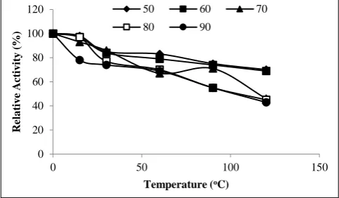
Activity was determined at different temperatures and at pH 8.0 using the standard assay method. Also, the thermal sta-bility was determined and related results were given in Fig-ure 7.
Figure 7 Effect of temperature on the activity and stability of purified β-mannanase of L. plantarum
3.3. The results of Vmax and Km values
Vmax and Km values of mannanase enzyme for 0.3 mM, locust bean gum was observed as 78.74 mol/min.mg and Km 0.592 mM, respectively.
3.4. Effects of some metal ions on the activity of the mannanase enzyme purified from L. plantarum
The effects of the metal ions, such as Mn2+, Co2+, Cu2+, Zn2+, Fe2+, Ca2+ ve Ni2+ on the activity of the mannanase enzyme purified from L. plantarum (ATCC 14917) were investigated.
The inhibition/activation effects obtained as the result of the studies and the activity values versus concentration are shown in Table 2. As seen in Table 2, all the metal ions enhanced the activity of the purified mannanase enzyme from L. palantarum. However, the highest activation was done by Co2+ and Fe2+ metal ions, which enhanced the mannanase activity by 375.3 % and 372.8 %, respectively. From the results, it was observed that not all the metal ions inhibited the mannanase enzyme purified from L. planta-rum (ATCC 14917) but that they even activated it, and due to such characteristic, it was considered to be quite conven-ient for industrial practices of this purified mannanase. It was determined that almost all of the metal ions increased the activity of purified mannanase enzyme from L. palanta-rum [26–29].
Table 2 The effect of some chemical compounds on β-mannanase activity.
4.
Conclusion
It was concluded from the acquired findings that the cata-lytic activities of the mannanase enzyme purified from L. plantarum were rather high, and that they were resistant to the metal ions and were highly stable against different pHs and temperatures.
References
[1] Rattanasuk S, Prasertsang K, Phiwphech S. Isolation of thermophilic mannanase-producing bacteria useful for mannooligosaccharide (MOS) production. In: 2015 International Conference on Science and Technology (TICST). p. 173–177.
[2] Mabrouk MEM, El Ahwany AMD. Production of β-mannanase by Bacillus amylolequifaciens10A1 cultured on potatopeels. African Journal of Biotechnology (2008) 7(8):1123–1128.
doi:10.5897/AJB08.047.
[3] Naganagouda K, Salimath PV, Mulimani VH. Purification and Characterization of Endo-beta-1,4 Mannanase from Aspergillus ni-ger gr for Application in Food Processing Industry. Journal of Mi-crobiology and Biotechnology (2009) 19(10):1184–1190. doi:10.4014/jmb.0901.029.
[4] Lin TC, Chen CS. Enhanced mannanase production by submerged culture of Aspergillus niger NCH-189 using defatted copra based media. Process Biochemistry (2004) 39(9):1103–1109. doi:10.1016/S0032-9592(03)00218-8.
[5] Titapoka S, Keawsompong S, Haltrich D, Nitisinprasert S. Selection and characterization of mannanase-producing bacteria useful for the formation of prebiotic manno-oligosaccharides from copra meal. World Journal of Microbiology & Biotechnology (2008) 24(8):1425–1433. doi:10.1007/s11274-007-9627-9.
[6] Gibson GR, Rastall ReA. Prebiotics: development and application. Chichester: John Wiley & Sons; 2006 (2006).
[7] Araujo A, Ward OP. Hemicellulases of Bacillus Species - Prelimi-nary Comparative-Studies on Production and Properties of Man-nanases and Galactanases. Journal of Applied Bacteriology (1990) 68(3):253–261. doi:10.1111/j.1365-2672.1990.tb02572.x. [8] Emi S, Yamamoto T, Fukumoto J. Studies of Hemicellulolytic
En-zymes of Bacillus-Subtilis .2. Crystallization and Some Properties of Mannanase. Agricultural and Biological Chemistry (1972) 36(6):991-&. doi:10.1080/00021369.1972.10860352.
[9] Ratto M, Poutanen K. Production of Mannan-Degrading Enzymes. Biotechnology Letters (1988) 10(9):661–664.
doi:10.1007/Bf01024721.
[10] Oda Y, Tonomura K. Characterization of β‐mannanase and β‐ mannosidase secreted from the yeast Trichosporon cutaneum JCM 2947 (2008). 173-178.
[11] Yamaura I, Nozaki Y, Matsumoto T, Kato T. Purification and some properties of an endo-1,4-beta-D-mannanase from a marine mollusc, Littorina brevicula. Bioscience Biotechnology and Biochemistry (1996) 60(4):674–676. doi:10.1271/bbb.60.674.
[12] Tamaru Y, Araki T, Amagoi H, Mori H, Morishita T. Purification and Characterization of an Extracellular Beta-1,4-Mannanase from a Marine Bacterium, Vibrio Sp Strain Ma-138. Applied and Environ-mental Microbiology (1995) 61(12):4454–4458.
[13] Takahashi R, Kusakabe I, Kobayashi H, Murakami K, Maekawa A, Suzuki T. Studies on Mannanase of Streptomyces .3. Purification and Some Properties of Mannanase from Streptomyces Sp. Agricul-tural and Biological Chemistry (1984) 48(9):2189–2195. doi:10.1080/00021369.1984.10866483.
[14] Soni H, Rawat HK, Pletschke BI, Kango N. Purification and charac-terization of beta-mannanase from Aspergillus terreus and its ap-plicability in depolymerization of mannans and saccharification of lignocellulosic biomass. 3 Biotech (2016) 6. doi:10.1007/s13205-016-0454-2.
[15] Oda Y, Komaki T, Tonomura K. Production of Beta-Mannanase and Beta-Mannosidase by Enterococcus-Casseliflavus Fl2121 Isolated from Decayed Konjac. Food Microbiology (1993) 10(4):353–358. doi:10.1006/fmic.1993.1041.
[16] Zahura UA, Rahman MM, Inoue A, Tanaka H, Ojima T. cDNA cloning and bacterial expression of an endo-beta-1,4-mannanase, 0
20 40 60 80 100 120
0 50 100 150
R
el
a
ti
v
e
A
ct
iv
it
y
(
%
)
Temperature (oC)
50 60 70
80 90
Chemical Compound
Concentration (mM)
Relative Activity (%)
Concentration (mM)
Relative Activity (%)
None - 100 ± 0.0 - 100 ± 0.0
Ca2+ 1 158.1 ± 0.62 5 136.8 ± 0.44
Mn2+ 1 213.4 ± 1.5 5 330.1 ± 1.7
Co2+ 1 326.5 ± 0.45 5 375.32 ± 2.1
Zn2+ 1 223.2 ± 1.3 5 180.7 ± 0.11
Cu2+ 1 210.0 ± 0.66 5 296.4 ± 0.81
Fe2+ 1 203.1 ± 1.7 5 372.8 ± 0.5
Some Properties of Endo-1,4-Beta-D-Mannanase from Pseudomo-nas Sp Pt-5. Agricultural and Biological Chemistry (1990) 54(9):2425–2427. doi:10.1080/00021369.1990.10870308. [18] Bradford MM. Rapid and Sensitive Method for Quantitation of
Microgram Quantities of Protein Utilizing Principle of Protein-Dye Binding. Analytical Biochemistry (1976) 72(1-2):248–254. doi:10.1006/abio.1976.9999.
[19] Miller GL. Use of Dinitrosalicylic Acid Reagent for Determination of Reducing Sugar. Analytical Chemistry (1959) 31:426–428. [20] Bailey MJ, Biely P, Poutanen K. Interlaboratory Testing of Methods
for Assay of Xylanase Activity. Journal of Biotechnology (1992) 23(3):257–270. doi:10.1016/0168-1656(92)90074-J.
[21] Laemmli UK. Cleavage of structural proteins during the assembly of the head of bacteriophage. Nature (1970) 22:680–685.
[22] Whitaker DR. Criteria for characterizing cellulases, In E. T Reese [ed.], Advances in enzymic hydrolysis of cellulose and related mate-rials. Pergamon Press, New York (1963):51–70.
[23] Setati ME, Ademark P, van Zyl WH, Hahn-Hagerdal B, Stalbrand H. Expression of the Aspergillus aculeatus endo-beta-1,4-mannanase encoding gene (man1) in Saccharomyces cerevisiae and characteri-zation of the recombinant enzyme. Protein Expression and Purifica-tion (2001) 21(1):105–114. doi:10.1006/prep.2000.1371.
[24] Ademark P, Lundqvist J, Hagglund P, Tenkanen M, Torto N, Tjer-neld F, et al. Hydrolytic properties of a beta-mannosidase purified from Aspergillus niger. Journal of Biotechnology (1999) 75 (2-3):281–289. doi:10.1016/S0168-1656(99)00172-8.
[25] Ademark P, Varga A, Medve J, Harjunpää V, Drakenberg T, Tjer-neld F, et al. Softwood hemicellulose-degrading enzymes from As-pergillus niger: purification and properties of a beta-mannanase. Journal of Biotechnology (1998) 63(3):199–210.
[26] Zhao SM, Xu W, Jiang WQ, Yu W, Lin Y, Zhang TF, et al. Regula-tion of Cellular Metabolism by Protein Lysine AcetylaRegula-tion. Science (2010) 327(5968):1000–1004. doi:10.1126/science.1179689. [27] Nadaroglu H, Adiguzel G, Adiguzel A, Sonmez Z. A
thermostable-endo-beta-(1,4)-mannanase from Pediococcus acidilactici (M17): purification, characterization and its application in fruit juice clarifi-cation. European Food Research and Technology (2017) 243(2):193–201. doi:10.1007/s00217-016-2735-8.
[28] Nadaroglu H, Adiguzel A, Adiguzel G. Purification and characteri-sation of beta-mannanase from Lactobacillus plantarum (M24) and its applications in some fruit juices. International Journal of Food Science and Technology (2015) 50(5):1158–1165.
doi:10.1111/ijfs.12739.