organic papers
Acta Cryst.(2005). E61, o2553–o2555 doi:10.1107/S1600536805022221 Athimoolamet al. C
6H6NO2+H2PO4
o2553
Acta Crystallographica Section E Structure Reports
Online
ISSN 1600-5368
Nicotinium dihydrogenphosphate
S. Athimoolam, K. Anitha and R. K. Rajaram*
Department of Physics, Madurai Kamaraj University, Madurai 625 021, India
Correspondence e-mail: [email protected]
Key indicators
Single-crystal X-ray study T= 293 K
Mean(C–C) = 0.003 A˚ Rfactor = 0.030 wRfactor = 0.084 Data-to-parameter ratio = 9.7
For details of how these key indicators were automatically derived from the article, see http://journals.iucr.org/e.
#2005 International Union of Crystallography
Printed in Great Britain – all rights reserved
In the title complex, C6H6NO2 +
H2PO4
, the nicotinium cations are sandwiched between layers of dihydrogen-phosphate anions. The structure is held together by extensive hydrogen bonding between the O and N atoms of nicotinium cations and the O atoms of the phosphate anions, and between the O atoms of the phosphate anions themselves. The nicotinium cations are packed in layers, with an interlayer distance of 3.116 (5) A˚ .
Comment
Nicotinic acid, a B vitamin also known as niacin, and its associated complexes have a variety of pharmacological properties. Principal among these is the lowering of serum cholesterol levels (Brutts & Lundholm, 1971). Niacin forms coordination complexes with Sn which have been found to have better antitumour activity than the well knowncis-platin or doxorubicin (Gielen et al., 1992). In addition, the enzyme nicotinic acid mononucleotide adenylyltransferase is essential for the synthesis of nicotinamide adenine dinucleotide in all living cells and is a potential target for antibiotics (Kimet al., 2004). The crystal structures of nicotinic acid (Wright & King, 1953; Kutoglu & Scheringer, 1983), isonicotinic acid (Taku-sagawa & Shimada, 1976), isonicotinic acid hydrazide (Bhatet al., 1974), 6-aminonicotinic acid hydrochloride (Giantsidis & Turnbull, 2000), 2-(methylsulfanyl)nicotinic acid (Basavojuet al., 2005), nicotinamide (Wright & King, 1954), 1-methyl nicotinamide iodide, chloride and picrate (Freeman & Bugg, 1974), and dinicotinamidium squarate (Bulutet al., 2003) have been reported previously. The crystal structure of nicotinic acid complexed with the protein Leghaemoglobin has also been studied (Ellis et al., 1997). The structure of the title compound, (I), is reported in the present investigation as part of a programme of work on pharmaceutical compounds.
The title compound (I), also known as 3-carboxypyridinium dihydrogenphosphate, has a nicotinium cation and a dihy-drogenphosphate anion in the asymmetric unit of the mono-clinic unit cell. One of the H atoms in the inorganic acid is transferred to the N atom of the nicotinic acid. This proton-ation is well confirmed by the C—N bond distances and C— N—C bond angle. The pyridinium ring is planar, with a mean
deviation of 0.0039 A˚ . The planar carboxyl group is twisted from the pyridinium plane, with a dihedral angle of 21.7 (2). The deviation from the pyridinium plane is slightly higher for atom O1B [0.427 (3) A˚ ] than for atom O1A [0.401 (3) A˚ ], probably as a result of hydrogen bonding, as in 2-aminonicotinic acid (Dobson & Gerkin, 1997). The C—O bond distances and C—C—O bond angles confirm that the H atom is connected to atom O1B(Table 1).
The P—O distances in the phosphate anions are as expected, with average single- and double-bonded P—O distances of 1.55 and 1.49 A˚ , respectively (Blessing, 1986). The lengthening of the P–O double bonds is due to the formation of strong O—H O hydrogen bonds. The tetrahedral geometry of the dihydrogenphosphate anion has also suffered due to the hydrogen-bonding interactions. All four phosphate O atoms are involved in hydrogen bonding, with two as donors and two as acceptors. The doubly-bonded O atoms of the anion act as acceptors in hydrogen bonds to both O and N donor atoms (Table 2). Two dihydrogenphosphate anions are linked by inversion-related O1—H1P O3 hydrogen bonds, while a glide-related hydrogen-bonded chain of O2P— H2P O3 bonds runs along the c axis, generating double chains along c. The cations are linked to these anion chains by
O1—H1B O4 hydrogen bonds and the classical hydrogen-bonding network is completed by N1—H1 O4 interactions. These serve to pack the nicotinium cations into layers sepa-rated by 3.116 (5) A˚ but not linked by any hydrogen bonding (Fig. 2).
Experimental
The title compound was crystallized from a mixture of nicotinic acid and orthophosphoric acid in a stoichiometric ratio of 1:1 at room temperature by slow evaporation from a solution in water.
Crystal data
C6H6NO2+H2PO4
Mr= 221.10
Monoclinic, P21=c
a= 12.158 (5) A˚ b= 10.593 (4) A˚ c= 7.001 (3) A˚
= 102.35 (3)
V= 880.8 (6) A˚3
Z= 4
Dx= 1.667 Mg m
3
Dm= 1.652 Mg m
3
Dmmeasured by flotation in a
mixture of CHBr3and CCl4
MoKradiation Cell parameters from 25
reflections
= 9.7–14.3 = 0.32 mm1 T= 293 (2) K Block, colourless 0.200.170.14 mm
Data collection
Nonius MACH3 four-circle diffractometer
!/2scans
Absorption correction: scan (Northet al., 1968) Tmin= 0.847,Tmax= 1.000
1880 measured reflections 1545 independent reflections 1317 reflections withI> 2(I)
Rint= 0.047
max= 25.0
h=14!14 k=12!1 l= 0!8
3 standard reflections frequency: 60 min intensity decay: none
Refinement
Refinement onF2 R[F2> 2(F2)] = 0.030
wR(F2) = 0.084 S= 1.05 1545 reflections 160 parameters
All H-atom parameters refined
w= 1/[2
(Fo2) + (0.0512P)2
+ 0.2334P]
whereP= (Fo2+ 2Fc2)/3
(/)max< 0.001
max= 0.25 e A˚
3
min=0.37 e A˚
3
Extinction correction:SHELXTL/ PC
[image:2.610.47.300.71.209.2]Extinction coefficient: 0.131 (7)
Table 1
Selected geometric parameters (A˚ ,).
N1—C6 1.334 (3)
N1—C2 1.340 (3)
C31—O1A 1.204 (2)
C31—O1B 1.317 (2)
P1—O4 1.5026 (13)
P1—O3 1.5135 (14)
P1—O2 1.5603 (16)
P1—O1 1.5640 (14)
C6—N1—C2 122.37 (18)
O1A—C31—C3 121.89 (17)
[image:2.610.314.567.555.621.2]O1B—C31—C3 113.40 (16)
Table 2
Hydrogen-bond geometry (A˚ ,).
D—H A D—H H A D A D—H A
N1—H1 O4i 0.83 (3) 1.92 (3) 2.746 (2) 171 (2) O1B—H1B O4ii
0.81 (3) 1.78 (3) 2.586 (2) 169 (3) O1—H1P O3iii
0.83 (3) 1.78 (3) 2.613 (2) 175 (3) O2—H2P O3iv
0.84 (4) 1.73 (4) 2.561 (2) 173 (3)
Symmetry codes: (i)x;y;zþ1; (ii)xþ1;y1 2;zþ
1
2; (iii)xþ2;yþ2;z; (iv)
x;yþ3 2;zþ
1 2.
organic papers
o2554
Athimoolamet al. C6H6NO2+H2PO4 Acta Cryst.(2005). E61, o2553–o2555
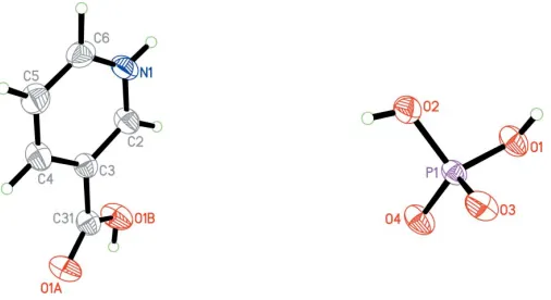
Figure 1
The molecular structure of the title compound, showing the atom-numbering scheme and 50% probability displacement ellipsoids.
Figure 2
[image:2.610.313.567.672.727.2]All H atoms were located and refined isotropically.
Data collection: CAD-4 EXPRESS (Enraf–Nonius, 1994); cell refinement: CAD-4 EXPRESS; data reduction:XCAD4 (Harms & Wocadlo, 1995); program(s) used to solve structure:SHELXTL/PC
(Bruker, 2000); program(s) used to refine structure:SHELXTL/PC; molecular graphics:SHELXTL/PC; software used to prepare mate-rial for publication:SHELXTL/PC.
The authors thank the Department of Science and Tech-nology, Government of India, for establishing the Single-Crystal Diffractometer Facility at the School of Physics, Madurai Kamaraj University, Madurai, through the FIST programme.
References
Basavoju, S., Reddy, C. M. & Desiraju, G. R. (2005).Acta Cryst.E61, o822– o823.
Bhat, T. N., Singh, T. P. & Vijayan, M. (1974).Acta Cryst.B30, 2921–2922.
Blessing, R. H. (1986).Acta Cryst.B42, 613–621.
Bruker (2000). SHELXTL. Version 6.10. Bruker AXS Inc., Madison, Wisconsin, USA.
Brutts, R. & Lundholm, L. (1971).J. Atheroscler. Res.pp. 14–19.
Bulut, A., Yesilel, O. Z., Dege, N., Icbudak, H., Olmaz, H. & Bujukgungor, O. (2003).Acta Cryst.C59, o727–o729.
Dobson, A. J. & Gerkin, R. E. (1997).Acta Cryst.C53, 1427–1429.
Ellis, P. J., Appleby, C. A., Guss, J. M., Hunter, W. N., Ollis, D. L. & Freeman, H. C. (1997).Acta Cryst.D53, 302–310.
Enraf–Nonius (1994). CAD-4 EXPRESS. Version 5.1/1.2. Enraf–Nonius, Delft, The Netherlands.
Freeman, G. R. & Bugg, C. E. (1974).Acta Cryst.B30, 431–443. Giantsidis, J. & Turnbull, M. M. (2000).Acta Cryst.C56, 334–335.
Gielen, M., Khloufi, A. E., Biesemans, M. & Willem, R. (1992).Polyhedron, 11, 1861–1868.
Harms, K. & Wocadlo, S. (1995).XCAD4. University of Marburg, Germany. Kim, H. L., Yoon, H. J., Ha, J. Y., Lee, B. I., Lee, H. H., Mikami, B. & Suh, W. S.
(2004).Acta Cryst.D60, 948–949.
Kutoglu, A. & Scheringer, C. (1983).Acta Cryst.C39, 232–234.
North, A. C. T., Phillips, D. C. & Mathews, F. S. (1968).Acta Cryst.A24, 351– 359.
Takusagawa, F. & Shimada, A. (1976).Acta Cryst.B32, 1925–1927. Wright, W. B. & King, G. S. D. (1953).Acta Cryst.6, 305–310. Wright, W. B. & King, G. S. D. (1954).Acta Cryst.7, 283–288.
organic papers
Acta Cryst.(2005). E61, o2553–o2555 Athimoolamet al. C
supporting information
sup-1
Acta Cryst. (2005). E61, o2553–o2555
supporting information
Acta Cryst. (2005). E61, o2553–o2555 [https://doi.org/10.1107/S1600536805022221]
Nicotinium dihydrogenphosphate
S. Athimoolam, K. Anitha and R. K. Rajaram
3-carboxypyridinium dihydrogenphosphate
Crystal data
C6H6NO2+·H2PO4−
Mr = 221.10 Monoclinic, P21/c
a = 12.158 (5) Å b = 10.593 (4) Å c = 7.001 (3) Å β = 102.35 (3)° V = 880.8 (6) Å3
Z = 4 F(000) = 456
Dx = 1.667 Mg m−3
Dm = 1.652 Mg m−3
Dm measured by flotation in CHBr3 and CCl4
Mo Kα radiation, λ = 0.71073 Å Cell parameters from 25 reflections θ = 9.7–14.3°
µ = 0.32 mm−1
T = 293 K Needle, colourless 0.20 × 0.17 × 0.14 mm
Data collection
Nonius MACH3 sealed-tube diffractometer
Radiation source: fine-focus sealed tube Graphite monochromator
ω/2θ scans
Absorption correction: ψ scan (North et al., 1968)
Tmin = 0.847, Tmax = 1.000
1880 measured reflections
1545 independent reflections 1317 reflections with I > 2σ(I) Rint = 0.047
θmax = 25.0°, θmin = 2.6°
h = −14→14 k = −12→1 l = 0→8
3 standard reflections every 60 min intensity decay: none
Refinement
Refinement on F2
Least-squares matrix: full R[F2 > 2σ(F2)] = 0.030
wR(F2) = 0.084
S = 1.05 1545 reflections 160 parameters 0 restraints
Primary atom site location: structure-invariant direct methods
Secondary atom site location: difference Fourier map
Hydrogen site location: inferred from neighbouring sites
All H-atom parameters refined w = 1/[σ2(F
o2) + (0.0512P)2 + 0.2334P]
where P = (Fo2 + 2Fc2)/3
(Δ/σ)max < 0.001
Δρmax = 0.25 e Å−3
Δρmin = −0.37 e Å−3
Extinction correction: SHELXTL/PC (Bruker, 2000), Fc*=kFc[1+0.001xFc2λ3/sin(2θ)]-1/4
supporting information
sup-2
Acta Cryst. (2005). E61, o2553–o2555 Special details
Geometry. All e.s.d.'s (except the e.s.d. in the dihedral angle between two l.s. planes) are estimated using the full covariance matrix. The cell e.s.d.'s are taken into account individually in the estimation of e.s.d.'s in distances, angles and torsion angles; correlations between e.s.d.'s in cell parameters are only used when they are defined by crystal symmetry. An approximate (isotropic) treatment of cell e.s.d.'s is used for estimating e.s.d.'s involving l.s. planes.
Refinement. Refinement of F2 against ALL reflections. The weighted R-factor wR and goodness of fit S are based on F2,
conventional R-factors R are based on F, with F set to zero for negative F2. The threshold expression of F2 > σ(F2) is used
only for calculating R-factors(gt) etc. and is not relevant to the choice of reflections for refinement. R-factors based on F2
are statistically about twice as large as those based on F, and R- factors based on ALL data will be even larger.
Fractional atomic coordinates and isotropic or equivalent isotropic displacement parameters (Å2)
x y z Uiso*/Ueq
N1 0.72318 (13) 0.57178 (16) 0.9647 (2) 0.0357 (4) H1 0.7311 (19) 0.649 (2) 0.989 (3) 0.040 (6)* C2 0.62653 (16) 0.53386 (18) 0.8483 (3) 0.0345 (5) H2 0.569 (2) 0.595 (2) 0.800 (3) 0.044 (6)* C3 0.61118 (15) 0.40873 (17) 0.7980 (3) 0.0290 (4) C4 0.69727 (16) 0.32384 (19) 0.8715 (3) 0.0359 (4) H4 0.687 (2) 0.236 (3) 0.839 (4) 0.059 (7)* C5 0.79443 (18) 0.3665 (2) 0.9921 (3) 0.0424 (5) H5 0.854 (2) 0.315 (2) 1.044 (3) 0.051 (6)* C6 0.80657 (17) 0.4919 (2) 1.0363 (3) 0.0386 (5) H6 0.877 (2) 0.530 (2) 1.127 (4) 0.056 (7)* C31 0.50543 (15) 0.36374 (17) 0.6669 (3) 0.0310 (4) O1A 0.50164 (11) 0.26655 (13) 0.5767 (2) 0.0419 (4) O1B 0.41945 (12) 0.44044 (14) 0.6607 (3) 0.0446 (4) H1B 0.366 (3) 0.405 (3) 0.593 (5) 0.069 (9)* P1 0.87510 (3) 0.88988 (4) 0.05786 (6) 0.0245 (2) O1 0.85483 (12) 1.03547 (12) 0.0364 (2) 0.0349 (4) H1P 0.916 (2) 1.074 (3) 0.055 (4) 0.058 (8)* O2 0.93597 (11) 0.87378 (13) 0.2765 (2) 0.0354 (4) H2P 0.943 (3) 0.800 (3) 0.318 (5) 0.084 (10)* O3 0.95263 (11) 0.84746 (12) −0.07246 (19) 0.0351 (3) O4 0.76205 (10) 0.82621 (12) 0.0151 (2) 0.0359 (4)
Atomic displacement parameters (Å2)
U11 U22 U33 U12 U13 U23
supporting information
sup-3
Acta Cryst. (2005). E61, o2553–o2555
O1 0.0295 (7) 0.0239 (7) 0.0500 (8) 0.0014 (5) 0.0054 (6) 0.0013 (5) O2 0.0353 (7) 0.0317 (7) 0.0355 (8) −0.0059 (6) −0.0011 (6) 0.0030 (6) O3 0.0335 (7) 0.0299 (7) 0.0436 (8) −0.0046 (5) 0.0122 (6) −0.0084 (6) O4 0.0219 (6) 0.0306 (7) 0.0516 (8) −0.0037 (5) −0.0004 (5) 0.0004 (6)
Geometric parameters (Å, º)
N1—C6 1.334 (3) C6—H6 1.03 (3)
N1—C2 1.340 (3) C31—O1A 1.204 (2)
N1—H1 0.83 (3) C31—O1B 1.317 (2)
C2—C3 1.374 (3) O1B—H1B 0.81 (3)
C2—H2 0.96 (2) P1—O4 1.5026 (13)
C3—C4 1.392 (3) P1—O3 1.5135 (14)
C3—C31 1.489 (3) P1—O2 1.5603 (16)
C4—C5 1.373 (3) P1—O1 1.5640 (14)
C4—H4 0.96 (3) O1—H1P 0.83 (3)
C5—C6 1.364 (3) O2—H2P 0.84 (4)
C5—H5 0.91 (3)
C6—N1—C2 122.37 (18) N1—C6—C5 119.73 (19)
C6—N1—H1 119.8 (16) N1—C6—H6 116.8 (14)
C2—N1—H1 117.8 (16) C5—C6—H6 123.5 (14)
N1—C2—C3 119.89 (18) O1A—C31—O1B 124.71 (18)
N1—C2—H2 119.5 (14) O1A—C31—C3 121.89 (17)
C3—C2—H2 120.6 (14) O1B—C31—C3 113.40 (16)
C2—C3—C4 118.57 (18) C31—O1B—H1B 106 (2)
C2—C3—C31 121.25 (17) O4—P1—O3 114.08 (8)
C4—C3—C31 120.18 (17) O4—P1—O2 111.58 (8)
C5—C4—C3 119.64 (19) O3—P1—O2 109.45 (8)
C5—C4—H4 120.8 (15) O4—P1—O1 107.75 (8)
C3—C4—H4 119.6 (15) O3—P1—O1 109.92 (8)
C6—C5—C4 119.79 (19) O2—P1—O1 103.52 (8)
C6—C5—H5 116.9 (16) P1—O1—H1P 110.2 (19)
C4—C5—H5 123.3 (16) P1—O2—H2P 116 (2)
C6—N1—C2—C3 −0.4 (3) C2—N1—C6—C5 −0.4 (3)
N1—C2—C3—C4 0.5 (3) C4—C5—C6—N1 1.1 (3)
N1—C2—C3—C31 −179.18 (17) C2—C3—C31—O1A 158.11 (19) C2—C3—C4—C5 0.3 (3) C4—C3—C31—O1A −21.5 (3) C31—C3—C4—C5 179.91 (18) C2—C3—C31—O1B −21.6 (3) C3—C4—C5—C6 −1.1 (3) C4—C3—C31—O1B 158.73 (18)
Hydrogen-bond geometry (Å, º)
D—H···A D—H H···A D···A D—H···A
N1—H1···O4i 0.83 (3) 1.92 (3) 2.746 (2) 171 (2)
supporting information
sup-4
Acta Cryst. (2005). E61, o2553–o2555
O1—H1P···O3iii 0.83 (3) 1.78 (3) 2.613 (2) 175 (3)
O2—H2P···O3iv 0.84 (4) 1.73 (4) 2.561 (2) 173 (3)