Otoacoustic Emissions and Evoked Potentials in Infants
after Breast-Feeding Jaundice
——Hearing Dysfunction in Breast-Feeding Jaundice
Adrián Poblano1*, Norma Ballesteros1, Carmina Arteaga1, Blanca Flores2, Teodoro Flores2
1Laboratory of Cognitive Neurophysiology, National Institute of Rehabilitation, Mexico City, Mexico; 2Laboratory of Clinical
Neu-rophysiology, National Institute of Rehabilitation, Mexico City, Mexico. Email: *[email protected]
Received November 11th, 2011; revised May 28th, 2012; accepted June 29th, 2012
ABSTRACT
We study hearing in a group of infants with Breast-feeding jaundice (BFJ) by means of Transient-evoked otoacoustic emissions (T-EOE) and Brainstem auditory evoked potentials (BAEP) searching for relationship between bilirubin se-rum levels and auditory dysfunction. Eleven infants born at-term with BFJ were selected for the study. We studied also 11 control age- and gender matched healthy at-term infants without signs of jaundice. T-EOAE studies were performed between 5 - 7 days after birth, and 3 months later. BAEP studies were performed once. BFJ group infants exhibited lower amplitudes in T-EOE than infants in the control group. These differences disappear at the 3-month evaluation. In BAEP, we observed a significant latency delay of waves I and V in Breast-feeding jaundice group infants. All infants in both groups demonstrated reproducible wave V response at 30 decibels. No significant correlation values were observed between bilirubin serum levels and T-EOE and BAEP variables. Our data suggest that BFJ can result in transient pe- ripheral and central auditory dysfunction. Dysfunction is reversible after treatment of infants with BFJ.
Keywords: Breast-Feed Jaundice; Hyperbilirubinemia; Hearing; Neonates; Transient-Evoked Otoacoustic Emissions; Brainstem Auditory Evoked Potentials
1. Introduction
Previous research has shown the relationship between neonatal hyperbilirubinemia and sensorineural hearing loss [1,2]. The majority of investigations have been per-formed by means of Brainstem auditory evoked re-sponses (BAEP) in High-risk newborns (HR-NB). These infants have been followed throughout several years after birth to confirm hearing loss [3-6]. Observations have suggested repeatedly that hearing dysfunction is a mani-festation of injury situated in the auditory neural pathway along the brainstem. However, one paper had situated the injury also in the peripheral cochlear system [7].
Breast-feeding jaundice (BFJ) provides a fair model to study the peripheral and central auditory alterations after pure neonatal hyperbilirubinemia, because this is differ-ent from hyperbilirubinemia in HR-NB. The majority of infants with BFJ are born at term with adequate weight and without other risk factors for auditory damage, such as asphyxia, intracranial hemorrhage, or others. Thus, the aim of our study was to study hearing function in a group of infants with BFJ by means of Transient-evoked oto-
acoustic emissions (T-EOE) and BAEP with the hypothesis that there is a relationship between bilirubin serum levels and peripheral and central auditory dysfunction. In order to test our hypothesis, we employed T-EOE as an index for cochlear toxicity, and BAEP as an index of auditory brainstem neurotoxicity.
2. Materials and Methods
2.1. Subjects
Healthy infants born at General Hospital “Dr. Darío Fernández” readmitted with jaundice to the Department of Pediatrics during their first week of life with diagnoses of BFJ were selected for the study. Inclusion criteria in-cluded the following: jaundice in Kramer zone ≥ 2; bilirubin levels > 15 mg/dL; age at birth between 37 and 41 of gestational weeks; weight > 2750 g; 5-min Apgar score > 7, and without other risk factors for hearing damage. All of the infants were exclusively breastfed. Mothers of the patients noted jaundice at home, and it was not accompanied by decreased activity or poor feed-ing. None newborn had any laboratory evidence of hemolytic disease as evidenced by anemia,
sis, or another abnormality. Infants were screened for neurological, and auditory dysfunction [8,9], and for perinatal factors associated with an increased risk of hy-perbilirubinemia including, maternal diabetes mellitus, high blood pressure, administration of oxytocin during labor, as well as central nervous system bleeding or in-jury, or infection. At time of readmission to the hospital, the infants were evaluated for clinical signs of acute bilirubin encephalopathy manifested as changes in level of consciousness, tone/movement, poor feeding, abnor- mal cry, opisthotonus and/or seizures, and alterations in brainstem function. Other causes of hyperbilirubinemia including scalp bruising, dehydration and sepsis were investigated. Laboratory studies included complete blood counts, electrolytes, glucose, and cultures. Cranial Ultra- sonographic studies were performed in all newborns to rule-out other brain pathologies. Bilirubin determinations and T-EOE were performed in the first days after jaun-dice was recognized to identify and treat possible neuro-development deviations; T-EOE were repeated 3 months later. BAEP determinations were performed rapidly after T-EOE examination once. We studied 11 infants with BFJ (four males and seven females) and 11 control heal- thy newborn infants who were age- and gender matched at the time of study without signs of jaundice. Results of each study were blinded to the researchers who revised BAEP and T-EOE determinations for group pertinence. The Research and Ethics Committees of the Hospital and of the Institute approved the protocol. Informed consent was obtained from parents of infants and is contained in signed forms according to the principles expressed in the Declaration of Helsinki.
2.2. Bilirubin Determinations
Samples were obtained by peripheral venipuncture. All specimens were protected from light after they were drawn, and these were analyzed immediately. Total bilirubin concentration was determined spectrophotomet-rically, and unbound bilirubin concentration was meas-ured by the peroxidase method using a clinical analyzer (UB analyzer, Arrows Co., Osaka, Japan). Samples with high unbound bilirubin > 0.5 µg/dL were measured for concentration of direct bilirubin by diazo method [10].
2.3. Transient-Evoked Otoacoustic Emissions (T-EOE)
Responses were recorded with the ILO-88 (Otodynamic Ltd, London, UK), during a sleep session. Studies were conducted in a sound-proofed room. Prior to the Oto- acoustic emission study, ear canals were checked with an otoscope to confirm permeability of external auditory channel. We use a miniature acoustic probe fitted into the infant ear channel. The probe incorporates a small micro-
phone and ear-speaker. Stimuli consisted of 80 decibel (dB) HL (Hearing level) click stimulation of 100 µsec delivered at a repetition rate of 11/sec. Signal was ampli-fied and filtered between 350 and 6400 Hz. Two hundred fifty stimuli were averaged. Analysis time was 20 milli-sec. Contralateral ear masking with white noise 20 dB below stimulus intensity was administrated simultane-ously. Adequate T-EOAE responses were identified based on reproducibility of ≥75%. The detection thresh-old of T-EOAE was measured by stimulating the ear with decreasing click intensities in 20-dB steps. Each ear was tested separately [11].
2.4. Brainstem Auditory Evoked Potentials (BAEP)
BAEP were recorded and analyzed following standard international recommendations [12]. BAEP determina- tions were performed after a feeding sleep in a sound- proof room. Each subject was tested with BAEP using an ATI system (Buenos Aires, Argentina). Three gold-disk electrodes were placed on the scalp, with negative elec- trode on ipsilateral mastoid, positive on vertex, and neu- tral on contralateral mastoid. Interelectrode impedances were ≤2 kilo-ohms. Electrical activity among electrodes was amplified and averaged over a 10 millisec time base. Stimulus was administrated through a Telephonics TDH- 49 earphone (Telephonics Co., Huntington, NY, USA). Stimuli were presented monoaurally at a rate of 11/sec. Initial presentation intensity was 70-dB HL and decreas- ed by 20-dB steps to determine neurophysiologic thresh- old of response. Contralateral ear masking with white noise 20 dB below stimulus intensity was administrated simultaneously. Stimuli were delivered mono-aurally and consisted of 100 µsec alternating clicks. Band pass filters were set between 100 and 3000 Hz, and stimulus average was 2024 clicks. The process was repeated at least once to ensure reproducibility of response. Latencies of waves I, III, and V were measured by manual cursor placement at left and right ear recordings separately; I-III, III-V, and I-V interpeak intervals were calculated automatically by computer software; a normal hearing threshold was de- termined if the reproducible wave V response was pre- sent at last at 30 dB level.
2.5. Data Analysis
t test. We performed Pearson correlation analysis be- tween total, indirect, and direct bilirubin serum levels and T-EOE and BAEP variables. The level of statistical significance a-priori chosen was p ≤ 0.05. Calculations were performed with the SPSS 14.0 (SPSS Inc., Chicago, IL, USA) computer program [15].
3. Results
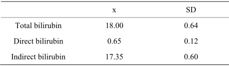
Clinical characteristics of infants in each group are shown in Table 1; no significant differences in compared variables between groups were observed. The infants age at which mother requested attention ranged between 5 and 7 days after birth (median, 6 days). Average bilirubin determinations in BFJ-group infants are presented in Ta-ble 2.
Averages of response amplitude by ear determined by T-EOE at birth and 3 months later are shown in Table 3; in the neonatal period, there were significant differences in both ear responses between groups, with BFJ-group infants exhibiting lower amplitudes than infants in the control group. However, these differences disappear at the 3-month evaluation when hyperbilirubinemia was resolved.
[image:3.595.309.536.111.188.2]Average of main waves (I, III, and V) and interwave intervals (I-III, III-V, and I-V) in BAEP determinations are presented in Table 4; we observed a significant la-tency delay of waves I and V in BFJ-group infants. However, all infants in both groups demonstrated repro-ducible wave V response at 30 dB, suggesting adequate hearing and thus no second BAEP study was performed in infants. We did not observe cases of auditory neu-ropathy. No significant correlation values were observed between total, indirect, and direct bilirubin serum levels and T-EOE, and BAEP variables.
Table 1. Clinical characteristics of control (n = 11) and in-fants with Breast-feeding jaundice (BFJ) (n = 11).
Control (x ± SD) BFJ (x ± SD)
Weight at birth (g) 2897 ± 371 2863 ± 350
Age at birth (weeks) 37.9 ± 1.23 38.3 ± 0.84
1 min Apgar score 7.6 ± 1.21 8.0 ± 0.52
5 min Apgar score 9 ± 0 9 ± 0
[image:3.595.309.538.258.377.2]x = average; SD = Standard deviation.
Table 2. Average of maximum bilirubin serum levels (mg/dL) in infants with Breast-feeding jaundice (BFJ).
x SD
Total bilirubin 18.00 0.64
Direct bilirubin 0.65 0.12
Indirect bilirubin 17.35 0.60
x = average; SD = Standard deviation.
Table 3. Average ± standard deviation of amplitude re-sponses (dB) in T-EOE in control and BFJ-group infants.
Right ear 1 week
Left ear 1 week
Right ear 3 m
Left ear 3 m Control
group 17.96 ± 7.93 19.08 ± 9.38 16.10 ± 6.02 18.60 ± 7.01 BFJ
group 12.48 ± 8.74 13.02 ± 7.57 16.60 ± 8.00 19.14 ± 6.25
p 0.05 0.04 ns ns
T-EOE = Transient-Evoked otoacoustic emissions; BFJ = Breast feeding jaundice; not significant.
Table 4. Average ± standard deviation of latencies (msec) of main waves and interval interwaves of BAEP at 80 dB in control infants and in infants with BFJ.
Wave/Interval (msec) Controls BFJ p
I 1.70 ± 0.13 1.83 ± 0.25 0.04
III 4.41 ± 0.18 4.49 ± 0.23 ns
V 6.64 ± 0.26 6.79 ± 0.29 0.05
I-III 2.71 ± 0.14 2.66 ± 0.21 ns
III-V 2.23 ± 0.23 2.30 ± 0.25 ns
I-V 4.94 ± 0.26 4.96 ± 0.29 ns
x = average; SD = standard deviation; BAEP = Brainstem auditory evoked potentials; dB = decibels; BFJ = Breast feeding jaundice; ns = not signifi-cant.
4. Discussion
In this paper, we showed that BFJ in low-risk at-term infants results in both auditory peripheral and central dysfunctions as reflected in T-EOE and BAEP alterations. We also observed that these auditory and neurophysi- ologic alterations could be reversible after rapid man- agement and resolution of the hyperbilirubinemia. How- ever, we could not demonstrate our hypothesis about sig- nificant correlation between bilirubin serum levels and peripheral and central auditory dysfunction.
Several in-vitro studies showed that bilirubin-induced neuronal toxicity involves changes in energy metabolism, alteration in membrane function, decreased membrane potential, alteration in enzyme function, and inhibition of protein synthesis [16,17]. It also appears from these pre- viously cited in-vitro studies that immature cells are more sensitive to bilirubin toxicity than differentiated cells, supporting clinical experience demonstrating that premature neonates are more susceptible to bilirubin- induced neurotoxicity than at-term infants. However, our data also suggest that the auditory responses of otherwise healthy at-term infants could be injured by hyper- bilirubinemia.
[image:3.595.58.288.531.610.2] [image:3.595.57.286.659.725.2]ex-plain the occurrence of bilirubin neurotoxicity at much lower levels of serum total bilirubin [1]. These factors are thought to increase the risk of kernicterus by affect-ing serum bilirubin-albumin bindaffect-ing, bilirubin entry into the brain, or tissue uptake of bilirubin. For example, one study identified a degree of acidosis with low serum al-bumin levels that occurred prior to the maximum un-bound bilirubin level and that was present in newborns with kernicterus [18]. On the other hand, a study of fol-low-up of at-term or near-term infants with BFJ reported transient neurologic abnormalities. These abnormalities resolved following management with phototherapy and exchange transfusions, and bilirubin level did not corre-late with the long-term prognosis [19]. Results from this study are partially in agreement with our data.
One of the tools commonly utilized to investigate bilirubin-induced neurotoxicity in neonates is BAEP test. BAEP has been studied in high-risk pre-term newborns as a non-invasive auditory neurophysiologic assessment of bilirubin neurotoxicity. Many observations confirm the higher frequency of auditory alterations in newborns after hyperbilirubinemia, such a delay of wave I, III and V latencies or the absence of response at higher intensi- ties of auditory stimulation [3-6]. This is in indirect agreement with our observations, because we observed a wave I and V delay. Other studies have shown correla- tion between indirect bilirubin serum levels and BAEP alterations [20]; however we are unable to confirm this correlation in our observations. Other studies have dem- onstrated that BAEP alterations are reversible after blood exchange [21]. However, we were unable to confirm this observation, because our infants with BFJ were not blood ex-changed and all our infants in both, control and BFJ groups presented response at 30 dB. Thus, there was no reason for subsequent appointment 3 months later for an additional BAEP study.
Otoacoustic emissions have been used to screen hear- ing alterations after neonatal hyperbilirubinemia. T-EOE has been employed as a first step in rapid screening of infant auditory performance. Study investigators usually carry out a BAEP examination after an abnormal T-EOE [2,7]. Researchers have observed a decrease in the re- sponse amplitude of T-EOE such as that which we ob- served in our BFJ-group infants, or an increased number in absences of response after auditory stimulation.
Recently, Jangaard et al. did not observed an increased frequency in cerebral palsy, developmental delay, autism, and hearing loss in a population of infants born at-term or near-term with other neurologic risk-factors other than hyperbilirubinemia (≥19 mg/dL) [22]. This result is also in indirect agreement with our observations, because we observed no case of hearing loss. However, because we measured T-EOE and BAEP variables quantitatively, we were able to detect subclinical effects of transient
hyper-bilirubinemia in the peripheral and central auditory sys-tem of infants with BFJ, that were not observed by the other work team.
We did not observe cases of auditory neuropathy in our sample, but this may be explained of our small sam- ple size [23]. An alternative explanation may be that auditory neuropathy after neonatal hyperbilirubinemia is a complex alteration that requires other risk-factors than only high bilirubin serum levels for the production of auditory alterations. However, our infants must be fol- lowed, because language delay has been reported as a possible complication after neonatal hyperbilirubinemia [24].
Our research possesses some limitations; thus, our re- sults must be considered as tendencies, not strong con- clusions. The sample is small, despite its being highly selected and age-gender-matched to a control group. Therefore, in the future a greater number of at-term in- fants with BFJ must be studied. Moreover, follow-up time was brief, notwithstanding follow-up of these in- fants will continue with these as out-patients in the Pedi- atrics Department to determine whether minimal auditory dysfunction or language alteration during child develop- ment can be a result of the BFJ episode during the neo- natal period of life.
5. Conclusion
Our data suggest that BFJ can result in peripheral and central auditory dysfunction. The dysfunction is reverse- ble; thus, early identification and treatment of infants with BFJ is mandatory to prevent possible auditory com- plications.
REFERENCES
[1] A. Gkoltsiouk, M. Tzoufi, S. Counsell, M. Rutherford and F. Cowan, “Serial Brain MRI and Ultrasound Findings: Relation to Gestational Age, Bilirubin Level, Neonatal Neurologic Status, and Neurodevelopmental Outcome in Infants at Risk of Kernicterus,” Early Human Develop- ment, Vol. 84, No. 12, 2008, pp. 829-838.
doi:10.1016/j.earlhumdev.2008.09.008
[2] N. Y. Boo, A. S. Rohani and A. Asma, “Detection of Sensorineural Hearing Loss Using Automated Auditory Brainstem-Evoked Response and Transient-Evoked Oto- acoustic Emissions in at Term Neonates with Severe Hy- perbilirubinemia,” Singapore Medical Journal, Vol. 49, No. 3, 2008, pp. 209-214.
[3] S. Garza-Morales, A. Poblano, A. Robledo-Galván and L. A. Fernández-Carrocera, “Auditory Evoked Potentials in Children at Neonatal Risk for Hypoacusis,” Panamerican
Journal of Public Health, Vol. 2, No. 4, 1997, pp. 232-
237. doi:10.1590/S1020-49891997001000002
Vol. 107, No. 4, 2001, pp. 664-670. doi:10.1542/peds.107.4.664
[5] Y. Yilmaz, S. Degirmenci, F. Akdas, S. Kulekci, A. Ciprut, S. Yuksel, F. Yildiz, L. Karadeniz and A. Say, “Prognostic Value of Auditory Brainstem Response for Neurologic Outcome in Patients with Neonatal Indirect Hyperbilirubinemia,” Journal of Child Neurology, Vol. 16, No. 10, 2001, pp. 772-775.
doi:10.1177/088307380101601014
[6] P. Sharma, N. P. Chhangani, K. R. Meena, R. Jora, N. Sharma and B. D. Gupta, “Brainstem Evoked Response Audiometry (BAER) in Neonates with Hyperbilirubine-mia,” Indian Journal of Pediatrics, Vol. 73, No. 5, 2006, pp. 413-416. doi:10.1007/BF02758564
[7] C. Oysu, I. Aslan, A. Ulubil and N. Baserer, “Incidence of Cochlear Involvement in Hyperbilirubinemic Deaf-ness,” Annals of Otology, Rhinology and Laryngology, Vol. 111, No. 11, 2002, pp. 1021-1025.
[8] A. Poblano, I. Chayo, J. Ibarra and E. Rueda, “Electro-physiological and Behavioral Methods in Early Detection of Hearing Impairment,” Archives of Medical Research, Vol. 31, No. 1, 2000, pp. 75-80.
doi:10.1016/S0188-4409(99)00060-0
[9] C. L. Pereira, A. M. Aguilar, F. Rivera, L. Cruz, I. Santos and A. Poblano, “Concordance between Neurologic Screen- ing Test and Neurologic Examination in Newborns,” Cli-
nical Pediatrics (Philadelphia), Vol. 45, No. 4, 2006, pp.
315-323.
[10] S. P. Harrison and I. M. Barlow, “Three Direct Spectro- photometric Methods for Determination of Total Bili- rubin in Neonatal and Adult Serum, Adapted to the Tech- nicon RA-1000 Analyzer,” Clinical Chemistry, Vol. 35, No. 9, 1989, pp. 1980-1986.
[11] J. How and M. E. Lutman, “Transient Evoked Otoacous-tic Emissions Input-Output Function Variation in a Large Sample of Neonates and Implications for Hearing Screen-ing,” International Journal of Audiology, Vol. 46, No. 11, 2007, pp. 670-679. doi:10.1080/14992020701438813 [12] M. R. Nuwer, M. Aminoff, D. Goodin, S. Matsuoka, F
Mauguiere, A. Star A and J. F. Vibert, “IFCN Recom-mended Standards from Brain-Stem Auditory Evoked Potentials. Report of IFCN Committee,” Electroence-
phalography and Clinical Neurophysiology, Vol. 91, No.
1, 1994, pp. 112-117. doi:10.1016/0013-4694(94)90013-2 [13] A. Poblano, A. Belmont, J. Sosa, J. Ibarra, Y. Rosas, V.
López and S. Garza, “Effects of Prenatal Exposure to Carbamazepine on Brainstem Auditory Evoked Potentials in Infants of Epileptic Mothers,” Journal of Child Neu-
rology, Vol. 17, No. 5, 2002, pp. 364-368.
doi:10.1177/088307380201700510
[14] A. Poblano, A. Belmont, J. Sosa, J. Ibarra, A. M. Vargas,
G. Limón and C. Martínez, “Amikacin Alters Auditory Brainstem Conduction Time in Newborns,” Journal of
Perinatal Medicine, Vol. 31, No. 3, 2003, pp. 237-241.
doi:10.1515/JPM.2003.032
[15] M. Ferrán-Aranaz, “Statistical Analyses in SPSS for Win-dows,” Osbourne-McGraw Hill, Madrid, 2001, pp. 19-28. [16] M. F. Notter and J. W. Kending, “Differential Sensitivity
of Neural Cells to Bilirubin Toxicity,” Experimental
Neu-rology, Vol. 94, No. 3, 1986, pp. 670-682.
doi:10.1016/0014-4886(86)90246-3
[17] A. S. Falcao, C. Bellarosa, A. Fernández, M. A. Brito, R. F. Silva, C. Tiribelli and D. Brites, “Role of Multidrug Ressistence-Associated Protein 1 Expression in the in Vi- tro Susceptibility of Rat Nerve Cell to Unconjugated Bili- rubin,” Neuroscience, Vol. 144, No. 3, 2007, pp. 878-888. doi:10.1016/j.neuroscience.2006.10.026
[18] P. Govaert, M. Lequin, R. Swarte, S. Robben, R. de Coo, N. Weisglas-Kuperus, Y. de Rijke, M. Sinassappel and J. Barkovich, “Changes in Globus Pallidus with (pre) Term Kernicterus,” Pediatrics, Vol. 112, No. 6, 2003, pp. 1256- 1263. doi:10.1542/peds.112.6.1256
[19] M. C. Harris, J. C. Bernbaum, J. Polin, R. Zimmerman and R. Polin, “Developmental Follow-Up of Breastfed Term and Near-Term Infants with Marked Hyperbiliru- binemia,” Pediatrics, Vol. 107, No. 5, 2001, pp. 1075- 1080. doi:10.1542/peds.107.5.1075
[20] C. E. Ahlfors, S. B. Amin and A. E. Parker, “Unbound Bilirubin Predicts Abnormal Automated Auditory Brain- stem Responses in a Diverse Newborns Population,” Jour-
nal of Perinatology, Vol. 29, No. 4, 2009, pp. 305-309.
doi:10.1038/jp.2008.199
[21] C. G. Nwaesei, J. van Aerde, M. Boyden and M. Peerl- man, “Changes in Auditory Brainstem Responses in Hy- perbilirubinemic Infants before and after Exchange Trans- fusion,” Pediatrics, Vol. 74, No. 5, 1984, pp. 800-803. [22] K. A. Jangaard, D. B. Fell, L. Doods and A. C. Allen,
“Outcomes in a Population of Healthy Term and Near- Term Infants with Serum Bilirubin Levels of ≥325 µmol/L (≥19 mg/dL) Who Were Born in Nova Scotia, Canada, between 1994 and 2000,” Pediatrics, Vol. 122, No. 1, 2008, pp. 119-124. doi:10.1542/peds.2007-0967 [23] K. Xionis, Y. Weirather, H. Mavoori, S. H. Shaha and L.
M. Iwamoto, “Extremely Low Birth Weight Infants Are at High Risk for Auditory Neuropathy,” Journal of
Peri-natology, Vol. 27, No. 11, 2007, pp. 718-723.
doi:10.1038/sj.jp.7211803
[24] S. B. Amin, D. Prinzing and G. Myers, “Hyper-bilirubinemia and Language Delay in Premature Infants,”
Pediatrics, Vol. 123, No. 1, 2009, pp. 327-321.