Evaluation of poor performance in racehorses
using a high-speed treadmill
P. Melkova, P. Jahn, S. Bodecek, O. Dobesova, J. Hanak
Faculty of Veterinary Medicine, University of Veterinary and Pharmaceutical Sciences, Brno, Czech Republic
ABSTRACT: The study reports the results of a comprehensive clinical evaluation of 18 racehorses with a history of inadequate athletic performance. The poor performance evaluation included general physical examination and laboratory screening, respiratory examination including upper airway endoscopy at rest and during maximal treadmill exercise, cardiac examination, post-exercise serum biochemistry and bronchoalveolar lavage cytology. A definitive diagnosis was established in 15 horses (83.3%). The most frequent findings were upper respiratory tract disorders, with dorsal displacement of the soft palate being most often diagnosed (eight horses) followed by axial deviation of the aryepiglottic folds (two horses) and recurrent laryngeal neuropathy (two horses). Inflam-matory airway disease was diagnosed in two horses and exercise-induced pulmonary haemorrhage in one horse. Subclinical myopathy was detected in two horses. Ten horses (55.6%) had multiple concomitant problems that may have contributed to their inadequate athletic performance; therefore, a comprehensive approach is required to diagnose the cause of decreased athletic capability.
Keywords: exercise testing; upper airway obstruction; endoscopy; horse List of abbreviations
ADAF = axial deviation of aryepiglottic fold, BAL = bronchoalveolar lavage, CK = creatine kinase, EIPH = exercise-induced pulmonary haemorrhage, IAD = inflammatory airway disease, PLH = pharyngeal lymphoid hyperplasia, RLN = recurrent laryngeal neuropathy, SET = standardised exercise test
Horses are considered to be elite athletes because of their unique physiology. Even subtle changes in their health can significantly affect performance. These abnormalities are often challenging to detect because many of the problems that cause poor per-formance are manifested only at medium or high speed exercise and horses with poor performance may have multiple concurrent problems (Morris and Seeherman 1991; Martin et al. 2000). In the study by Morris and Seeherman (1991) describing the results of the evaluation of 275 racehorses with a history of poor racing performance, 84% of the horses were diagnosed as suffering from more than one problem.
An accurate history, a complete physical exami-nation, a thorough lameness evaluation and routine clinicopathological tests are essential initial steps
in attempting to identify the causes of inadequate athletic performance. Evaluation of equine athletes often requires additional special diagnostic meth-ods including exercise testing because findings in resting horses may be unremarkable or equivocal. Several studies have documented that, in particu-lar, dynamic upper respiratory tract obstructions cannot be diagnosed accurately during resting en-doscopic examination and that endoscopy during exercise is required to make a definitive diagnosis (Lane et al. 2006; Allen and Franklin 2010; Garret et al. 2011; Kelly et al. 2013). Exercise testing can be performed on a high-speed treadmill or in the field using overground endoscopy. Conducting clinical exercise tests on a treadmill allows standardisa-tion of environmental condistandardisa-tions and exercise test
parameters; thus, the speed and duration of each phase of an exercise test can be controlled. There is also good access to horses at suitable times during and after exercise for cardiorespiratory measure-ments and blood collections.
The aim of this study was to report the findings of clinical evaluation and exercise testing using a high-speed treadmill to ascertain the reasons for inadequate athletic performance in Thoroughbred racehorses referred to the Equine Clinic, University of Veterinary and Pharmaceutical Sciences Brno, the Czech Republic.
MATERIAL AND METHODS
Horses. Eighteen Thoroughbred racehorses with a history of poor performance were evaluated in the Equine Clinic, Brno, between January 2011 and April 2014. The group comprised 10 stallions, four mares and four geldings; the average age was 3.6 years (2–5 years). All of the horses were actively involved in training or racing at the time of clinical evaluation. Horses suffering from lameness were not included in the study.
Before the exercise tests were undertaken, a de-tailed history was obtained for all the horses and each horse underwent a thorough physical exami-nation of all body systems including haematology and serum biochemistry screening to ensure that they had no significant clinical abnormalities.
Cardiac examination. Cardiac examination included detailed auscultation and electrocardio-graphic analysis at rest and during an exercise test using a telemetry ECG system (Televet 100, Kruuse, Denmark). A continuous base-apex electrocardio-gram was recorded before, during and for 5 min after exercise on the high-speed treadmill. If these tests indicated a cardiac abnormality, echocardio-graphic evaluation was performed.
Resting upper and lower airway endoscopy. Complete resting endoscopic examination was per-formed without sedation using a flexible endoscope (1.4 m × 9 mm, Karl Storz, Germany) and included visual inspection of the nasal cavity, ethmoidal area, pharynx, soft palate, larynx and trachea to the level of the carina. Scoring on the basis of a previously described system was used for grading of pharyn-geal lymphoid hyperplasia (PLH) (Holcombe and Ducharme 2007) and laryngeal function (Robinson 2004).
Standardised exercise test. The tests were per-formed on a high speed treadmill (Säto I, Sato AB, Knivsta, Sweden). Before testing, each horse was given two to three acclimation sessions on the treadmill. After a warm-up phase of 5 min at walk (1.6 m/s) and 5 min at trot (3.6 m/s), the treadmill was set at a 6% slope and the horses performed the standardised incremental exercise test (SET) com-prising three stages at canter and gallop (7.0, 8.0 and 9.0 m/s). The first stage lasted 120 s and the following stages 60 s. The treadmill was then stopped to allow positioning of the endoscope in the upper airway. A flexible videoendoscope was inserted through the left nostril and advanced until the tip was just caudal to the pharyngeal openings of the auditory tubes. Thereafter, the horses performed a fourth phase at a speed from 10 to 12 m/s according to the individ-ual horse’s fitness. The exercise test was terminated when the horse showed signs of exhaustion or when a diagnosis had been established. This was followed by a recovery period at trot and walk.
All endoscopic examinations were recorded and analysed afterwards in real speed as well as in slow motion. Laryngeal function during exercise was scored (Robinson 2004).
Collection of samples post exercise. Broncho- alveolar lavage (BAL) samples for cytology were col-lected one hour after completion of the SET. Horses were sedated with xylazine (0.4 mg/kg of bwt i.v.) and butorphanol (0.01 mg/kg of bwt i.v.); then a nose twitch was applied. Lavage was performed using 300 ml of sterile saline and a commercially available BAL tube as previously described (Taylor et al. 2010). Five aliquots of 60 ml were instilled by syringe and aspirated immediately. The samples were pooled and centrifuged. Slides were stained using Hemacolor (Merck, Germany) and differential cell count was determined by the evaluation of 500 cells. A diagnosis of inflammatory airway disease (IAD) was established when the bronchoalveolar lavage fluid contained > 5% neutrophils or > 2% mast cells or > 0.5% eosinophils, or a combination of these criteria (Robinson 2003; Couetil et al. 2007; Bedenice et al. 2008; Wilson and Robinson 2015).
RESULTS
Eighteen Thoroughbred racehorses were includ-ed in the study. The initial presenting complaints, in addition to decreased athletic performance, were abnormal respiratory noise during exercise (17 horses; 94.4%), exercise intolerance resulting in loss of pace or slowing towards the end of a race (six horses), occasional coughing (two horses) and nasal discharge (one horse). One horse had a his-tory of inappropriate fatigue after exercise. All the horses were in good body condition, clinical exami-nation of all organ systems was unremarkable, and none of the horses showed signs of systemic illness.
Results of resting airway endoscopy
In 17 horses (94.4%) pharyngeal lymphoid hy-perplasia was found. In the majority of cases, mild grade PLH (1 and 2) was recognised and only two horses were more severely affected (grades 3 and 4 respectively). The mean age of horses diagnosed with PLH grade 1 was 4.5 years (range: 4–5 years); in horses with PLH 2 the mean age was 3.6 years (2–5); one horse with PLH 3 was three years old and one horse with PLH 4 was two years old. In three horses the presence of mucus in the trachea was noted. Eight horses (44.4 %) had evidence of recurrent laryngeal neuropathy (RLN). Six horses had grade II – subgrade 1 (grade II-1) RLN, and in two horses RLN was assessed as grade III-3.
Results of exercise videoendoscopy
Twelve horses (66.6%) had some form of dynamic obstruction of the airway detected during videoen-doscopy while exercising on the high-speed treadmill. Dorsal displacement of the soft palate (DDSP) was detected in eight horses and axial deviation of the aryepiglottic folds (ADAF) in two horses. In all the horses with grade II-1 RLN no obstruction was pre-sent during exercise (grade A); in the two horses with level III-3 RLN, one was classified as grade B and the other as C with respect to the degree of obstruction.
Results of cardiac examination
Mild heart murmur was noted on auscultation in four horses (22.2%), and these horses were then subjected to echocardiographic examination. Three of them had a mild mitral regurgitation, and one
horse had a functional murmur. Valvular regurgita-tion was not considered to be sufficiently severe to affect performance in any of the horses and cardiac chamber size and myocardial function were normal.
Dysrhythmia was detected at rest in five horses (27.8%). In all of these cases bradyarrhythmia (sec-ond-degree AV-block, sinoatrial block) was diag-nosed. None of the horses developed arrhythmia during exercise, but in five horses arrhythmia was noted during the recovery period immediately after exercise. Three of them had sinus arrhythmia, one horse had second-degree AV-block and two horses had single supraventricular premature depolarisa-tion (SVPD). Only in one horse were arrhythmias detected both at rest and during recovery.
Post-exercise BAL cytology and serum CK activity
The bronchoalveolar lavage fluid was abnormal in three horses. In two horses an elevated percentage of neutrophils was found (10% and 7.8%) and one horse exhibited evidence of previous intrapulmo-nary haemorrhage (11% of alveolar macrophages contained hemosiderin).
[image:3.595.304.532.528.698.2]Serum CK activity > 1000 IU/l after exercise was detected in two horses. In one horse a serum CK activity increase of more than five fold from the basal level was noted, although the post-exercise CK value was below 1000 IU/l. This horse also had
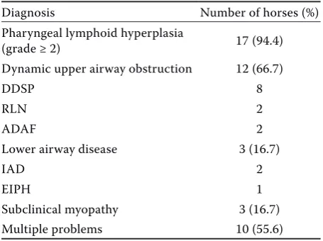
Table 1. Results of the clinical evaluation of 18 race-horses with poor performance
Diagnosis Number of horses (%)
Pharyngeal lymphoid hyperplasia
(grade ≥ 2) 17 (94.4)
Dynamic upper airway obstruction 12 (66.7)
DDSP 8
RLN 2
ADAF 2
Lower airway disease 3 (16.7)
IAD 2
EIPH 1
Subclinical myopathy 3 (16.7)
Multiple problems 10 (55.6)
elevated serum AST activity pre- and post-exercise. None of these three horses exhibited clinical signs of rhabdomyolysis.
All findings are summarised in Table 1.
DISCUSSION
All 18 horses completed the SET and the exami-nation was well tolerated by all of them.
A definitive cause of poor performance was ob-tained in 12 horses (66.7%). Intermittent dorsal dis-placement of the soft palate was the most common abnormality and was detected in eight horses; two horses had RLN during exercise (grade B and C); and two horses had ADAF. In two previous studies which evaluated poor performance problems, more than 40% of the horses examined had some type of upper airway obstruction during exercise (Morris and Seeherman 1991; Martin et al. 2000). In the present study the percentage of horses with URT obstruction was higher (80%). This could be influ-enced by case selection; owners requested exercise testing mostly in horses producing an abnormal respiratory noise during exercise.
PLH was a frequent finding in the URT and was detected in 17 horses. The impact of PLH on athletic performance is questionable. PLH is an inflammato-ry reaction secondainflammato-ry to immunological stimulation, which is usually physiological in young horses and is not regarded to cause reduced athletic performance unless present in a severe form (Rush and Mair 2004; Van Erck-Westergren et al. 2013). Nevertheless, some clinical evidence suggests that regional inflam-mation of the upper airway may predispose animals to obstructive upper airway disease, such as naso-pharyngeal collapse, DDSP and aryepiglottic fold collapse (Holcombe and Ducharme 2007).
Idiopathic RLN was found in eight horses during resting endoscopic examination. Stress testing is highly important in horses with a diagnosis of RLN because resting endoscopy is unreliable in the pre-diction of airway obstruction during exercise (Lane 2004). In the study by Martin et al. (2000), dynamic collapse of the left arytenoid cartilage was detect-ed most frequently in horses with grade III and IV RLN, but was also detected in one horse with grade II RLN. In the study by Lane (2004), 19 out of 338 horses referred for investigation of poor per-formance which had been graded I or II-1 at rest, showed dynamic collapse of the left arytenoid
carti-lage or vocal fold under exercise conditions. In the present study, none of the horses classified as grade II-1 exhibited any evidence of obstruction (grade II-A) during stress testing, but in two horses, both with III-3 RLN, different grades of obstruction were evident during exercise (B and C). On the basis of these results larynx evaluation based on resting endoscopy alone may be considered incomplete and the results obtained by dynamic endoscopy may be crucial for further clinical decisions regarding the necessity for surgical intervention.
Heart murmur was detected in four horses; however, none of them had clinically relevant valvular regurgitation. Several previous studies on racehorses found no negative association with performance despite the high prevalence of val-vular regurgitation (Young et al. 2008; Richard et al. 2010). Nevertheless, normal cardiac structure and function at rest may not exclude the possibil-ity of abnormalities induced by strenuous exercise (Martin et al. 2000; Jose-Cunilleras et al. 2006). Exercise-induced myocardial dysfunction can be detected by echocardiography immediately after exercise, but this was not performed in the present study. Continuous monitoring of the heart rhythm during exercise is vital for the diagnosis of dys-rhythmias which may impact on performance, or potentially even result in collapse during exercise (Jose-Cunileras et al. 2006; Marr and Reef 2010). In the present study no horse developed arrhythmia during exercise and dysrhythmia was noted only in resting horses or immediately after exercise. All of cases were assessed as either physiological bradyar-rhythmia (resting horses) or single supraventricular premature complex in two cases (after excercise), which was considered as clinically unimportant. Dysrhythmias are frequently found during the post-exercise period in normal horses and are caused by rapid changes in autonomic nervous system control (Marr and Reef 2010). In the present study the cause of inadequate performance was not considered to be of cardiac origin in any of the horses assessed.
per-formance. We can suppose that the finding of 11% of hemosiderophages in BAL fluid is suggestive of pre-vious mild pulmonary haemorrhage and is probably not associated with reduced athletic performance in this horse. However, post-exercise tracheoscopy was not performed in this case. The cases with an increase in post-exercise muscle enzyme activity without any signs of stiffness were classified as suf-fering from subclinical myopathy. Nevertheless, we admit that some increases in muscular enzymes may be linked to an individual muscular response to exercise test or local muscular trauma. Additional diagnostic steps should be performed in order to allow a definitive diagnosis.
Ten horses (55.6%) had more than one abnormal finding. Five horses had multiple URT disorders (DDSP, PLH, RLN), two horses had upper and lower respiratory tract involvement (DDSP and IAD), and three horses had URT abnormalities and subclinical myopathy. The major cause of reduced performance in the majority of horses was dynamic upper air-way obstruction, but in some horses other possible abnormal clinicopathological contributing factors were identified. This fact highlights the importance of a comprehensive analysis in identifying causes of inadequate performance in the equine athlete.
In three horses no definitive diagnosis was reached. All of these animals had a history of abnormal respiratory noise during exercise and were suspected of suffering from dynamic URT obstruction, although this was not confirmed by exercise testing. In these cases, the noise might not be indicative of an abnormality, or it could be caused by an abnormality that is either intermit-tent or so subtle that it cannot be identified by the exercise endoscopy. The treadmill exercise does not precisely simulate racing and all the related conditions experienced during a race (e.g. mental stress, exhaustion). Therefore, a URT obstruction (DDSP) may not always be revealed and care should be taken in interpretation of negative findings.
This study reports the results of a comprehensive clinical evaluation of 18 racehorses with a history of inadequate athletic performance. A thorough physical examination, haematological and serum chemistry analysis and resting endoscopic and electrocardio-graphic examination were combined with dynamic exercise testing. The combination of these diagnos-tic techniques enables detection of conditions which are not apparent at resting examination and manifest themselves only during strenuous exercise. Stress
testing is essential, particularly for diagnosing dynam-ic URT obstruction in whdynam-ich the endoscopy during exercise is required to make a definitive diagnosis. However, causes of poor performance in the horse are often multifactorial and comprehensive evaluation of the cardiorespiratory and musculoskeletal system at rest, during exercise, and after exercise is recom-mended for all poorly performing horses.
REFERENCES
Allen KJ, Franklin SH (2010): Assessment of the exercise tests used during overground endoscopy in UK Thor-oughbred racehorses and how these may affect the diag-nosis of dynamic upper respiratory tract obstructions. Equine Veterinary Journal 42, 587–591.
Bedenice D, Mazan MR, Hoffman AM (2008): Association between cough and cytology of bronchoalveolar lavage fluid and pulmonary function in horses diagnosed with inflammatory airway disease. Journal of Veterinary In-ternal Medicine 22, 1022–1028.
Couetil LL, Hoffman AM, Hodgson J, Buechner-Maxwell V, Viel L, Wood JLN, Lavoie JP (2007): Inflammatory air-way disease of horses. ACVIM Consensus Statement. Journal of Veterinary Internal Medicine 21, 356–361. Garret KS, Woodie JB, Embertson RM (2011): Association
of treadmill upper airway endoscopic evaluation with re-sults of ultrasonography and resting upper airway endo-scopic evaluation. Equine Veterinary Journal 43, 365–371. Hinchcliff KW, Jackson MA, Morley PS, Brown JA, Dredge
AE, OCallaghan PA, McCaffrey JP, Slocombe RE, Clarke AE (2005): Association between exercise-induced pul-monary haemorrhage and performance in Thoroughbred racehorses. Journal of the American Veterinary Medical Association 227, 768–774.
Holcombe SJ, Ducharme NG (2007): Disorders of the na-sopharynx and soft palate. In: McGorum BC, Dixon PM, Robinson NE, Schumacher J (eds.): Equine Respiratory Medicine and Surgery. 1st ed. Saunders, Philadelphia. 437–457.
Jose-Cunilleras E, Young LE, Newton JR, Marlin DJ (2006): Cardiac arrhythmias during and after treadmill exercise in poorly performing Thoroughbred racehorses. Equine Veterinary Journal 36 (Suppl.), 163–170.
Kelly PG, Reardon RJM, Johnston MS, Pollock PJ (2013): Comparison of dynamic and resting endoscopy of the upper portion of the respiratory tract in 57 Thoroughbred yearlings. Equine Veterinary Journal 45, 700–704. Lane JG (2004): Differences between resting and treadmill
Rob-Corresponding Author:
MVDr. Pavlina Melkova, University of Veterinary and Pharmaceutical Sciences Brno, Faculty of Veterinary Medicine, Equine Clinic, Palackeho 1/3, 612 42 Brno, Czech Republic
E-mail: [email protected]
inson NE, Wade JF (eds.): Proceedings of a Workshop on Equine Recurrent Laryngeal Neuropathy, Havemeyer Foun-dation Monograph Series N. 11, R&W Publications, 47–48. Lane JG, Bladon B, Little DRM, Naylor JR, Franklin SH (2006):
Dynamic obstructions of the equine upper respiratory tract. Part 2: Comparison of endoscopic findings at rest and dur-ing high-speed treadmill exercise of 600 Thoroughbred racehorses. Equine Veterinary Journal 38, 401–408. MacLeay J (2010): Disorders of the musculoskeletal system.
In: Reed SM, Bayly WM, Sellon DC (eds.): Equine Inter-nal Medicine. 3rd ed. Saunders, Philadelphia. 488–544. Marr CM, Reef VB (2010): Dysrhythmias: assessment and
medical management. In: Marr CM, Bowen M (eds.): Cardiology of the Horse. 2nd ed. Saunders, Philadelphia. 159–178.
Martin BB, Reef VB, Parente EJ, Sage AD (2000): Causes of poor performance of horses during training, racing, or showing: 348 cases (1992–1996). Journal of the American Veterinary Medical Association 216, 554–558.
McKane SA, Canfield PJ, Rose RJ (1993): Equine bronchoal-veolar lavage cytology: survey of thoroughbred racehorses in training. Australian Veterinary Journal 70, 401–404. Morris EA, Seeherman HJ (1991): Clinical evaluation of
poor performance in the racehorse: the results of 275 evaluations. Equine Veterinary Journal 23, 169–174.
Richard EA, Fortier GD, Pitel PH, Dupuis MC, Valette JP, Art T, Denoix JM, Lekeux PM, Van Erck E (2010): Sub-clinical diseases affecting performance in Standardbred trotters: Diagnostic methods and predictive parameters. Veterinary Journal 184, 282–289.
Robinson NE (2003): Inflammatory airway disease: defining the syndrome. Conclusions of the Havermeyer Workshop. October 2002, Michigan. Equine Veterinary Education 15, 61–63.
Robinson NE (2004): Consensus statements on equine re-current laryngeal neuropathy: conclusions of the Have-meyer Workshop. September 2003, Stratford-upon-Avon. Equine Veterinary Education 16, 333–336.
Rush B, Mair T (2004): The Pharynx. In: Rush B, Mair T (eds.): Equine Respiratory Diseases. Blackwell Science Ltd., Oxford. 81–103.
Taylor FGR, Brazil TJ, Hillyer MH (2010): Bronchoalveolar lavage. In: Taylor FGR, Brazil TJ, Hillyer MH (eds.): Di-agnostic Techniques in Equine Medicine. 2nd ed. Saun-ders, Philadelphia. 236–237.
Van Erck-Westergren E, Franklin SH, Bayly WM (2013): Respiratory diseases and their effects on respiratory func-tion and exercise capacity. Equine Veterinary Journal 45, 376–387.
Wilson ME, Robinson NE (2015): Recurrent airway obstruc-tion and inflammatory airway disease. In: Sprayberry KA, Robinson NE (eds.): Robinson’s Current Therapy in Equine Medicine. 7th ed. Elsevier, St. Louis. 257–261.
Young LE, Rogers K, Wood JLN (2008): Heart murmurs and valvular regurgitation in Thoroughbred racehorses: epi-demiology and associations with athletic performance. Journal of Veterinary Internal Medicine 22, 418–426.