Original Article
A novel method for astrocytes
isolation: compared with a classical method
Bingke Lv*, Weiyi Huang*, Feng Li, Yi Liu, Fanfan Chen, Limin Xu, Chengmei Sun, Qinghua Wang, Xiaodan
Jiang
The National Key Clinical Specialty; The Engineering Technology Research Center of Education Ministry of China, Guangdong Provincial Key Laboratory on Brain Function Repair and Regeneration, Department of Neurosurgery, Zhujiang Hospital, Southern Medical University, Guangzhou, China. *Equal contributors.
Received November 25, 2015; Accepted February 13, 2016; Epub April 15, 2016; Published April 30, 2016 Abstract: Astrocytes have attracted much attention due to their preferable functions since they have been isolated in vitro. This study aimed to explain a novel method for astrocytes isolation and to illustrate the influence of novel method on astrocytes biological characteristics compared to the traditional method. Neonatal Wistar rats were sacrificed for the cerebral cortices collection. Both the classical shaking method and the novel method were used to isolate Astrocytes. As for the novel method, rat brain tissues were digested with trypsin instead of shaking or other complicate steps as described in classical methods. Furthermore, immunofluorescence was used to observe the isolated astrocytes with the two kinds of methods. Moreover, cell proliferation ability, cell migration ability and secretion ability were measured with CCK-8 assay, Transwell chamber and ELISA assay, respectively. Compared with the traditional isolation method, astrocytes isolated with the novel method performed high purity, with phenotype marker of glial fibrillary acidic protein (GFAP). Moreover, astrocytes isolated with the novel method retain similar abilities on proliferation, migration, and secretion abilities compared to that obtained using the classical method (P>0.05). Our study suggested that the novel method resulted in a high-yield, easy, and high pure preparation for astrocytes isolation.
Keywords: Astrocytes, novel method, classic method, isolation, trypsin, in vitro
Introduction
Glial cells are some nervous system associated cells, and accounts about 90% among these kinds of cells [1]. Astrocytes are some glial cells that are abundant and are widely distributed in cell subsets, which are very important in the immune system associated brain diseases, including ischemia, neurodegenerative diseas-es, inflammatory demyelinating diseasdiseas-es, and neoplastic diseases [2, 3]. It has been demon-strated that astrocytes played variety of impor-tant functions in many biological processes, such as the central nervous system develop-ment and pathology, the intake, inactivated, and supplement of neurotransmitters, antioxi-dant and repair of nutrients, and inhibitory excited neurons transition [4, 5]. Therefore, to investigate the biological structure and biologi-cal functions of astrocytes will be of great sig-nificance for immune system associated with brain diseases in clinical.
Novel method for astrocytes isolation
method with easy and rapid steps for astro-cytes isolation and to obtain pure astroastro-cytes. In recent years, many researchers have focused on investigating some more simple and high-yield isolation methods for microglial and astro-cytes from CNS cells. For instance, Yoshihiro et al. expounded a novel simple and high-yield method for microglial cultures utilizing Aclar plastic film, which save the time cost and pro-duced high yield and purity of microglial [9]. Josep and his colleagues invented a high-yield isolation method for murine microglia by mild trypsinization, which was convenient and eco-nomic [10]. Wang et al. isolated high purity astrocytes from the newborn 1 day SD rats by combining the mechanical and trypsin diges-tion methods [11]. In this present study, we described a novel protocol for astrocytes isola-tion from postnatal rats based on reviewing the literatures that associated with astrocytes iso-lation methods. We prepared astrocytes from neonatal rat brain and isolated astrocytes with a novel method using trypsin digestion only. Comprehensive experimental methods were used to compare the cell abilities including cell proliferation, cell migration, and secretion abil-ity of the isolated astrocytes by the classical and novel methods. This study aimed to explore a new method for astrocytes purification, which is rapidly, efficiently, economically, and conve-niently. Our novel approach for astrocytes isola-tion may be beneficial for the treatment of brain correlated disease in clinical.
Materials and methods
Cell preparation
All the experimental procedures were approved by the relevant local research animal ethics committee. The newborn Wistar rats (pur-chased from Animal Center, Southern Medical University) aging at 1-3 days were sacrificed for astrocytes collection. Briefly, cerebral hemi-spheres from neonatal rat brain were dissected out, and brain regions such as meninges, hip-pocampus, basal ganglion and olfactory bulb were carefully removed using microsurgical instruments under a microscope (Olympus, Japan). After that, the remaining cerebral corti-cal cells were seeded in DMEM-F12 (Dulbecco’s modified Eagle medium, CellGro, Herndon, VA, USA) supplemented with 10% fetal bovine serum (FBS, Sigma, USA) at 37°C in humidified 5% CO2 to adjust cell density at 60,000 cells/ cm.
Classical method for astrocyte isolation
The collected cells were seeded in medium on 75 cm2 flasks with medium changed for one
time in every 3 days. After 3 weeks cultivation, cell cultures were rinsed gently with complete medium for 3 times to move the floating cells. Then 10 mL fresh medium was added into each flask for 2 h at the condition of 5% CO2. Followed by removing flasks from chamber and tighten-ing the caps completely, and securely fixtighten-ing the flasks onto the surface of orbital shaker. The flasks were shaken for 15-18 h at 37°C (250 rpm, stroke diameter of 1.5 in) if cells were secured. After that, flasks were rinsed for 3 times to remove the suspended cells, and then 10 mL fresh medium was added into each flask. Finally, flasks were shaken vigorously by hand until the total oligodendrocytes had detached from the surface (observed using a light microscopy [12]), then cells were rinsed for 5 times with fresh medium supplemented with trypsin. Then astrocytes preparation was obtained.
Novel method for astrocyte isolation
Total collected cells were seeded in 75 cm2
flasks. The medium was changed every 2-3 days and after for 2 weeks cultivation, each flask was subjected to gentle agitation for 2 min and then rinsed with fresh medium for 5 times to remove cells with low adhere ability. After that, the diluted trypsin (trypsin 0.25%: DMEM-F12=1:3, GIBCO, USA) was added into each flask, followed by putting them into a cul-ture chamber for 40 min. Finally, suspended cells from all flasks were collected, and an enriched astrocytes preparation was obtained.
Immunofluorescence
Cells isolated with the classical method were cultivated for 3 weeks while cells isolated using the novel method were cultured for 2 weeks. Total cells isolated with two kinds of methods were seeded in 24-well plates to produce cell density of 1 × 10⁵ cells/well. After 3 days of
(4’,6’-diamidino-2-phenylindole, Invitrogen, USA) was performed 5 min at RT. The ratio of stained cells/number of total cells was calculated for 3-5 fields to evaluate the percentage of positive cells.
Cell proliferation
The astrocytes isolated with the two kinds of methods were seeded in the 96-well plates at a density of 1 × 10⁴ cells/well within fresh
DMEM-F12 medium supplemented with 10% FBS. After 4 days of culture, cell growth of astrocytes was measured using Cell Counting Kit-8 (cck-8; Dojindo Molecular Technology, Gaithersburg, MD) each day according to man-ufacturer’s protocols [13]. Briefly, 10 μL of CCK-8 was added into each well, and cells were incubated at 37°C for 2 h. Absorbance of cells in each well was detected using a multiwall spectrophotometer (Multiskan MK3, Thermo Scientific Company) at wavelength of 450 nm. The experiment was performed in 3 indepen-dent experiments.
Cell migration
The isolated astrocytes were re-suspended to obtain the suspensions and then were digested using 0.25% trypsin-EDTA solution (0.25% tryp-sin, 1 mM EDTA in HBSS). The Transwell
cham-ber was prepared to detect the migration ability of astrocytes isolated using the two kinds of methods. Astrocytes were rinsed for 3 times to remove the cell suspensions and then digested using trypsin as previously described. At the end of digestion, mixtures were centrifuged at 12,000 rpm at room temperature and re-sus-pended with PBS buffer (PH 7.4) for 2 times. Followed with re-suspended the harvested astrocytes in serum-free medium supplement-ed with bovine serum albumin (BSA) to adjust cell density of 5 × 10⁵/mL. After that, 100 μL of
cell suspension were added into the Transwell chamber, and then 600 μL of DMEM-F12 medi-um supplemented with 10% FBS was added into the 24-well plates. After 4-12 h of culture (time which was mainly depended on the migrate ability of astrocytes), the upper side fil-ter was scraped with a cotton tip to eliminate cells without migration ability. Cells that migrat-ed to the lower side of filter were calculatmigrat-ed using a microscope. The experiments were per-formed in triplicate and ≥10 fields were count-ed in each experiment.
Enzyme-linked immunosorbent assay
[image:3.612.94.522.74.329.2]In order to analyze the secretion capacity of iso-lated astrocytes with the two kinds of methods,
Novel method for astrocytes isolation
cells were seeded in 6-well plates. Then 2 μg/ mL of lipopolysaccharide (LPS) was mixed with cells to stimulate astrocytes. After 24 h of cul-ture, supernatant of astrocytes was collected for the analysis of the amount of IL-1β using the enzyme-linked immunosorbent assay (ELISA) kit according to the manufacturer’s instruction [14]. The experiments were conducted for three times independently.
Statistical analysis
All data were expressed as mean ± SEM (stan-dard error of mean). Statistical analysis was completed using SPSS 16.0 (IL, CA, USA). Significance among groups was calculated
using ANOVA (one-way analysis of variance).
P<0.05 was considered as the statistically significant.
Results
Cell culture
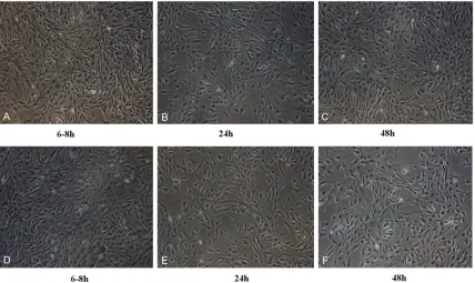
Astrocytes isolated by the two kinds of meth-ods were observed under the inverted phase contrast microscope (Figure 1). The photos showed that during the process of astrocytes purification, most of the cells have been adher-ent after 6-8 h, become oval, spindle shaped, and irregular shaped (Figure 1A and 1D). After 24 h of cultivation, cells become are adherent, with an obvious halo, and mixed cultures of astrocytes have cluster shaped growth (Figure 1B and 1E). At the time of 48 h for culture, cells have completely fused and astrocytes increased with more slender branches (Figure 1C and 1F).
Immunocytofluorescence
[image:4.612.91.523.69.290.2] [image:4.612.91.289.344.468.2]Immunocytofluorescence was used to detect astrocytes under a fluorescence microscope (Figure 2). Astrocytes with GFAP-positive mark-er could present green florescence [15]. In this study, positive astrocytes in four visual fields (top, bottom, left, and right) were observed. The photos showed that about 95% astrocytes had
Figure 2. Immunocytofluorescence analysis of astrocytes isolated by the classical and novel methods. A-C: Astro-cytes isolated by the novel method; D-F: AstroAstro-cytes isolated by the classical method.
GFAP marker in the whole light microscope. Also, there was no difference of GFAP-marked astrocytes isolated by the classical and novel methods.
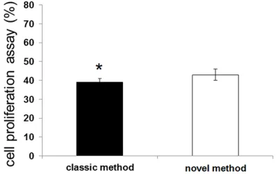
Cell proliferation
CCK-8 assay was used to detect the cell prolif-eration ability of astrocytes isolated by the two kinds of methods (Figure 3). The results showed that there was no significant difference of astrocytes proliferation that isolated with
the classical and with the novel methods (P>0.05).
Cell migration
Transwell chamber was used to assess the cell migration ability of astrocytes isolated by two kinds of methods (Figure 4). The photos show- ed that there was no significant difference for migrated cell number of astrocytes isolated by two kinds of methods. Also, migrate cells from the two isolated methods performed no differ-ence at different time points (P>0.05).
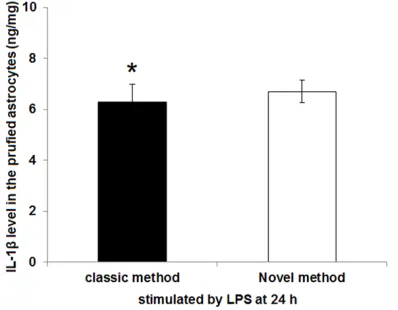
ELISA assay
ELISA assay was used to measure the proteins or cytokines secreted by astrocytes that isolat-ed with the classical and novel method (Figure 5). Our results displayed that the level of IL-1β secreted by astrocytes isolated with the novel method was 6.31 ± 0.68 ng/mg, while IL-1β level of the purified astrocytes in novel method group was 6.72 ± 0.45 ng/mg. Also, there was no significant difference of the IL-1β level secreted by strocytes between the two groups (P>0.05).
Discussion
[image:5.612.93.522.72.246.2]The glial cells were one of the two kinds of nerve tissue cells, and play pivotal roles in sup-porting neurons and promoting regeneration post-injuries [16]. Astrocytes were some most widespread and with largest number in nerve
Figure 4. Changes of astrocytes migration level of acquired by Transwell assay for different old and new methods. A-E: The number of astrocytes going across the membrane with a new method; F-J: The number of astrocytes going across the membrane with the classical method. Count 5 field for observing cells under microscope with × 400.
[image:5.612.91.290.312.467.2]Novel method for astrocytes isolation
tissues, and play crucial roles in immune asso-ciated brain diseases [17]. Although many stud-ies have devoted to the exploration of astro-cytes isolation, the inventive methods used for astrocytes were not so good which often result in high economic cost and low purity [6, 11]. In this study, we reviewed the previous studies that aimed on astrocytes isolation and described a novel method for astrocytes isola-tion with trypsin. Our data revealed that cell cul-ture character of astrocytes isolated by our novel method was the same as that isolated by the classical method (P>0.05). Moreover, bio-logical characters such as proliferation, cell migration, and secretion ability of astrocytes isolated by our novel method were similar as that isolated with the classical method (P>0.05).
Studies have demonstrated that astrocytes were characterized with huge volume and large nucleus, lots of cell number, star or ovoid dur-ing development [18, 19]. The astrocytes iso-lated with the two kinds of method performed adherent ability after cultured for about 6-9 h, and cells become oval, spindle shaped, and irregular shaped. Consequently, cells become are adherent, with an obvious halo, and mixed cultures of astrocytes have cluster shaped growth at time point of 24 h. Moreover, cells have completely fused and astrocytes incre- ased with more slender branches, implying that our novel method could not change astrocytes biological characters.
In our study, immunofluorescence analysis showed that astrocytes isolated by the two kinds of methods had GFAP-marker. GFAP, a kind of intermediate filament protein, exists in cytoplasm of astrocytes, which is crucial for maintaining the steady of astrocytes structure and the growth and extension of synapse and is considered to be the characteristic symbol for astrocytes [20]. Several papers that devoted to the useful exploration of astrocytes isolation methods and other biological processes had all experimented the GFAP marker for cells [21, 22]. Therefore, our data suggested that there was no significant difference of astrocytes iso-lated by the classic and novel method. On the other hand, when we measured the biological characters for astrocytes isolated by classic and novel method, we observed that there was no significant difference of cell migration and proliferation between the two kinds of methods
(P>0.05), indicating that novel method could not change the cell proliferation and migration ability during isolation.
Meanwhile, IL-1β is one of the strongest inflam-matory mediators that secreted by the activat-ed astrocytes, and can not only promote amy-loid precursor protein expression but also accelerate entanglement of nerve fibers [23, 24]. Release of IL-1β contributes the cell adhe-sion, leukocyte migration, and reactive oxygen species (ROS) generation, which results in inducing cascade reactions of cytokines [25, 26]. Abundant IL-1β secretion would lead to inflammatory damage and malnutrition axons in nervous system associated diseases such as AD [27, 28]. Our data presented that the isolat-ed astrocytes purifiisolat-ed with the novel could secret 6.72 ± 0.45 ng/mg IL-1β, implying the cell viability for astrocytes. Besides, the IL-1β secreted by astrocytes isolated with classical method was 6.31 ± 0.68 ng/mg, and there was no significant difference of IL-1β yield secreted by astrocytes purified with two methods. Thus, we speculated that novel method could not change the cell viability and secretion ability of astrocytes.
In conclusion, the data presented that there were no significant differences between char-acteristics of astrocytes isolated by two meth-ods such as cell biological character, migration, and proliferation ability, suggesting that novel isolation method would save the tedious opera-tion steps for astrocytic isolaopera-tion compared to the classic method. Our study suggested that the novel isolation method on astrocytes is high-yield, easy operation, and high pure prepa-ration. This study may provide basis for the method exploration on astrocytes in vitro and may reduce the economic cost for clinical application.
Acknowledgements
Collaborative Innovation of Guangzhou (No. 201400000003-2).
Disclosure of conflict of interest
None.
Address correspondence to: Dr. Xiaodan Jiang, The National Key Clinical Specialty; The Engineering Technology Research Center of Education Ministry of China, Guangdong Provincial Key Laboratory on Brain Function Repair and Regeneration, Depart- ment of Neurosurgery, Zhujiang Hospital, Southern Medical University, 253 Gongye Road, Guangzhou 510282, China. E-mail: [email protected]
References
[1] Kiernan J and Rajakumar R. Barr’s the human nervous system: an anatomical viewpoint. Lippincott Williams & Wilkins; 2013.
[2] Parpura V, Heneka MT, Montana V, Oliet SH, Schousboe A, Haydon PG, Stout RF, Spray DC, Reichenbach A, Pannicke T, Pekny M, Pekna M, Zorec R, Verkhratsky A. Glial cells in (patho) physiology. J Neurochem 2012; 121: 4-27. [3] Morrens J, Van Den Broeck W and Kempermann
G. Glial cells in adult neurogenesis. Glia 2012; 60: 159-174.
[4] Murphy S. Astrocytes: pharmacology and func-tion. Academic Press; 2012.
[5] Oberheim NA, Goldman SA and Nedergaard M. Heterogeneity of astrocytic form and function. In: editors. Astrocytes. Springer; 2012. pp. 23-45.
[6] Silva GA, Feeney C, Mills LR and Theriault E. A novel and rapid method for culturing pure rat spinal cord astrocytes on untreated glass. J Neurosci Methods 1998; 80: 75-79.
[7] Crocker SJ, Frausto RF, Whitton JL and Milner R. A novel method to establish microglia-free astrocyte cultures: Comparison of matrix me-talloproteinase expression profiles in pure cul-tures of astrocytes and microglia. Glia 2008; 56: 1187-1198.
[8] Sawada M, Suzumura A, Yamamoto H and Marunouchi T. Activation and proliferation of the isolated microglia by colony stimulating factor-1 and possible involvement of protein kinase C. Brain Res 1990; 509: 119-124. [9] Seki Y, Suzuki SO, Masui K, Harada S,
Nakamura S, Kanba S and Iwaki T. A simple and high-yield method for preparation of rat microglial cultures utilizing Aclar plastic film. Neuropathology 2011; 31: 215-222.
[10] Saura J, Tusell JM and Serratosa J. High-yield isolation of murine microglia by mild trypsiniza-tion. Glia 2003; 44: 183-189.
[11] Wang X, Huang S, Chen X and Jiang Y. The im-provement on the primary culture and the puri-fication of astrocytes from cerebral cortex of SD rats. Journal of Shanxi Medical 2009; 1107-1110.
[12] Bolte S and Cordelieres F. A guided tour into subcellular colocalization analysis in light mi-croscopy. J Microsc 2006; 224: 213-232. [13] Kinoshita M, Nakatsuji Y, Moriya M, Okuno T,
Kumanogoh A, Nakano M, Takahashi T, Fujihara K, Tanaka K and Sakoda S. Astrocytic necrosis is induced by aquaporin-4 anti-body-positive serum. Neuroreport 2009; 20: 508-512.
[14] Paulie S, Perlmann H and Perlmann P. Enzyme-linked Immunosorbent Assay. eLS 2003; 77: 980-8.
[15] Dyer CA, Kendler A, Jean-Guillaume D, Awatramani R, Lee A, Mason LM and Kamholz J. GFAP-positive and myelin marker-positive glia in normal and pathologic environments. J Neurosci Res 2000; 60: 412-426.
[16] Malatesta P, Hartfuss E and Gotz M. Isolation of radial glial cells by fluorescent-activated cell sorting reveals a neuronal lineage. Development 2000; 127: 5253-5263.
[17] Furnari FB, Fenton T, Bachoo RM, Mukasa A, Stommel JM, Stegh A, Hahn WC, Ligon KL, Louis DN and Brennan C. Malignant astrocytic glioma: genetics, biology, and paths to treat-ment. Genes Dev 2007; 21: 2683-2710. [18] Verkhratsky A and Butt A. Morphology of glial
cells. Glial Neurobiology: A Textbook 2007; 21-28.
[19] Trivin A, Ramirez JM, Ramirez AI, Salazar JJ and Garcia-Sanchez J. Retinal perivascular as-troglia: an immunoperoxidase study. Vision Res 1992; 32: 1601-1607.
[20] Mayer CA, Brunkhorst R, Niessner M, Pfeilschifter W, Steinmetz H and Foerch C. Blood levels of glial fibrillary acidic protein (GFAP) in patients with neurological diseases. PLoS One 2013; 8: e62101.
[21] Wong SY, Chan SJ, Wong WF, Wong PTH and Lai MK. Andrographolide attenuates interleukin-1β-stimulated upregulation of che-mokine CCL5 and glial fibrillary acidic protein in astrocytes. Neuroreport 2014; 25: 881-886. [22] Cano V, Valladolid-Acebes I, Hernández-Nuño
F, Merino B, del Olmo N, Chowen JA and Ruiz-Gayo M. Morphological changes in glial fibril-lary acidic protein immunopositive astrocytes in the hippocampus of dietary-induced obese mice. Neuroreport 2014; 25: 819-822. [23] Akin D, Ravizza T, Maroso M, Carcak N, Eryigit
ab-Novel method for astrocytes isolation
sence epilepsy at the onset of spike-and-wave discharges, and contributes to their occur-rence. Neurobiol Dis 2011; 44: 259-269. [24] Andersson U and Tracey KJ. Neural reflexes in
inflammation and immunity. J Exp Med 2012; 209: 1057-1068.
[25] Yang CM, Hsieh HL, Yu PH, Lin CC and Liu SW. IL-1β Induces MMP-9-Dependent Brain Astro- cytic Migration via Transactivation of PDGF Receptor/NADPH Oxidase 2-Derived Reactive Oxygen Species Signals. Mol Neurobiol 2015; 52: 303-17.
[26] Ransohoff RM and Brown MA. Innate immunity in the central nervous system. J Clin Invest 2012; 122: 1164-1171.
[27] Parajuli B, Sonobe Y, Horiuchi H, Takeuchi H, Mizuno T and Suzumura A. Oligomeric amyloid β induces IL-1β processing via production of ROS: implication in Alzheimer’s disease. Cell Death Dis 2013; 4: e975.