HPLC METHOD FOR THE DETERMINATION OF TRANSDERMAL
DRUG DIFFUSION OF KETOPROFEN
Deep Kotal* and Dr. Rama Bukka
Nargund College of Pharmacy, II Main, Dattatreya Nagar, BSK III stage, Bangalore, India.
ABSTRACT
A simple, isocratic and sensitive high performance liquid
chromatographic method was developed for the estimation of
Ketoprofen during transdermal diffusion studies across porcine ear
epithelium. A reversed-phase (C18) column was used with UV detector
at 260 nm. Selected mobile phase contained a mixture of Acetonitrile
and double distilled water (40:60 v/v), pH adjusted to 3 using 1%
ortho-phosphoric acid solution. Calibration curve showed good
linearity over the concentration range of 5-50 µg/ml in ear epidermis
permeates of pH 7.4 phosphate buffer (Blank permeates). The
applicability of the method was demonstrated by the analysis of
porcine ear epithelium permeated samples of Ketoprofen in a diffusion study. The steady
state permeability Flux (J) of Ketoprofen through ear epidermis using 5mg and 10mg of
Ketoprofen solution in donor cell of diffusion study was found to be 0.0002 µg/sqcm/hr and
0.0006 µg/sqcm/hr respectively.
KEYWORDS: RP-HPLC, Flux, Diffusion study, Ketoprofen.
INTRODUCTION
The aim of the study was to develop a simple, isocratic and sensitive HPLC method for the
analysis of Ketoprofen during transdermal permeation studies. Ketoprofen,
(RS)-2-(3-benzoylphenyl)-propionic acid (chemical formula C16H14O3) is one of the propionic acid
class, a nonsteroidal anti-inflammatory drug (NSAID)[1,2] has been extensively utilized for treatment of rheumatism.[3] It is associated with oral side effects including gastrointestinal irritation when administered by oral route. The adverse effects may worsen to renal and
Volume 6, Issue 9, 725-732. Research Article ISSN 2277– 7105
*Corresponding Author
Deep Kotal
Nargund College of
Pharmacy, II Main,
Dattatreya Nagar, BSK III
stage, Bangalore, India. Article Received on 28 June 2017,
Revised on 18 July 2017, Accepted on 08 August 2017
DOI: 10.20959/wjpr20179-9249
deliver the drug via skin. Therefore, an eventual need has emerged to develop a transdermal
dosage form of Ketoprofen to minimize the oral side-effects and to provide relatively
consistent drug levels for prolonged periods.[4] The major problem associated with transdermal drug delivery is barrier properties of stratum corneum which is considered one of
the most impermeable epithelia of the human body to exogenous substances. These
[image:2.595.192.408.219.314.2]permeation problems can be minimized by use of chemical permeation enhancers.[5,6]
Figure 1: Chemical structure of Ketoprofen.
The present study is planned to estimate the Ketoprofen during transdermal permeability
study.
MATERIALS AND METHODS
Ketoprofen was purchased from BMR Pharma and Chemicals, Hyderabad. HPLC grade
solvents were purchased from Ranbaxy Fine Chemicals Ltd., New Delhi. Phosphoric acid
was purchased from Karnataka Fine Chem. Triple distilled water was prepared in laboratory.
Porcine ear tissue obtained from local slaughter house.
Chromatographic conditions[7,8,9]
The instrument used for the HPLC analysis was Shimadzu LC equipped with LC 20 AT
prominence liquid chromatograph pump, and a Shimadzu SPD-20A UV prominence UV/Vis
detector. An Inertsil ODS-3V (4.6 mm ×250mm; 5 μm particle size) column was employed
during the analysis. An isocratic method was used with a mobile phase containing a mixture
of Acetonitrile (ACN) and triple distilled water (40:60v/v). The pH of the used triple distilled
water was adjusted to pH 3 with 1% ortho-phosphoric acid solution. Flow rate of the mobile
phase was 1.0 ml per minute. The mobile phase was vaccum-filtered through 0.45 μm
The injection volume was 20 μL. After equilibration with the solvent to obtain a stable
baseline, aliquots of calibration solutions containing Ketoprofen and internal standard were
injected.
The total run time was 10 minutes. The absorbance of the eluent was monitored at 260nm
with a detection sensitivity of 0.250 aufs. All the analysis was performed at room
temperature.
Preparation of Calibration Standards
Preparation of Blank Permeates
Porcine ear tissue was obtained from a local slaughter house immediately after pigs were
slaughtered. Ear tissue of freshly slaughtered pigs was immediately transferred to our
laboratory. The dorsal hair was removed with a clipper and full thickness skin was surgically
removed with the help of micro- dissecting scissor and blunt forceps. Then the ear skin was
dipped into the hot water (60ºC) and then fat adhering to epidermis was carefully removed to
get epidermis without any damage to stratum corneum. Washed with water and used in
permeation studies. Then the permeation studies were initiated within 1hr of isolating ear
epithelium. The tissue permeates of the pH 7.4 Phosphate buffer (blank permeates) was
prepared by placing the porcine ear epidermis in Franz diffusion cell between donor and
receptors and clamped with the help of rubber bands and pH 7.4 Phosphate buffer was added
in the donor (5 ml) as well as receptor compartment (35 ml) of the diffusion cells and taking
care to avoid the entry of air bubbles. Magnetic bead was added in the receptor to maintain
stirring conditions. The entire set up was placed over magnetic stirrer and temperature was
maintained at 370C by placing the diffusion cell in a water bath for 24 hrs at 370C. The Permeates collected after 24 hrs were used as diluent for the preparation of calibration
standards. This will eliminate any interference of the components which were eluted during
diffusions study from the porcine ear epidermis.
Preparation of Calibration Standards for HPLC Estimation
In this method, Internal Standard was also added in a constant amount to calibration samples
which will correct for the loss of analyte during sample preparation and sample inlet.
Propyphenazone was selected as Internal Standard. Standard stock solution of Ketoprofen
A secondary stock solutions (100 μg/ml) were prepared by dilution of the primary stock
solution with pH 7.4 Phosphate buffer for both Ketoprofen and internal standard solutions.
From secondary stock solutions aliquots (0.5, 1, 2, 3, 4, 5 ml) of Ketoprofen and 1 ml of
internal standard were further diluted to 10 ml with blank permeates to obtain six calibration
standards (5, 10, 20, 30, 40 and 50 μg/ml) of Ketoprofen and 10 μg/ml of internal standard
using 10 ml volumetric flask. The prepared calibration standards were analyzed after filtering
through 0.45 μm Millipore membrane filter.
As some of the tissue components will also diffuse into the receptor compartment during the
study and which are found to be eluted in a time range of 1–4 minutes, the mobile phase was
selected to give a retention time of greater than 6 minutes for the drug.
For every calibration sample ratio of peak areas of Ketoprofen and internal standard were
calculated. Standard graph was plotted by taking concentration on X axis and ratio of areas
on Y axis. R2 and slope were calculated to estimate the linearity.
Ex-vivo permeation study of Ketoprofen from solution through Porcine ear tissue
The Franz diffusion cells are thoroughly cleaned with triple distilled water and as previously
discussed separated porcine ear epidermis are placed between the donor and receptor
compartments of the diffusion cell. The permeation studies were initiated within 1hr of
isolating ear epithelium. The pH 7.4 Phosphate buffer was filled into the receptor without any
air bubbles. 1mg/ml of pure Ketoprofen solution was prepared by dissolving Ketoprofen in
little amount of Ethanol and volume make up with pH 7.4 phosphate buffer. 5 ml of this
solution was added into the donor compartment. Then the entire set up was placed over
magnetic stirrer and temperature was maintained at 370C by placing the diffusion cell in a water bath for 24 hrs at 370. Then 1ml of sample was withdrawn from the bottom portion of receptor using injection syringe fitted with a catheter. Samples were withdrawn at 1hr, 2 hrs,
4 hrs, 6 hrs, 8 hrs, 10 hrs 12 hrs, 17 hrs and 24 hrs and replaced with 1 ml of pH 7.4
phosphate buffer after every sample withdrawal.
For estimation of Ketoprofen in the permeation, 0.1 ml of permeate was added with 0.1 ml of
I.S. solution (100 μg/ml). The volume was made upto 1 ml with mobile phase. This sample was filtered using 0.2 μm Millipore membrane filter before injected into HPLC. Similar study
In both studies, graph is plotted by taking cumulative amount permeated per square on Y axis
and time interval on X axis. Flux was calculated from the slope of the straight line of the
graph.
RESULTS AND DISCUSSION
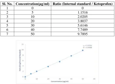
Calibration Standard of Ketoprofen by HPLC
The standard graph was found to be linear in the range of 5-50 μg/ml. The R2 and the slope(Y) were found to be 0.9983 and 0.1936 respectively. The values were presented in
Table no. 1 and Fig no. 2.
Table 1: Calibration curve of pure drug solution by HPLC.
Sl. No. Concentration(μg/ml) Ratio (Internal standard / Ketoprofen)
1 0 0
2 5 1.1516
3 10 2.0205
4 20 3.8037
5 30 5.6146
6 40 7.7489
[image:5.595.108.488.281.559.2]7 50 9.7895
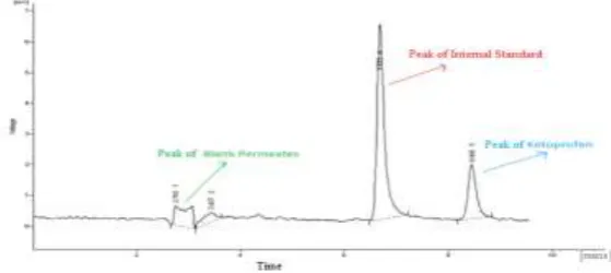
[image:5.595.161.438.601.745.2]Fig. 4: Chromatogram of Calibration Solution Containing Ketoprofen and IS.
Ex-vivo permeation study of Ketoprofen from solution through Porcine ear tissue
The diffusion study of Pure Ketoprofen drug solution was done by HPLC and the blank peaks
due to tissue components showed retention time around 2-3 mins. Ketoprofen’s retention
time was observed at 8.4 mins and peaks due to Propyphenazone (I.S) was observed at 6
mins. As some of the tissue components will also diffuse into the receptor compartment
during the diffusion study, and many interference with the drug and internal standard peaks.
To avoid such interference blank tissue permeates were used in standard graph preparation.
The mobile phase was selected to give a retention time of greater than 4 minutes for the drug.
The mobile phase and calibration standards were freshly prepared on the day of usage. The
model chromatogram of Blank permeates and a calibrated solution containing both drug and
IS were shown in the figures 3 and 4.
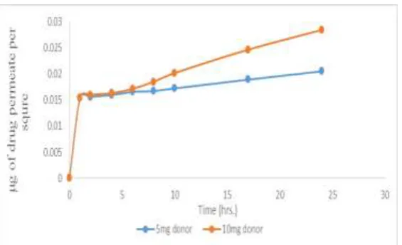
The values of Diffusion studies were tabulated in table no. 2 and Fig no 5. The flux (J)
observed when 5mg and 10mg of Ketoprofen was used in donor was to 0.0002 µg/sqcm/hr
and 0.0006 µg/sqcm/hr respectively, which indicates that increasing the drug quantity, in
donor has increased permeation.[10,11]
Table 2: Diffusion Study Curve of Pure Ketoprofen Solution by HPLC.
Time (Hrs.)
μg of drug permeated per sq.cm.
For 1mg/ml For 2mg/ml
0 0 0
1 0.0155 0.0155
2 0.0156 0.0160
4 0.0159 0.0163
6 0.0166 0.0172
8 0.0167 0.0185
10 0.0172 0.0202
17 0.0189 0.0247
Fig. 5: Diffusion curve of Ketoprofen solutions through porcine ear epithelium.
CONCLUSION
A simple, isocratic and sensitive HPLC method was developed to estimate the Ketoprofen
during Transdermal diffusion study through porcine ear epithelium using for 5mg and 10mg
of drug in donor and flux was found to be 0.0002 µg/sqcm/hr and 0.0006 µg/sqcm/hr
respectively.
This study can be used to estimate the concentration of Ketoprofen in any transdermal
formulation diffusion studies.
ACKNOWLEDGEMENTS
We are grateful to Professor DR. L.V.G. Nargund, Principal of Nargund College of Pharmacy
Mrs. Prachi Kabra, Professor of Nargund College of Pharmacy for their kind help and
inspiration for this experiment to publish this research articles.
REFERENCES
1. Ketoprofen Veterinary - Systemic, 2004; 1–21.
2. Tripathi K. Essentials Of Medical Pharmacology. Seventh Ed. New Delhi: Jaypee
Brothers Medical Publishers (P) Ltd, 2013; 192-209.
3. Deruiter J. Non-Steroidal Antiinflammatory Drugs (Nsaids). Princ Drug Action, 2002; 2:
1–26.
4. Hafeez A, Jain U, Singh J, Maurya A, Rana L. Recent Advances In Transdermal Drug
Delivery System (Tdds): An Overview. J Sci Innov Res, 2013; 2(3): 695–709.
6. Premjeet S, Bilandi A, Sahil K, Akanksha M. Transdermal Drug Delivery System
(Patches), Applications In Present Scenario. Int J Res Pharm Chem, 2011; 1(4): 2231–
781.
7. Bukka R, Prakasam K. Hplc Estimation Of Rasagiline Mesylate During Buccal
Permeation Studies. Int J Pharm Sci Rev Res., 2012; 16(1): 115–9.
8. Tsvetkova B, Peikova L. Hplc Determination Of Ketoprofen In Tablet Dosage Forms.
Trakia J Sci, 2013; 11(1): 55–9.
9. Patel Kc. Improving The Bioavailability Of Model Bcs Class Ii Drug Simvastatin By Self
Micro Emulsifying Drug Delivery System, 2013.
10.Gide Ps, Gidwani Sk, Kothule Ku. Enhancement Of Transdermal Penetration And
Bioavailability Of Poorly Soluble Acyclovir Using Solid Lipid Nanoparticles
Incorporated In Gal Cream. Indian J Pharm Sci, 2013; 75(2): 138-142.
11.Uner M, Karaman Ef, Aydogmus Z. Solid Lipid Nanoparticles And Nanostructure Lipid
Carriers Of Loratadine For Topical Application: Physicochemical Stability And Drug
Penetration Through Rat Skin. Topical Journal Of Pharmaceutical Research, 2014; 13(5):