MAGNETIZATION APPLICATION IN THE NON INVASIVE DRUG DELIVERY OF MAGNETIC
MICROSPHERES OF THE ANTI CANCER DRUGS
1,*
Kirupakar, B.R., DR.
1Vishwanath,
1
Department of Pharmaceutics, Aditya Bangalore Institute of Pharmaceutical Education and Research,
Rajiv Ghandhi Univerity of Helath
2
Research & Development, Gland Pharma, Hyderabad, AP, India
ARTICLE INFO ABSTRACT
A number of novel drug delivery systems have emerged by various routes of administration, to achieve controlled and targeted drug delivery, magnetic drug delivery system being one of them which include magnetic microspheres, magnetic liposomes, magnetic nanoparticles, magnetic resealed erythrocytes, magnetic emulsion and others. Magnetic microsphe
labels have been used for great number of application in various areas of biosciences, targeted drug delivery, imaging and in bioseparation technology. This review summarize about application of magnetism in targeted non invasive dr
magnetic drug delivery system have been investigated for targeted drug delivery especially magnetic targeted chemotherapy due to their better tumor targeting, therapeutic efficacy, lower toxicity flexibility to be tailored for varied desirable purposes.
Copyright © 2015 Kirupakar et al.This is an open access article distributed under the Creative Commons Att distribution, and reproduction in any medium, provided the original work is properly cited.
INTRODUCTION
Since the pioneering idea proposed by Freeman iron particles could be transported through the
and be concentrated at a particular point in the body with the aid of a magnetic field, the use of magnetic particles for the delivery of drugs or antibodies to organs or tissues altered by disease has become an active and attractive field
(1, 2)
In the chemotherapy of the cancer patients less than 0.1 to 1% of the drugs are taken up by tumor cancerous cells, with the remaining 99% going into healthy tissue.
prescribe combination drugs in the chemot
compound side effects and the dosage is finalized based on the patient’s tolerance to the toxic effect rather than dose required to kill all the tumor cells. (5, 6, 7) The ability totarget the drug, to physically direct and focus it to specific sites or organ in the body, would provide the better treatment to cancer as well as other diseases. (8-11) Hence a need exists to focus the drugs to the disease locations of the body such as head, neck, lungs, liver, kidney, breast, ovary, testis, intestine and cervical region, to mention few.
*Corresponding Author: Kirupakar, B.R
Department of Pharmaceutics, Aditya Bangalore Institute of Pharmaceutical Education and Research, Rajiv Ghandhi Univerity of Helath Sciences, Bangalore, Karnataka, India
ISSN: 0975-833X
Vol.
Article History:
Received 24th September, 2015 Received in revised form 10th October, 2015
Accepted 15th November, 2015 Published online 30th December,2015
Key words:
Magnet, Anti cancer drug, Magnetic force, magnetic field, Rare earth magnet.
Citation: Kirupakar, B.R., DR. Vishwanath, B.A., Padma Sree, M., Shwetha, R and Prasanna Sagar
of herbicides and Mgnrega in India”,International Journal of Current Research
RESEARCH ARTICLE
MAGNETIZATION APPLICATION IN THE NON INVASIVE DRUG DELIVERY OF MAGNETIC
MICROSPHERES OF THE ANTI CANCER DRUGS
Vishwanath, B.A.,
1Padma Sree, M.,
1Shwetha, R and
Department of Pharmaceutics, Aditya Bangalore Institute of Pharmaceutical Education and Research,
iv Ghandhi Univerity of Helath Sciences, Bangalore, Karnataka, India
Research & Development, Gland Pharma, Hyderabad, AP, India
ABSTRACT
A number of novel drug delivery systems have emerged by various routes of administration, to achieve controlled and targeted drug delivery, magnetic drug delivery system being one of them which include magnetic microspheres, magnetic liposomes, magnetic nanoparticles, magnetic resealed erythrocytes, magnetic emulsion and others. Magnetic microsphe
labels have been used for great number of application in various areas of biosciences, targeted drug delivery, imaging and in bioseparation technology. This review summarize about application of magnetism in targeted non invasive drug delivery system of magnetic microspheres. Over the years, magnetic drug delivery system have been investigated for targeted drug delivery especially magnetic targeted chemotherapy due to their better tumor targeting, therapeutic efficacy, lower toxicity flexibility to be tailored for varied desirable purposes.
is an open access article distributed under the Creative Commons Attribution License, which distribution, and reproduction in any medium, provided the original work is properly cited.
Since the pioneering idea proposed by Freeman et al. that fine iron particles could be transported through the vascular system and be concentrated at a particular point in the body with the aid of a magnetic field, the use of magnetic particles for the delivery of drugs or antibodies to organs or tissues altered by disease has become an active and attractive field of research.
In the chemotherapy of the cancer patients less than 0.1 to 1% of the drugs are taken up by tumor cancerous cells, with the remaining 99% going into healthy tissue. (3, 4) Physicians prescribe combination drugs in the chemotherapy that can compound side effects and the dosage is finalized based on the patient’s tolerance to the toxic effect rather than dose required The ability totarget the drug, to ific sites or organ in the body, would provide the better treatment to cancer as well as Hence a need exists to focus the drugs to the disease locations of the body such as head, neck, lungs, estine and cervical region,
Department of Pharmaceutics, Aditya Bangalore Institute of Pharmaceutical Education and Research, Rajiv Ghandhi Univerity of
Magnetic drug targeting (MDT) refers to the attachment of therapeutic drug to magnetizable particles such as iron oxide, and then applying magnetic fields to concentrate them to disease locations such as to solid tumors, regions of infection, or blood clots. (12-18) The magnetizable particles can also be introduced into the body outside the blood flow, e.g. as in magnetic treatment of the inner
containing nanoparticles is placed on the round window membrane or intranasally, usually fe
directly injected into the circulation by a vein or artery.
Injected drug loaded iron particles will circulate throughout the vasculature as the applied magnetic field with rare earth magnet directs the particles and re
locations. Depending on the vessel into which the particles were injected (vein or artery), MDT will occur before the particles pass through the liver (first pass method or after the particles pass through the liver, lung and heart
Magnetic fields more than light, electric fields, and ultrasound are desirable for directing therapeutics inside patients because they can penetrate deep into the body, also usually applied through the body in magnetic resonance imaging
are considered safe to very high strengths (8 Tesla in adults, 4 T in children).(36-44) Magnetic fields can both sense and actuate magnetic particles, although achieving both at once is an International Journal of Current Research
Vol. 7, Issue, 12, pp.24733-24742, December, 2015
INTERNATIONAL
Kirupakar, B.R., DR. Vishwanath, B.A., Padma Sree, M., Shwetha, R and Prasanna Sagar, 201
International Journal of Current Research, 07, (12), 24733-24742.
MAGNETIZATION APPLICATION IN THE NON INVASIVE DRUG DELIVERY OF MAGNETIC
R and
2Prasanna Sagar
Department of Pharmaceutics, Aditya Bangalore Institute of Pharmaceutical Education and Research,
Sciences, Bangalore, Karnataka, India
Research & Development, Gland Pharma, Hyderabad, AP, India
A number of novel drug delivery systems have emerged by various routes of administration, to achieve controlled and targeted drug delivery, magnetic drug delivery system being one of them which include magnetic microspheres, magnetic liposomes, magnetic nanoparticles, magnetic resealed erythrocytes, magnetic emulsion and others. Magnetic microspheres& molecular magnetic labels have been used for great number of application in various areas of biosciences, targeted drug delivery, imaging and in bioseparation technology. This review summarize about application of ug delivery system of magnetic microspheres. Over the years, magnetic drug delivery system have been investigated for targeted drug delivery especially magnetic targeted chemotherapy due to their better tumor targeting, therapeutic efficacy, lower toxicity and
ribution License, which permits unrestricted use,
Magnetic drug targeting (MDT) refers to the attachment of therapeutic drug to magnetizable particles such as iron oxide, and then applying magnetic fields to concentrate them to disease locations such as to solid tumors, regions of infection, magnetizable particles can also be introduced into the body outside the blood flow, e.g. as in magnetic treatment of the inner-ear where a small gel containing nanoparticles is placed on the round window membrane or intranasally, usually ferromagnetic particles are directly injected into the circulation by a vein or artery.(12, 19-31) Injected drug loaded iron particles will circulate throughout the vasculature as the applied magnetic field with rare earth magnet directs the particles and remains confined at target locations. Depending on the vessel into which the particles were injected (vein or artery), MDT will occur before the particles pass through the liver (first pass method or after the particles pass through the liver, lung and heart.(12, 25, 31-35)
Magnetic fields more than light, electric fields, and ultrasound are desirable for directing therapeutics inside patients because they can penetrate deep into the body, also usually applied through the body in magnetic resonance imaging (MRI), and are considered safe to very high strengths (8 Tesla in adults, 4 Magnetic fields can both sense and actuate magnetic particles, although achieving both at once is an INTERNATIONAL JOURNAL OF CURRENT RESEARCH
engineering challenge. In contrast, light and ultrasound have limited tissue penetration depths while strong electric fields (> 60 V/cm) are able to damage nerve and muscle cells. (40, 42,
45-51)
It is hypothesized that control of magnetic microspheres
in vivo is feasible given a strong magnetic field and gradient
space super positioned on target organ system. This approach triggered the desire for the non invasive surgery. Magnetic microspheres have potential use as magnetic seeds for drug delivery. Magnetic microspheres are paramagnetic and have been made to range in size from approximately one micron to greater then 600 microns.
Magnetic Microspheres Design
Magnetic microspheres incorporate magnetic materials with the drugs. Encapsulation of the drugs with magnetite shells or attaching the drugs to a functionalized outer coating may be performed. Functionalized outer coatings of microspheres are a well-established procedure.(52) Such modified magnetic microspheres may then be delivered to the target cells, where the microspheres will decompose due to degradable coating, and the drug will be delivered. Each of the microspheres contains multiple magnetite molecules. A magnetic domain is a volume of material whose magnetic field is aligned in a given direction. When magnetite is smaller than approximately 30 nm in diameter only single magnetic domains form. Magnetite clusters larger than 30 nm start to interact and form a multiple domain material. In single domain materials there is little to no hysteresis and the magnetic particles reach saturation faster compared to a multi domain material.
If multiple domains were formed a hysteresis loop in the magnetization curves is observed. Hysteresis from multi domain formation causes a decrease in the response of the system. The organic sheaths surrounding the magnetite clusters provide both the ability to functionally attach drugs as well as to keep the magnetic particles from aggregating, preserving single domain formation. FeRx (company) used milled 1 μm iron-activated carbon which has higher magnetic moment when compared to magnetite. Iron is injected into blood vessels near the target organ and then a single external permanent magnet pulls the particles. The field removed after approximately 15 minutes and an angiogram is performed to make sure blockage of the main arteries has not occurred. (53)
Physics of magnetic drug targeting
Rare Earth Magnet- Magnetic Field Generation
The magnetic fields generated by external magnets in in-vitro
studies have ranged anywhere from 70 m T to ≤ 1.5 T. (54-56) Animal trials have had ranges between 0.1 T and 1.5 T. (30, 35,
57-59)
While the FDA has approved magnetic strengths up to 8 T for use with humans and human clinical trials have utilized 0.2 to 0.8 T magnet field strengths. (12, 19, 29, 30,60)
Most often permanent magnets have been used with sizes ranging from tens of millimeters to tens of centimeters. (31, 33, 54,
56, 57, 60)
Occasionally electromagnets were utilized. (55, 61) The distance of particles from magnets has ranged from ≈ 1 mm to ≈ 12 cm in the literature. (12, 31, 35, 53)
Magnetic fields and forces acting upon magnetic Microspheres
Magnetic nanoparticles are small and experience small forces even under strong magnetic fields. In magnetic drug delivery experiments, magnet strengths have ranged from 70 milli-Tesla to 2.2 Tesla, and corresponding magnetic gradients have varied from 0.03 T/m to 100 T/m, a range that reflects magnet cost, complexity, safety, and ease-of-use versus desired depth of targeting site.(54, 62-64) Modern neodymium-iron-boron (Nd12Fe14B) permanent magnets can be purchased in strengths
of up to 1.48 T and the electromagnets used in magnetic resonance imaging systems create fields of 1 - 4.7T, with some commercially-available MRI systems going as high as 9.4T. (41,
65-67)
In the human trials, 0.2 - 0.8 T permanent magnets were used to target 100 nm diameter particles to 5 cm depths.(12,30)Targeting depths of up to 12 cm have been reported in animal experiments using larger 500 nm to 5 μm diameter particles and a 0.5 T permanent magnet. (31)
Both permanent and electromagnet designs can be optimized to extend magnetic fields and gradients further out, to increase the depth of magnetic forces. Permanent and electromagnet designs can be optimized to extend magnetic fields and gradients further out, to increase the depth of magnetic forces. Particles injected through vein or artery will circulate throughout the vasculature as the applied magnetic field attempt confinement at the target organ or site in the body. As a first consideration, the particles must be small (400 – 600 nm) enough to make it out from the blood vessels into surrounding tissue to extravasate out from even 'leaky' tumor vessels, and, more subtly and crucially, they must be small enough to have sufficiently long in vivo residence times (larger particles are removed faster by the mononuclear phagocyte system; in human clinical trials Chemicell’s 100 nm particles were shown to have 30 min plasma residence times.(19, 29, 60,
68-72)
Second, the magnetic force on these small particles is minimal. Magnetic force scales with particle volume, decreasing the size of a particle by a factor of 10 decreases the magnetic force on it by 1000. Even with strong magnetic fields (> 1 Tesla) and high magnetic gradients (≈ 0.5 T/cm), the forces on ferro-magnetic nanoparticles remain extremely small, in the range of pico-Newtons.(73-75) A key issue in magnetic drug delivery is whether the applied magnetic forces can compete with convective blood (drag) forces that tend to wash particles away.
Counter force on the magnetic microspheres in the blood
The force that counteracts the magnetic force on the particle in the bloodstream is due to blood flow. Stokes Law governs the hemodynamic forces on a particle in a flowing liquid.
F= 6 π η v r
requires more magnetic field force to pull the microspheres and such interfacial transport could cause damage to the tissue. Therefore, the microsphere size and forces needed for effective microsphere and dependent on the drug delivery area.
Magnetization and external magnetic field
All materials are magnetic to some extent, with the degree depending on their atomic structure and the temperature. Electrons circulating around atomic nuclei, electrons spinning on their axes and rotating positively-charged atomic nuclei are all magnetic dipoles, also called magnetons. Altogether, these effects may cancel out, so that a given type of atom may not be a magnetic dipole. If they do not fully cancel out, however, the atom is a permanent magnetic dipole, as in the case of iron atoms. The strength of a magnetic dipole is called the magnetic dipole moment and may be thought of as a measure of the capacity of the dipole to align itself with a given external magnetic field. When an external magnetic field (H) is applied to a material, the atomic dipoles tend to align themselves with the field, thereby causing a magnetic moment within the material. The quantity of magnetic moment per unit volume is defined as magnetization (M).
The relation between magnetization and the magnetic field is given by:
M=XH
Where χ is the volumetric magnetic susceptibility, which in SI units is dimensionless, and both M and H are expressed in A·m−1. Magnetic materials may be conveniently classified in terms of their χ. (76) when the materials exhibit weak repulsion (negative susceptibility, with χ in the range −10−6 to −10−3), they are termed diamagnets. If the materials show small positive susceptibility (χ in the range 10−1 to −10−6), they are paramagnets whereas a ferromagnet is a material that exhibits a large positive susceptibility. The magnetic properties of the diamagnet and paramagnet do not persist if the external magnetic field is removed whereas ferromagnetic materials have stable magnetic properties even after removal of the external field.
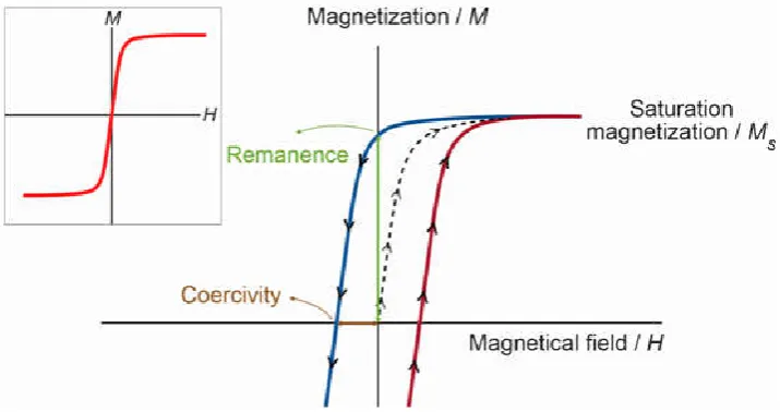
For diamagnets and paramagnets, the relationship M = χ H is usually linear. In contrast, for ferromagnets, there is no one-to-one correspondence between H and M, and this relationship is not linear. If a paramagnet is demagnetized (H = M = 0) and the relationship between M and H is plotted for increasing levels of H, then M follows the initial magnetization curve (see figure dashed line). This curve increases rapidly at first and then becomes asymptotic as it approaches magnetic saturation (Ms). If H values are reduced monotonically, M follows a different curve (see figure blue line). At H = 0, M is offset from the origin by an amount called the remanent magnetization, Mr, which indicates the level of residual magnetism in the material. Therefore, the curve, of a sigmoidal shape, tends to a point where M = 0. This is called the point of coercivity on the curve. Therefore, the coercivity is the magnitude of the field that must be applied in the negative direction to bring the magnetization of the sample backto zero. As H increases in the negative direction, the material will again become magnetically saturated, but in the opposite direction. Increasing H in the positive direction again will return H to zero, and the curve returns to the saturation point (see figure red line), where it completes the hysteresis loop. The width of the middle section is twice the coercivity of the material. The area of the hysteresis loop is related to the amount of energy dissipated upon reversal of the field.
[image:3.595.121.479.556.745.2]In ferromagnetic materials, magnetons are associated in groups called domains. A magnetic domain refers to a volume of ferromagnetic material in which all magnetonsare aligned in the same direction by exchanging forces. A bulk ferromagnet spontaneously subdivides into a multidomain structure to reduce the magnetostatic energy associated with a large stray field. (77)Within each domain, the magnetization does not vary; but between domains, there are relatively thin domain walls in which the direction of magnetization rotates from the direction of one domain to that of the other. The formation of the domain walls is a process driven by the balance between the magnetostatic energy, which increases proportionally to the volume of material, and the domain wall energy, which increases proportionally to the interfacial area between domains.
When the size of a ferromagnetic material is reduced below a critical value, the so-called critical diameter, more energy is required to create a domain wall than to support the external magneto static energy of the single domain state; the material becomes a single domain. Critical diamateris few tens of nanometers and depends on the material. The critical diameter of a spherical particle is reached when the magnetostatic energy equals the interfacial energy. A single domain particle is uniformly magnetized with all of the spins aligned in the same direction. The magnetization will be reversed by spin rotation, since there are no domain walls to move. Reduction in size causes the thermal energy to exceed the energy barrier, which separates the two energetically-equivalent easy directions of magnetization and the direction of the magnetization fluctuates randomly. Such a system is named a super paramagnet.
The magnetic moments of individual crystallites compensate for each other, and the overall magnetic moment becomes null. When an external magnetic field is applied, the behavior is similar to paramagnetism, except that, instead of each individual atom being independently influenced by an external magnetic field, the magnetic moment of the crystallite aligns itself with the field. Consequently, super paramagnetic particles become magnetic in the presence of an external magnet, but revert toa non-magnetic state when the external magnet is removed.
This is of paramount importance when these particles are introduced into living systems (e.g., in drug delivery), because, once the externalmagnetic field is removed, the magnetization disappears (they have negligible remanent magnetizationand coercivity; see Figure), and thus, agglomeration (and the possible embolization ofcapillary vessels) is avoided. (78) The coercivity is zero for super paramagnets, but it increases inthe single domain regimen and shows a peak with the development of multiple magnetic domains, as the particles reach the micrometer scale, the coercivity essentially becomes thesame as that of bulk iron. The shape of the loops (see Figure) is determined in part by particle size,in larger particles, witha multidomain ground state, the hysteresis loop is narrow, since it takes relatively little field energyto make the domain walls move.while in smaller particles, there is a single domain ground state,which leads to a broad hysteresis loop.
Magnetic Force
In order to analyze the movement of the magnetic microparticle in a static magnetic field Senyei et al(79) assumed that (1) there is no interaction between particles, (2) the particles are perfect spheres, (3) the gravity force does not affect the movement of the analyzed microspheres, (4) the product
dH
H --- in the measurement area is constant across the
capillary, (5) the magnetic field in x-direction
dy
dH
(along the capillary,) is constant --- 0 and (6) the particle’s Reynold number is less than 1
Dx
(i.e., the friction force becomes the Stoke's force). The particle movement in this hypothetic situation thus becomes one-dimensional and constant and is equal to
The movement of a magnetic microsphere in a well defined magnetic field is determined by (1) the magnetic properties of the particle (volume of the magnetic component, Vm; and magnetic susceptibility, ), (2) the hydrodynamic properties of the medium (viscosity, ), and (3) the dimensions (diameter, D) and physical properties (mass, m). When analyzing magnetic microspheres of the same type with identical shape and homogeneous distribution of the magnetic component, but different particle diameter, then the velocity v can be expressed as a function of the changing radius r or diameter D as
In order to effectively overcome the influence of blood flow, and in order to achieve desired external magnetic field-controlled guidance, the magnetic force due to the external field must be larger than the drag force.
To a first approximation, the magnetic force on the microsphere is governed by
⃗ = ∇⃗( ⃗• ⃗) (Newtons)
whereFis the magnetic force, m is the total magnetic moment of the material in the microsphere, ∇ is the gradient, assumed in our modeling to be derived from characteristics of the Bfield alone, and Bis the magnetic flux density, also known simply as the Bfield. Each of these quantities thus influences the degree to which an external magnetic field may be used to guide internal microspheres.
Magnetic guided drug targeting- Invivo administration &
retention Method
The method of magnetically-guided drug targeting (MGDT) involves the immobilization of a drug in Magnetic particles then,the injection of the drug/carrier complex into the subject, either via intravenous (i.v.) or intra-arterial(i.a.) injection; and, finally, the use of high-gradient external magnetic fields generated by rare-earthpermanent NdFeB magnets with a maximum surface flux density of a little overone Tesla to guide the complex and concentrate it at the desired locations. Once the complex is concentratedat the target in vivo, the therapeutic agent is then released from the magnetic carrier, either via enzymeactivity or through changes in physiological conditions, such as pH, osmolality or temperature.
This results in increased uptake of the drug by the tumor cells at the target sites and a limited systemicdrug concentration. (80,
81)
magnetic field, as well as the route of injection and the vascular supply tothe targeted tissues will all influence the effects. The physiological parameters of the patient, such asbody weight, blood volume, cardiac output, peripheral resistance of the circulatory system and organfunction, will also affect the efficiency of the external magnet; apart from the possibility of placing themagnet in close vicinity to the target location. (82)
Moreover, the administration route play role for the success of the therapy, sincei.a. delivery avoids, or at least minimizes, the particle clearance by the mononuclear phagocyte system (MPS) in liver and spleen in comparison to intravenously-applied particles for most magnetic carriers, the field strength (flux density) at the target site should be ofthe order of 200–700 mT with gradients along the z-axis of approximately 8–100 T/m, depending on theflow rate (higher blood flow rates require either stronger fields or higher gradients) (83-85) As a generalrule, the model indicates that when the magnetic forces exceed the linear blood flow rates in arteries(10 cm·s−1) or capillaries (0.05 cm·s−1), the MNPs will be retained at the target site and may beinternalized by the endothelial cells of the target tissue.(86)
Magnetic force and blood drag force invitro /invivo contrast
outcome
Past animal experiments (20, 25,27,30,31,33,35,87,57,58,88-109 ) and phase I human clinical trials (12, 30, 59, 110) have observed the accumulation of magnetic nanoparticles by visual inspection, magnetic resonance imaging, and histology studies. These have shown that magnetic forces can concentrate micro- and nanoparticles in vivo near magnets, but the details of that concentration cannot be seen experimentally. MRI and visual inspection do not have the resolution to show in which vessels magnetic forces have exceeded blood drag forces, and they certainly cannot show where in the vessel accumulation is occurring.
Equally, histology studies are carried out after the animal has been sacrificed and blood flow stopped; they speak only partially to where in the blood vessels the particles might have been. Alexander nacev et al. did simulation study to map the parameter space and characterize what should happen in an idealized blood vessel in terms of applied magnetic force strength and blood flow velocity. Lübbe and Bergemann et al used a 0.5 Tesla, 5 cm long, 5 mm wide permanent magnet to focus 250 nm diameter iron-oxide nanoparticles. Even for a particle at a distance of just 1 mm away from the magnet (just below skin depth), the magnetic force on this particle, including the effect of particle magnetic saturation and using an exact solution for the magnetic field around the magnet, is only about 1 x 10-13 N. (73,74,111) By comparison, the Stokes blood drag force on the same particle, for the slowest measured 0.1 mm/s blood-flow velocities in rat capillaries, is 7 x 10-13 N, a factor of x 7 greater. (112-115)
This simple comparison suggests that the field gradient near the magnet cannot capture a 250 nm particle against even the weakest blood flow in a rat. Dark spots of the particles were seen in the rats. The study was carried out while the rats were
alive and their blood was flowing, and it has been repeated even with 100 nm diameter particles where the magnetic forces are 2.53 = 15.625 times smaller. Clearly, a crude comparison of magnetic forces per particle to Stokes drag is insufficient to match in vivo behavior. This mismatch is also apparent in the literature both for in vitro and in vivo experiments. In in-vitro
studies, (108,56) particles were focused even when centerline stokes drag forces exceeded magnetic forces.
In the in vivo cases, (25, 35, 99) Stokes drag due to the slowest blood flow in the animals/humans exceeded maximum magnetic forces yet particle focusing was still observed. Above deficient is due to two main reasons. One, the blood flow drag forces on the particle vary with its position in the blood vessel with high velocity at the center of the vessel hence a higher drag force, but a particle near the blood vessel wall will have zero blood velocity. This decrease in velocity is due to the flow resistance provided by the vessel wall, the 'no-slip' boundary condition. (69,116, 117) Thus a particle near the vessel wall will experience a much smaller drag force and can potentially be held by a much smaller magnetic force. Second, the particles might agglomerate to some degree even though they are typically engineered to minimize agglomeration. (19, 118,
71)
This will increase the magnetic force, which grows with volume, much faster than the Stokes drag, which grows with diameter, thus increasing trapping. The magnetic moments of microspheres can be increased in three ways: - By clustering magnetite at the center of each sphere to produce large macro domains. By magnetizing the spheres to saturation levels prior to vascular targeting. By substituting one of the newer ferromagnetic materials that has high susceptibility than Fe3O4. (119)
Conclusion
Strong magnetic field required for the ferrofluid and deposition of magnetite the magnetic microcarriers still play an important role in the selective targeting, and the controlled delivery of various drugs. It is a challenging area for future research in the drug targeting so more researches, long term toxicity study, and characterization will ensure the improvement of magnetic drug delivery system. Huge progress in the technology has led to the development of manetic materials with properties that promise breakthroughs in a vast number of potential applications such as gene therapy, destroying built up plaque in arteries, image and extract foreign metallic and ferric objects from the body, and affect cancer therapies of in-vitro vesicular blockage, targeted radiation therapy, and hyperthermia.
REFERENCES
1. H. Weber and G. Landwehr, Ophthalmic Res. 14, 326 1982. 2. H Weber, G.Landwehr, H.Kilp, and H.Neubauer,
Ophthalmic Res., 14, 335 (1982).
3. R. Reszka, P. Beck, I.Fichtner, M.Hentschel, J.Richter, and J. Kreuter, “ Body distribution of free, liposomal and nanoparticle-associated mitoxantrone in B16-melanoma-bearing mice,” J. Pharmacol. Exp. Ther., vol. 280, no. 1, pp. 232–237, Jan. 1997.
4. A.A.M. Veldt, N.H. Hendrikse, E.F. Smit, M.P.J. Mooijer, A.Y. Rijnders, W.R. Gerritsen, J.J. M.Hoeven, A.D.Windhorst, A.A. Lammertsma, and M.Lubberin , “ Biodistribution and radiation dosimetry of 11C-labelled docetaxel in cancer patients,” Eur. J. Nucl. Med. Mol.
Imaging, vol. 37, no. 10, pp. 1950–1958, May 2010.
5. M. Perry, The chemotherapy source book, 2nd ed. Baltimore: Williams & Wilkins, 1996.
6. S.B. Horwitz, “Taxol (paclitaxel): Mechanisms of action,”
Ann. Oncol. Off. J. Eur. Soc. Med. Oncol. Esmo, vol. 5
Suppl 6, pp. S3–6, 1994.
7. D.Lorusso, A.Pietragalla, S.Mainenti, V.Masciullo, G.DiVagno, and G.Scambia, “Review role of topotecan in gynaecological cancers: Current indications and perspectives,” Crit. Rev. Oncol. Hematol., vol. 74, no. 3, pp. 163–174, Jun. 2010.
8. S. B. Duffull and B.A.Robinson, “Clinical pharmacokinetics and dose optimisation of carboplatin,”
Clin. Pharmacokinet., vol. 33, no. 3, pp. 161–183, Sep.
1997.
9. S. Roc well and G.B.Grindey, “Effect of 2’,2’-difluorodeoxycytidine on the viability and radio sensitivity of EMT6 cells in vitro,” Oncol. Res., vol. 4, no. 4–5, pp. 151–155, 1992.
10. E. Mini, S.Nobili, B.Caciagli, I.Landini, and T.Mazzei, “Cellular pharmacology of gemcitabine,” Ann. Oncol. Off.
J. Eur. Soc. Med. Oncol. Esmo, vol. 17 Suppl 5, pp. v7–
12, May 2006.
11. T.De Vita Jr., S.Hellman, and S.A. Rosenberg, Cancer,
principles and practice of oncology, 8th ed. Philadelphia
PA: Lippincott Williams and Wilkins, 2008.
12. A.S. Lubbe, C.Bergemann, H.Riess, F.Schriever, P.Reichardt, K.Possinger, M.Matthias, B. Dorken, F.Herrmann,R.Gurtler, P.Hohenberger, N. Haas, R.Sohr, B.Sander, A. J. Lemke, D.Ohlendorf, W.Huhnt, andD.Huhn, “Clinical experiences with magnetic drug targeting: A phase I study with 4’-epidoxorubicin in 14 patients with advanced solid tumors,” Cancer Res., vol. 56, no. 20, pp. 4686–4693, 1996.
13. J. Dobson, “Magnetic micro- and nano-particle-based targeting for drug and gene delivery,” Nanomed., vol. 1, no. 1, pp. 31–37, 2006.
14. M. Johannsen, U.Gneveckow, BThiesen, K. Taymoorian, C.H.Cho, N.Waldöfner, R.Scholz, A.Jordan, S.A.Loening, and P., “Thermotherapy of prostate cancer using magnetic nanoparticles: feasibility, imaging, and three-dimensional temperature distribution,” Eur. Urol., vol. 52, no. 6, pp. 1653–1661, Dec. 2007.
15. K. Maier-Hauff, R.Rothe, R.Scholz, U.Gneveckow, P.Wust, B.Thiesen, A.Feussner, A.vonDeimling, N.Waldoefner, R.Felix, and A.Jordan, “Intracranial thermotherapy using magnetic nanoparticles combined
with external beam radiotherapy: results of a feasibility study on patients with glioblastomamultiforme,” J.
Neurooncol., vol. 81, no. 1, pp. 53–60, Jan. 2007.
16. N.M. Orekhova, R.S.Akchurin, A.A.Belyaev, M.D.Smirnov, S.E.Ragimov, and A.N. Orekhov, “Local prevention of thrombosis in animal arteries by means of magnetic targeting of aspirin-loaded red cells,” Thromb. Res., vol. 57, no. 4, p. 611, 1990.
17. P.K. Stoimenov, R.L.Klinger, G.L.Marchin, and K.J.Klabunde, “Metal Oxide Nanoparticles as Bactericidal Agents,” Langmuir, vol. 18, no. 17, pp. 6679–6686, Aug. 2002.
18. P. Gong, H.Li, X.He, K.Wang, J.Hu, W.Tan, S.Zhang, and X.Yang, “Preparation and antibacterial activity of Fe3O4@Ag nanoparticles,” Nanotechnology, vol. 18, no. 28, p. 285604, Jul. 2007.
19. M. Arruebo, R.Fernandez-Pacheco, M.R.Ibarra, and J.Santamaria, “Magnetic nanoparticles for drug delivery,”
Nano Today, vol. 2, no. 3, 2007.
20. R.D. Kopke, R.A.Wassel, F.Mondalek, B.Grady, K.Chen, J.Liu, D.Gibson, and K.J.Dormer, “Magnetic nanoparticles: inner ear targeted molecule delivery and middle ear implant,” AudiolNeurotol, vol. 11, no. 2, pp. 123–133, 2006.
21. B. Shapiro, I.Rutel, and K.Dormer, “A System to Inject Therapeutically-coated Magnetic Nano-particles into the Inner Ear: Design and Initial Validation,” in Proceedings of the 3rd International Conference on Micro- and
Nanosystems, IDETC 2009, San Diego, CA, 2009.
22. L. Danielyan, R.Schäfer, A.VonAmeln-Mayerhofer, M.Buadze, J.Geisler, T.Klopfer, U.Burkhardt, B.Proksch, S.Verleysdon , and M.Ayturan, “Intranasal delivery of cells to the brain,” Eur. J. Cell Biol., vol. 88, no. 6, pp. 315–324, 2009.
23. R.A. Guerrero, J.M.Ball, S.S.Krater, S.E.Pacheco, J.D.Clements, and M.K.Estes, “Recombinant Norwal virus-like particles administered intranasally to mice induce systemic and mucosal (fecal and vaginal) immune responses,” J Virol, vol. 75, no. 20, pp. 9713–22, 2001. 24. M. Shinkai, “Functional magnetic particles for medical
application,” J. Biosci.Bioeng., vol. 94, no. 6, pp. 606– 613, 2002.
25. C. Alexiou, W.Arnold, R.J.Klein, F.G.Parak, P.Hulin, C.Bergemann, W.Erhardt, S.Wagenpfeil, and A.S.Lubbe, “Locoregional cancer treatment with magnetic drug targeting,” Cancer Res., vol. 60, no. 23, pp. 6641–6648, 2000.
26. C. Alexiou, R.Jurgons, R.Schmid, W.Erhardt, F.Parak, C.Bergemann, and H.Iro, “Magnetic Drug Targeting - A new approach in locoregionaltumortherapy with chemotherapeutic agents. Experimental animal studies,”
Hno, vol. 53, no. 7, pp. 618–622, 2005.
27. J.W. Barry, J.J.Bookstein, and J.F.Alksne, “Ferromagnetic embolization. Experimental evaluation,”
Radiology, vol. 138, no. 2, pp. 341–349, 1981.
28. A.A. Kuznetsov, V.I.Filippov, R.N.Alyautdin, N.L.Torshina, and O.A.Kuznetsov, “Application of magnetic liposomes for magnetically guided transport of muscle relaxants and anti-cancer photodynamic drugs,” J.
29. A.S.Lubbe, C.Bergemann, J.Broc, and D.G.McClure, “Physiological aspects in magnetic drug-targeting,” J.
Magn. Magn.Mater., vol. 194, no. 1–3, pp. 149–155,
1999.
30. A.S.Lubbe, C.Bergemann, W.Huhnt, T.Fricke, H.Riess, J.W.Brock, and D.Huhn, “Preclinical experiences with magnetic drug targeting: Tolerance and efficacy,” Cancer Res., vol. 56, no. 20, pp. 4694–4701, 1996.
31. S.Goodwin, C.Peterson, C.Hoh, and C.Bittner, “Targeting and retention of magnetic targeted carriers (MTCs) enhancing intra-arterial chemotherapy,” J. Magn.
Magn.Mater.,vol. 194, no. 1–3, pp. 132–139,1999.
32. T.Wu, H.-P.Lin, C.-N.Hu, J.-P.Chen, Y.-J.Chang, S.-T.Lee, and Y.-H.Ma, “Intra-Arterial Application of Magnetic Nanoparticles for Targeted Thrombolytic Therapy: A Rat Embolic Stro e Model,” in International Conference on the Scientific and Clinical Applications of
Magnetic Carriers, Rostock, Germany, 2010.
33. S.C.Goodwin, C.A.Bittner, C.L.Peterson, and G.Wong, “Single-dose toxicity study of hepatic intra-arterial infusion of doxorubicin coupled to a novel magnetically targeted drug carrier,” Toxicol. Sci., vol. 60, no. 1, pp. 177–183, 2001.
34. C.Alexiou, R.Jurgons, C.Seliger, S.Kolb, B.Heubeck, and H.Iro, “ Biodistribution of mitoxantrone after magnetic drug targeting: Fluorescence microscopic investigations on VX2 squamous cell carcinoma cells,” Z. Phys.
Chem.-Int. J. Res. Phys. Chem. Chem. Phys., vol. 220, no. 2, pp.
235–240, 2006.
35. K.J.Widder, R.M.Morris, G.Poore, K.P.Howard, and A.E.Senyei, “Tumor remission in {Y}oshida sarcoma-bearing rats by selective targeting of magnetic albumin microspheres containing doxorubicin,” PNAS, vol. 78, no. 1, pp. 579–581, 1981.
36. W.G.Pitt, G.A.Husseini, and B.J.Staples, “Ultrasonic drug delivery-a general review,” Expert Opin. Drug Deliv., vol. 1, no. 1, pp. 37–56, 2004.
37. Z.-G.Gao, H.D.Fain, and N.Rapoport, “Controlled and targeted tumor chemotherapy by micellar-encapsulated drug and ultrasound,” J. Control. Release Off. J. Control.
Release Soc., vol. 102, no. 1, pp. 203–222, Jan. 2005.
38. B.W.Barry, “Novel mechanisms and devices to enable successful transdermal drug delivery,” Eur. J. Pharm. Sci., vol. 14, no. 2, pp. 101–114, 2001.
39. A.R.Denet, R.Vanbever, and V.Preat, “Skin electroporation for transdermal and topical delivery,” Adv.
Drug Deliv. Rev., vol. 56, no. 5, pp. 659–674, 2004.
40. T.J.Dougherty, C.J.Gomer, G.Jori, D.Kessel, M.Korbelik, J.Moan, and Q.Peng, “Photodynamic therapy,” J. Natl.
Cancer Inst., vol. 90, no. 12, pp. 889–905, 1998.
41. J.F.Schenc, “Safety of Strong, Static Magnetic Fields,” J.
Magn. Reson. Imaging, vol. 12, no. 1, pp. 2–19, Jul. 2000.
42. D.J.Schaefer, J.D.Bourland, and J.A.Nyenhuis, “Review of patient safety in time-varying gradient fields,” J. Magn.
Reson.Imaging Jmri, vol. 12, no. 1, pp. 20–29, Jul. 2000.
43. E.D.Allen and J.H.Burdette, Questions and Answers in MRI, 2nd Ed. St. Louis, Missouri: Mosby, 2001.
44. E.Andersen, “Magnetic resonance imaging - safety and health issues,” Aaohn J. Off. J. Am. Assoc. Occup. Heal.
Nurses, vol. 55, no. 4, pp. 137–139, Apr. 2007.
45. S.Martel, O.Felfoul, J.B.Mathieu, A.Chanu, S.Tamaz, M.Mohammadi, M.Mankiewicz, and N.Tabatabaei, “MRI-based medical nanorobotic platform for the control of magnetic nanoparticles and flagellated bacteria for target interventions in human capillaries,” Int. J. Robot. Res., vol. 28, no. 9, pp. 1169–1182, 2009.
46. C.Catana, Y.Wu, M.S.Judenhofer, J.Qi, B.J.Pichler, and S.R.Cherry, “Simultaneous acquisition of multislice PET and MR images: {I}nitial results with a MR-compatible PET scanner,” J. Nucl. Med., vol. 47, no. 12, pp. 1968–76, 2006.
47. W.T.Katsiyiannis, D.P.Melby, J.L. Matelski, V.L.Ervin, K.L.Laverence, and C.C.Gornic, “Feasibility and safety of remote-controlled magnetic navigation for ablation of atrial fibrillation,” Am. J. Cardiol., vol. 102, no. 12, pp. 1674–1676, 2008.
48. A.Haake and J.Dual, “Contactless micromanipulation of small particles by an ultrasound field excited by a vibrating body,” J. Acoust. Soc. Am., vol. 117, no. 5, pp. 2752–2760, May 2005.
49. S.Oberti, A.Neild,D.Möller, and J.Dual, “Strategies for single particle manipulation using acoustic radiation forces and external tools,” Phys. Procedia, vol. 3, no. 1, pp. 255–262, Jan. 2010.
50. C.Polk and E.Postow, Handbook of Biological Effects of
Electromagnetic Fields. CRC Pr I Llc, 1996.
51. Raphael C.Lee, “Cell Injury by Electric Forces,” Ann. N.
Y. Acad. Sci., vol. 1066, no. Cell Injury: Mechanisms,
Responses, and Repair, pp. 85–91, 2006.
52. Tech Notes. Bang Labs, Inc. http://www.bangslabs. com/support/index.php
53. Jacqueline Johnson, Thomas Kent, Joy Koda, Caryn Peterson, Scott Rudge, GillesTaposlky “The MTC Technology: A Platform Technology for the Site-Specific Delivery of Pharmaceutical Agents.” Fourth International Conference on the Scientific and Clinical Applications of Magnetic Carriers.22-25, 2002.
54. D.L.Holligan, G.T.Gillies, and J.P.Dailey, “Magnetic guidance of ferrofluidic nanoparticles in an in vitro model of intraocular retinal repair,” Nanotechnology, vol. 14, no. 6, pp. 661–666, 2003.
55. R.Jurgons, C.Seliger, A.Hilpert, L.Trahms, S.Odenbach, and C.Alexiou, “rug loaded magnetic nanoparticles for cancer therapy,” J. Phys.-Condens. Matter, vol. 18, no. 38, pp. S2893–S2902, 2006.
56. R.Ganguly, A.P.Gaind, S.Sen, and I.K.Puri, “Analyzing ferrofluid transport for magnetic drug targeting,” J. Magn.
Magn.Mater., vol. 289, pp. 331–334, 2005.
57. C.Alexiou, R.Jurgons, R.Schmid, A.Hilpert, C.Bergemann, F.Parak, and H.Iro, “In vitro and in vivo investigations of targeted chemotherapy with magnetic nanoparticles,” J. Magn. Magn.Mater., vol. 293, no. 1, pp. 389–393, 2005.
58. K.Schulze, A.Koch, B.Schopf, A.Petri, B.Steitz, M.Chastellain, M.Hofmann, H. \Hofmann, and B.vonRechenberg, “Intraarticular application of superparamagnetic nanoparticles and their uptake by synovial membrane-an experimental study in sheep,” J.
Magn. Magn.Mater., vol. 293, no. 1, pp. 419–432, 2005.
Carcinoma: Regional Therapy with a Magnetic Targeted Carrier Bound to Doxorubicin in a Dual MR Imaging/ Conventional Angiography Suite–Initial Experience with Four Patients,” Radiology, vol. 230, no. 1, pp. 287–293, 2004.
60. A.S.Lubbe, C.Alexiou, and C.Bergemann, “Clinical applications of magnetic drug targeting,” J. Surg. Res., vol. 95, no. 2, pp. 200–206, 2001.
61. C.S.Lee, H.Lee, and R.M.Westervelt, “Microelectromagnets for the control of magnetic nanoparticles,” Appl. Phys. Lett., vol. 79, no. 20, pp. 3308–3310, 2001.
62. C.Alexiou, D.Diehl, P.Henninger, H.Iro, R.Rockelein, W. Schmidt, and H. Weber, “A High Field Gradient Magnet for Magnetic rug Targeting,” Ieee Trans. Appl.
Supercond., vol. 16, no. 2, pp. 1527–1530, Jun. 2006.
63. J.Ally, B.Martin, M.B.Khamesee, W.Roa, and A.Amirfazli, “Magnetic targeting of aerosol particles for cancer therapy,” vol. 293, pp. 442–449, 2005.
64. P.Dames, B.Gleich, A.Flemmer, K.Hajek, N.Seidl, F.Wiekhorst, D.Eberbeck, I.Bittmann, C.Bergemann, T.Weyh, L.Trahms, J.Rosenecker, and C.Rudolph, “Targeted delivery of magnetic aerosol droplets to the lung,” Nat Nano, vol. 2, no. 8, pp. 495–499, 2007. 65. M.Mahmoudi, V.Serpooshan, and S.Laurent, “Engineered
nanoparticles for biomolecular imaging,” Nanoscale, 2011.
66. K.Magnetics, “K&J Magnetics - Neodymium Magnet Specifications,” Neodymium Magnet Physical Properties, 2011. [Online]. Available: http://www.kjmagnetics.com/ specs.asp. [Accessed: 15-Nov-2011].
67. M.Sagawa, S.Fujimura, N.Togawa, H.Yamamoto, and Y.Matsuura, “New material for permanent magnets on a base of Nd and Fe,” J. Appl. Phys., vol. 55, no. 6, pp. 2083–2087, 1984.
68 R.L.Fournier, Basic Transport Phenomena in Biomedical
Engineering. New York: Taylor & Francis, 2007.
69. W.M.Saltzman, Drug Delivery: Engineering Principles
for Drug Therapy. New York, NY: Oxford University
Press, 2001.
70. S.K.Hobbs, W.L.Monsky, F.Yuan, W.G.Roberts, L.Griffith, V.P.Torchilin, and R.K.Jain, “Regulation of transport pathways in tumor vessels: Role of tumor type and microenvironment,” PNAS, vol. 95, no. 8, pp. 4607– 4612, 1998.
71. Y.Okuhata, “Delivery of diagnostic agents for magnetic resonance imaging,” Adv. Drug Deliv. Rev., vol. 37, no. 1–3, pp. 121–137, 1999.
72. P.Decuzzi, R.Pasqualini, W.Arap, and M.Ferrari, “Intravascular Delivery of Particulate Systems: Does Geometry Really Matter?,” Pharm. Res., vol. 26, no. 1, pp. 235–243, 2009.
73. R.E.Rosensweig, Ferrohydrodynamics. Mineola, NY: Dover Publications, Inc., 1985.
74. B.Shapiro, R.Probst, H.E.Potts, D.A.Diver, and A.Lubbe, “Control to concentrate drug-coated magnetic particles to deep-tissue tumors for targeted cancer chemotherapy,” in
46th IEEE Conference on Decision and Control, New
Orleans, LA, 2007, pp. 3901–3906.
75. C.I.Mikkelsen, “Magnetic separation and hydrodynamic interactions in microfluidic systems,” Technical University of Denmark, 2005.
76. Pankhurst, Q.; Connolly, J.; Jones, S.; Dobson, J. Applications of magnetic nanoparticles in biomedicine. J.
Phys. D Appl. Phys. 2003, 36, R167–R181.
77. Bogart,L.K.; Pourroy,G.; Murphy,C.J.; Puntes,V.; Pellegrino,T.; Rosenblum,D.; Peer, D.; Levy,R. Nanoparticles for imaging, sensing, and therapeutic intervention. ACS Nano 2014, 8, 3107–3122.
78. Arruebo, M.; Fernández-Pacheco,R.; Ibarra,M.; Santamaría, J. Magnetic nanoparticles for drug delivery.
NanoToday 2007, 2, 22–32.
79. A. Senyei, K.Widder, G.Czerlinski (1978) J. Appl.
Physiol. 49, 3578-83.
80. D.R.May, F.G.Noll, and R.Munoz, Arch. Ophthalmol. 107, 281(1989).
81. S.Burke, B.Laufer, E.Leibzon, A.Schwartz, M.Strugach, R.Van Allen, and C.Warren, Magnetically Assisted Cataract Surgery: Overview (Aura Systems, Inc., El Segundo, California, October 27, 1992), 2 pp.
82. H. Tillander, ActaRadiol. 35, 62 (1951). 83. H. Tillander, ActaRadiol. 45, 21 (1956).
84. H. Tillander, IEEE Trans. Magn. MAC-4 355 (1970). 85. M.T. Modny, G. Ridge, and J.P. Bambara, U.S. Patent
No. 2 863 458 (9 December 1958). 86.R.J. Amalong, Am. J. Ophthal. 70, 10 (1970).
87. H.-L.Liu, M.-Y.Hua, H.-W.Yang, C.-Y.Huang, P.-C.Chu, J.-S. Wu,I.-C.Tseng, J.-J.Wang, T.-C.Yen, P.-Y.Chen, and K.-C.Wei, “Magnetic resonance monitoring of focused ultrasound/magnetic nanoparticle targeting delivery of therapeutic agents to the brain,” Proc. Natl.
Acad. Sci., vol. 107, no. 34, pp. 15205–15210, Aug. 2010.
88. C.Alexiou, W.Arnold, P.Hulin, R.J.Klein, H.Renz, F.G.Parak, C.Bergemann, and A.S. Lubbe, “Magnetic mitoxantrone nanoparticle detection by histology, {X}-ray and MRI after magnetic tumor targeting,” J. Magn.
Magn.Mater., vol. 225, no. 1–2, pp. 187–193, 2001.
89. C.Alexiou, R.Jurgons, R.J.Schmid, C.Bergemann, J.Henke, W.Erhardt, E.Huenges, and F.Para, “Magnetic drug targeting - Biodistribution of the magnetic carrier and the chemotherapeutic agent mitoxantrone after locoregional cancer treatment,” J. Drug Target., vol. 11, no. 3, pp. 139–149, 2003.
90. C.Alexiou, A.Schmidt, R.Klein, P.Hulin, C.Bergemann, and W.Arnold, “Magnetic drug targeting: biodistribution and dependency on magnetic field strength,” J. Magn.
Magn.Mater., vol. 252, no. 1–3, pp. 363–366, 2002.
91. N.A. Brusentsov, T.N. Brusentsova, E.Y. Filinova, N.Y. Jurchenko, D.A. Kupriyanov, Y.A. Pirogov, A.I.Dubina, M.N.Shumskikh, L.I.Shumakov, and E.N.Anashina, “Magnetohydrodynamicthermochemotherapy and MRI of mouse tumors,” J. Magn. Magn.Mater., vol. 311, no. 1, pp. 176–180, 2007.
92. B.Chertok, B.A. Moffat, A.E. David, F.Yu, C.Bergemann, B. D.Ross, and V. C.Yang, “Iron oxide nanoparticles as a drug delivery vehicle for MRI monitored magnetic targeting of brain tumors,” Biomaterials, vol. 29, no. 4, pp. 487–496, 2008.
magnetic field based drug delivery to magnetizable implants under flow,” Ieee Trans. Biomed. Eng., vol. 55, no. 2 Part 1, pp. 643–649, 2008.
94. U.O.Hafeli, S.M.Sweeney, B.A.Beresford, J.L.Humm, and R.M.Macklis, “Effective targeting of magnetic radioactive 90Y-microspheres to tumor cells by an externally applied magnetic field. {P}reliminary in vitro and in vivo results,” Nucl. Med. Biol., vol. 22, no. 2, pp. 147–155, 1995.
95. M.G.Krukemeyer, V.Krenn, M.Jakobs, and W.Wagner, “Mitoxantrone-Iron Oxide Biodistribution in Blood, Tumor, Spleen, and Liver-Magnetic Nanoparticles in Cancer Treatment,” J Surg Res, 2012.
96. T.Kubo, T.Sugita, S.Shimose, Y.Nitta, Y.Ikuta, and T.Murakami, “Targeted delivery of anticancer drugs with intravenously administered magnetic liposomes in osteosarcoma-bearing hamsters.,” Int. J. Oncol., vol. 17, no. 2, pp. 309–315, 2000.
97. T.Matsuo, T.Sugita, T.Kubo, Y.Yasunaga, M.Ochi, and T.Murakami, “Injectable magnetic liposomes as a novel carrier of recombinant human BMP-2 for bone formation in a rat bone-defect model,” J. Biomed. Mater. Res., vol. 66A, no. 4, pp. 747–754, Sep. 2003.
98 O.Mykhaylyk, A.Cherchenko, A.Ilkin, N.Dudchenko, V.Ruditsa, M.Novoseletz, and Y. Zozulya, “Glial brain tumor targeting of magnetite nanoparticles in rats,” J.
Magn.Magn.Mater., vol. 225, no. 1–2, pp. 241–247, Jan.
2001.
99. H.Nobuto, T.Sugita, T.Kubo, S.Shimose, Y.Yasunaga, T.Murakami, and M.Ochi, “Evaluation of systemic chemotherapy with magnetic liposomal doxorubicin and a dipole external electromagnet,” Int. J. Cancer, vol. 109, no. 4, pp. 627–635, 2004.
100. B. Polyak, I.Fishbein, M.Chorny, I.Alferiev, D.Williams, B.Yellen, G.Friedman, and R. J. Levy, “High field gradient targeting of magnetic nanoparticle-loaded endothelial cells to the surfaces of steel stents,” Proc.
Natl. Acad. Sci., vol. 105, no. 2, pp. 698–703, 2008.
101. P.Pouponneau, J.-C.Leroux, G.Soulez, L.Gaboury, and S.Martel, “Co-encapsulation of magnetic nanoparticles and doxorubicin into biodegradable microcarriers for deep tissue targeting by vascular MRI navigation,”
Biomaterials, vol. 32, no. 13, pp. 3481–3486, May 2011.
102. S.K.Pulfer and J.M.Gallo, “Enhanced Brain Tumor Selectivity of Cationic Magnetic Polysaccharide Microspheres,” J. Drug Target., vol. 6, no. 3, pp. 215– 227, Jan. 1998.
103. S.L.Raut, B.Kirthivasan, M.M.Bommana, E. Squillante, and M. Sadoqi, “The formulation, characterization and in vivo evaluation of a magnetic carrier for brain delivery of NIR dye,” Nanotechnology, vol. 21, no. 39, p. 395102, Oct. 2010.
104. G.R.Reddy, M.S.Bhojani, P.McConville, J.Moody, B.A.Moffat, D.E. Hall, G.Kim, Y.-E.L. Koo, M.J. Woolliscroft, J.V.Sugai, T.D.Johnson, M.A.Philbert, R.Kopelman, A. Rehemtulla, and B.D.Ross, “Vascular Targeted Nanoparticles for Imaging and Treatment of Brain Tumors,” Clin. Cancer Res., vol. 12, no. 22, pp. 6677–6686, Nov. 2006.
105. H.Tanaka, T.Sugita, Y.Yasunaga, S.Shimose, M.Deie, T.Kubo, T.Murakami, and M.Ochi, “Efficiency of
magnetic liposomal transforming growth factor-beta 1 in the repair of articular cartilage defects in a rabbit model,”
J. Biomed. Mater.Res.A, vol. 73A, no. 3, pp. 255–263,
2005.
106. T.Tang, J.-W.Zheng, B.Chen, H.Li, X.Li, K.-Y.Xue, X.Ai, and S.-Q.Zou, “Effects of targeting magnetic drug nanoparticles on human cholangiocarcinomaxenografts in nude mice,” HepatobiliaryPancreatDist, vol. 6, no. 3, pp. 303–307, 2007.
107. R.Tietze, R.Jurgons, S.Lyer, E.Schreiber, F.Wiekhorst, D.Eberbeck, H.Richter, U.Steinhoff, L.Trahms, and C.Alexiou, “Quantification of drug-loaded magnetic nanoparticles in rabbit liver and tumor after in vivo administration,” J. Magn. Magn.Mater., vol. 321, no. 10, pp. 1465–1468, May 2009.
108. H.Xu, T.Song, X.Bao, and L.Hu, “Site-directed research of magnetic nanoparticles in magnetic drug targeting,” J.
Magn. Magn.Mater., vol. 293, no. 1, pp. 514–519, 2005.
109. P.O.Vaccaro, K.Kubota, and T.Aida, “Strain-driven self-positioning of micromachined structures,” ApplPhysLett, vol. 78, no. 19, pp. 2852–4, 2001.
110. A.J.Lemke, M.I.S.vonPilsach, A.Lubbe, C.Bergemann, H.Riess, and R.Felix, “MRI after magnetic drug targeting in patients with advanced solid malignant tumors,” Eur.
Radiol., vol. 14, no. 11, pp. 1949–1955, 2004.
111. R.Engel-Herbert and T.Hesjedal, “Calculation of the magnetic stray field of a uniaxial magnetic domain,” J.
Appl. Phys., vol. 97, no. 7, pp. 74504–74505, 2005.
112. P.C. Hiemenz, Principles of colloid and surface
chemistry, 3rd ed., rev. and expanded. New York: Marcel
Dekker, 1997.
113. R.D.Braun, A.Abbas, S.O.Bukhart, and W.Willson-III, “Hemodynamic Parameters in Blood Vessels in Choroidal Melanoma Xenografts and Rat Choroid,” Invest.
Ophthalmol. Vis. Sci., vol. 43, pp. 3045–3052, 2002.
114. A.J.Fisher, N.W.Schrader, and B.Klitzman, “Effects of chronic hypoxia on capillary flow and hematocrit in rat skeletal muscle,” Am. J. Physiol.- Heart Circ. Physiol., vol. 262, no. 6, pp. 1877–1883, 1992.
115. J.W.Roy and H.N.Mayrovitz, “Microvascular blood flow in the normotensive and spontaneously hypertensive rat,”
Hypertens. J. Am. Heart Assoc., vol. 4, pp. 264–271,
1982.
116. J.P.Woodcock , “Physical properties of blood and their influence on blood-flow measurement.,” Reports Prog.
Phys., vol. 39, pp. 65–127, 1976.
117. W.W.Nichols and M.F.O’Rourke, McDonald’s Blood Flow in Arteries: Theoretical, Experimental and Clinical
Principles. London, UK: Hodder Arnold, 2005.
118. Q.A.Pankhurst, J.Connolly, S.K.Jones, and J.Dobson, “Applications of magnetic nanoparticles in biomedicine,”
J. Phys. Appl. Phys., vol. 36, no. 13, p. R167, 2003.
119. Jawed Akhtar, Rajeev Chaturvedi, Jyoti Sharma, Deepak Mittal, PankajPardhan Magnetized carrier as novel drug delivery system International Journal of Drug Delivery Technology 2009; 1(1): 28-35
121. Joan Estelrich , Elvira Escribano, JosepQueralt and Maria AntòniaBusquets. Iron Oxide Nanoparticles for Magnetically-Guided and Magnetically-Responsive Drug Delivery.Int. J. Mol. Sci. 2015, 16, 8070-8101; doi:10.3390/ijms16048070
122. Jeffrey H. Leach, Magnetic Targeted Drug Delivery submitted in the partial fulfillment of requirement of degree of master of science in Electrical Engineering, February 2003.
123. U.O.Häfeli, R. Ciocan, J.P. Dailey.Characterization of magnetic particles and microspheres and Theirmagnetophoretic mobility using a digital microscopy Method. European Cells and Materials, Vol. 3. Suppl. 2, 2002 (pages 24-27).