Original Article
Increased expression of oncogene-induced senescence
markers during cervical squamous cell
cancer development
Yongsheng Zhang1*, Liangsheng Guo2*, Pengfei Xing3, Yuanyuan Chen3, Feng Li1, Weipei Zhu2, Xueguan Lu3
1Department of Pathology, The Second Affiliated Hospital of Soochow University, Suzhou, China; 2Department of
Gynecology, The Second Affiliated Hospital of Soochow University, Suzhou, China; 3Department of Oncology &
Radiotherapy, The Second Affiliated Hospital of Soochow University, Suzhou, China.
Received October 14, 2014; Accepted December 1, 2014; Epub December 1, 2014; Published December 15, 2014
Abstract: Purpose: To investigate the expression of p15INK4b, p16INK4a and p21Waf1/Cip1 in specimens from cases of
normal cervical epithelium (NCE), cervical intraepithelial neoplasia (CIN) and squamous cell carcinoma (SCC), and to evaluate whether there is evidence implicating oncogene-induced senescence (OIS) in cervical squamous cell cancer development. Methods: The immunohistochemical expression of p15INK4b, p16INK4a and p21Waf1/Cip1 were
in-vestigated in formalin-fixed paraffin-embedded specimens from 19 NCE, 51 CIN and 21 SCC cases, respectively.
Comparisons among different groups for each marker were performed with Chi-square test. Results: The expression of p15INK4b, p16INK4a and p21Waf1/Cip1 were significantly higher in both CIN and SCC compared to NCE. Furthermore,
the expression of p15INK4b and p21Waf1/Cip1 was significantly higher in CIN П compared to CIN І, and these expressions
were statistically higher in CIN Ш compared to CIN П, respectively. The p16INK4a expression was significantly higher in
CIN Ш compared to CIN І. Conclusions: The results suggested that the senescence programs mediated by p15INK4b,
p16INK4a and p21Waf1/Cip1 were activated during the stage of CIN and SCC, and demonstrated that senescence may
play important role in preventing from NCE to SCC.
Keywords: Cervical cancer, senescence, carcinogenesis
Introduction
Cervical cancer is the second only to breast cancer in women as the most common of gyne-cologic malignancies, and it remains one of the most important causes of mortality in women worldwide [1]. More than 90% of cervical
can-cer are SCC in pathologic classification. The
direct precursor of cervical SCC is represented by CIN, that is usually detected and managed through the Papanicolaou (Pap) test cytological screening and/or high-risk human
papillomavi-rus (HPV) DNA testing [2]. Most of CIN І has
complete regression during the 2-year
follow-up period. In contrast, high-grade CIN (CIN П and CIN Ш) carries a significant risk of progres -sion to invasive carcinoma. So one of the focus-es on cervical cancer rfocus-esearch has always been the mechanism of the initiation and develop-ment of CIN and SCC.
It was recently demonstrated that cellular apop-tosis and senescence are assumed to be two main mechanisms that prevent from cancer development for cells with accumulated
somat-ic mutations. Senescence is defined by a pro -cess that keeps the stable form of cell cycle arrest at G1 phase [3], which can be subdivided into two distinct categories: replicative and pre-mature senescence [4, 5]. OIS, as one type of stress-induced senescence, has emerged as a barrier to carcinogenesis [6]. Senescent cells
are characterized by a flat and large morpholo
-gy with vacuoles, and with an increase in
p21Waf1/Cip1 have been identified to be important
in maintaining senescence [7, 9]. The p16INK4a
negatively regulates the cell cycle through com-petitive binding of CDK4 and 6, thereby inhibit-ing their bindinhibit-ing to cyclin D1. The p15INK4b is
located centromeric to the p16/p14 gene locus p14ARF, which is a tumor suppressor and causes
cell cycle arrest through transforming growth
factor β [10]. The p21Waf1/Cip1 is involved in
con-trolling CDKs activity, and results in cell cycle arrest at the G1- to S-phase transition. Its effec-tor functions are predominantly induced by p53 and it is considered to be a mediator of the tumor-suppressor activity of p53. However, p21Waf1/Cip1 can also be induced in a
p53-inde-pendent manner [11].
More recent evidence has revealed that senes-cence markers p15INK4b, p16INK4a and p21Waf1/Cip1
had different expression level in many types of premalignant lesions and cancers, indicating senescence may play important role in cancer development. However, these studies have
reported conflicting results of senescence
markers expression in different cancers [10, 12, 13]. In the present study, we investigate the expression of p15INK4b, p16INK4a and p21Waf1/Cip1
in specimens from cases of NCE, CIN (including
CIN І, CIN П and CIN Ш) and SCC, and evaluate
whether there is evidence implicating OIS in cervical squamous cell cancer development.
Materials and methods
The pathology database in the department of
pathology, the Second Affiliated Hospital of
Soochow University, was retrospectively revie-
paraffin-embedded tissue. One slide was used
to give HE staining again. The remaining 3 slides was used to give immunohistochemical staining. The staining was performed by using the two-step procedure. The anti-human p15INK4b rabbit polyclonal antibody (ab53034)
(Abcam, Cambridge, MA; diluted 1:500), anti-human p16INK4a rabbit monoclonal antibody
(ab108349) (Abcam, Cambridge, MA; diluted 1: 250), and anti-human p21Waf1/Cip1 rabbit
mono-clonal antibody (2947) (Cell Signaling, Cambridge, MA; diluted 1:50) were used. After
de-paraffinization and hydration, the slides
were subjected to antigen retrieval by pressure-cooking for 30 minutes. Endogenous peroxi-dase activity was neutralized using peroxide block placement on the slides for 15 minutes at room temperature. The slides were then incubated with anti-p15INK4b, anti-p16INK4a, and
anti-p21 Waf1/Cip1 antibody for 30 minutes at 4°C,
respectively. This was followed by incubation with peroxidase-conjugated polymer (Chem- Mate EnVision/HRP; Gene Tech, Shanghai, China) for 30 minutes at room temperature. The chromogen reaction was developed in 3, 3’-diaminobenzidine (DAB; Gene Tech, Shang- hai, China) tetrahydrochloride for 10 minutes. Finally, hematoxylin was used as a light nuclear counterstain.
Assessment of p15INK4b, p16INK4a, and p21Waf1/ Cip1 expression and statistical analysis
All slides were evaluated independently by two experienced pathologist (Zhang Y and Li F), and
five high-power fields were selected randomly
for each slide. The percentage of positive-stain-Table 1. Expression of OIS markers in cases of NCE, CIN and SCC
No. p15INK4b p16INK4a p21Waf1/Cip1
Low (%) High (%) Low (%) High (%) Low (%) High (%)
NCE 19 19 (100.0) 0 (0) 13 (68.4) 6 (31.6) 19 (100.0) 0 (0)
CIN 51 24 (47.1) 27 (52.9) 16 (31.4) 35 (68.6) 21 (41.2) 30 (58.8)
SCC 21 0 (0) 21 (100.0) 4 (19.0) 17 (81.0) 5 (23.8) 16 (76.2)
wed. All investigations were approved by the local ethics committee, and waived the need for written informed con-sent. We recruited speci-mens from 19 cases of NCE, 51 cases of CIN and 21 cases of SCC. Furthermore, there were
18 CIN І, 16 CIN П, and 17 CIN Ш in total of 51
specimens of CIN.
Immunohistochemical staining
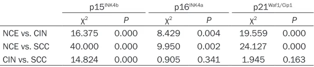
[image:2.612.90.407.84.152.2]Four serial slides, each 5 um thick, were cut from Table 2. Statistical results of expression differences of OIS markers
among NCE, CIN and SCC
p15INK4b p16INK4a p21Waf1/Cip1
χ2 P χ2 P χ2 P
NCE vs. CIN 16.375 0.000 8.429 0.004 19.559 0.000
NCE vs. SCC 40.000 0.000 9.950 0.002 24.127 0.000
[image:2.612.90.404.201.268.2]ing cells were graded on a scale of 0-3, with
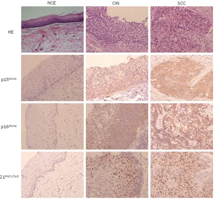
[image:3.612.98.524.68.464.2] [image:3.612.91.399.515.582.2]less than 5% positive-staining cells as grade 0, gher than 2 was determined as high expr- ession. Figure 1. Expression of OIS markers in NCE, CIN and SCC (magnification × 200).
Table 3. Expression of OIS markers in cases of CIN І, CIN П and CIN Ш
No. p15INK4b p16INK4a p21Waf1/Cip1
Low (%) High (%) Low (%) High (%) Low (%) High (%)
CIN І 18 18 (100.0) 0 (0) 8 (44.4) 10 (55.6) 14 (77.8) 4 (22.2)
CIN П 16 6 (37.5) 10 (62.5) 6 (37.5) 10 (62.5) 6 (37.5) 10 (62.5)
CIN Ш 17 0 (0) 17 (100.0) 2 (11.8) 15 (88.2) 1 (5.9) 16 (94.1)
Table 4. Statistical results of expression differences of OIS markers
among CIN І, CIN П and CIN Ш
p15INK4b p16INK4a p21Waf1/Cip1
χ2 P χ2 P χ2 P
CIN І vs. CIN П 15.938 0.000 0.423 0.515 5.673 0.017
CIN І vs. CIN Ш 35.000 0.000 4.575 0.032 18.453 0.000
CIN П vs. CIN Ш 7.792 0.005 2.169 0.141 4.930 0.026
5-25% as grade 1, 26-50% as grade 2, and more than 50% as grade 3. The intensity of stain-ing also graded on a scale of 0-2, with negative to weak intensity as grade 0, weak-moderate inten-sity as grade 1, and mod-erate to strong intensity as grade 2. For each marker, the score of per-centage and intensity
was multiplied. The final
[image:3.612.91.394.624.691.2]Comparisons among different groups for each marker were performed with Chi-square test. For all tests, a two-sided P < 0.05 was
consid-ered significant.
Results
Expression differences of p15INK4b, p16INK4a,
and p21Waf1/Cip1 among NCE, CIN, and SCC
The expression of p21Waf1/Cip1 was
predominant-ly within the nucleus, while the expression of p15INK4b and p16INK4a was predominantly within
the cytoplasm. The p15INK4b expression level
was low in all of NCE, and its expression was high in CIN (52.9%) and SCC (100.0%), respec-tively. The expression p16INK4a and p21Waf1/Cip1 were significantly higher in CIN and SCC com
-pared to NCE. However, this expression was no statistically differences between CIN and SCC (Tables 1 and 2; Figure 1).
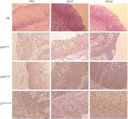
Expression differences of p15INK4b, p16INK4a,
and p21Waf1/Cip1 among CIN І, CIN П, and CIN Ш The expression of p15INK4b and p21Waf1/Cip1 was significantly higher in CIN П (62.5% and 62.5%) compared to CIN І (0% and 22.2%), and these expression were statistically higher in CIN Ш (100.0% and 94.1%) compared to CIN П,
respectively. The p16INK4a expression was no significantly difference between CIN І (55.6%) and CIN П (62.5%) group, and between CIN П and CIN Ш (88.2%) group. However, its expres
[image:4.612.101.524.73.465.2]-sion was significantly higher in CIN Ш compared to CIN І (Tables 3 and 4; Figure 2).
Discussion
Recent studies have revealed that OIS plays important role in limiting the progression of premalignant lesions to invasive cancer during tumor initiation [6]. Elucidation of a number of potential biomarkers for detecting senescent cells has facilitated to evaluate the role of OIS
in cancer development. Now SAβ-gal seems to
be a reliable marker of senescent cells in cul-ture [5, 14], but it fails to demonstrate senes-cent cells in vivo models [15, 16]. Other mark-ers of senescence involving signaling pathway were studied.
Previous studies have revealed that the ARF/ p53/p21 and p16/Rb/E2F pathways play important role in inducing cellular senescence [8]. The senescent-associated genes, including p15INK4b, p16INK4a and p21Waf1/Cip1, involve into
these processes. Several studies showed that p15INK4b, p16INK4a and p21Waf1/Cip1 are
upregulat-ed in premalignat lesions and early stage of cancer, but widely downregulated in the corre-sponding cancers, including thyroid, hepatocel-lular, breast, pancreatic carcinoma and glioma [7, 17, 18]. However, Bai et al [10] found that the expression of p15INK4b and p16INK4a were
almost completely negative in the normal esophageal epithelium. The p15INK4b and
p16INK4a was found to be expressed in 73% and
73% of the esophageal intraepithelial dysplasia (EID), and 92% and 88% of the esophageal squamous cell carcinoma (ESCC). Similarly, Feng et al [13] found that p15INK4b and p16INK4a
were also overexpressed in both CIN and cervi-cal SCC. Van de Putte et al [12] found that p21Waf1/Cip1 had no expression in normal
cervi-cal squamous epithelium, while its high expres-sion were detected in 20% cervical SCC. In the present study, p15INK4b, p16INK4a and p21Waf1/Cip1 expression were significantly higher in both CIN
and SCC compared to NCE. Furthermore, the expression of p15INK4b and p21Waf1/Cip1 was sig-nificantly higher in CIN П compared to CIN І, and
these expression were statistically higher in
CIN Ш compared to CIN П, respectively. The
p16INK4a expression was significantly higher in CIN Ш compared to CIN І. These results sug -gested that the senescence programs mediat-ed by p15INK4b, p16INK4a and p21Waf1/Cip1 were also activated as reflected in the overexpres -sion of these markers in cervical dysplasia and SCC, and the ARF/p53/p21 and p16/Rb/E2F pathways were activated during the dysplasia
stage of cervical carcinogenesis and remained intact in most cervical SCC. In addition, these results suggested that the expression of these
senescence markers may exist tissue-specific,
and different cancer tissues have different expression level.
In conclusion, the results showed that the senescence programs mediated by p15INK4b,
p16INK4a and p21Waf1/Cip1 were activated during
the stage of CIN and SCC, and demonstrated that senescence may play important role in pre-venting from NCE to SCC. However, the exact mechanism is still unclear, and the further study is needed.
Acknowledgements
This study was supported by grants from Jiangsu Natural Science Funding (BK20141185) and Jiangsu Province’s Key Medical Person (RC2011144).
Disclosure of conflict of interest
None.
Address correspondence to: Dr. Xueguan Lu, De- partment of Oncology & Radiotherapy, The Second
Affiliated Hospital of Soochow University, 1055
Sanxiang Road, Suzhou 215004, Jiangsu Province, P. R. China. Tel: 67784823; Fax: 86-512-68284303; E-mail: [email protected]
References
[1] Sun Y, Liu JH, Jin L, Lin SM, Yang Y, Sui YX, Shi H. Over-expression of the Beclin1 gene upregu-lates chemosensitivity to anti-cancer drugs by enhancing therapy-induced apoptosis in cervix squamous carcinoma CaSki cells. Cancer Lett 2010; 294: 204-210.
[2] Origoni M, Salvatore S, Perino A, Cucinella G, Candiani M. Cervical intraepithelial neoplasia (CIN) in pregnancy: the state of the art. Eur Rev Med Pharmacol Sci 2014; 18: 851-860. [3] Stein GH, Dulic V. Origins of G1 arrest in
senes-cent human fibroblasts. Bioessays 1995; 17:
537-543.
[4] Flores JM, Martin-Caballero J, Garcia-Fernan- dez RA. p21 and p27 a shared senescence his-tory. Cell Cycle 2014; 13: 1-2.
[5] Larsson L. Oncogene- and tumor suppressor gene-mediated suppression of cellular senes-cence. Semin Cancer Biol 2011; 21: 367-376. [6] Caldwell ME, DeNicola GM, Martins CP,
DA. Cellular features of senescence during the evolution of human and murine ductal pancre-atic cancer. Oncogene 2012; 31: 1599-1608. [7] Vizioli MG, Possik PA, Tarantino E, Meissl K,
Borrello MG, Miranda C, Anania MC, Pagliardini S, Seregni E, Pierotti MA, Pilotti S, Peeper DS,Greco A. Evidence of oncogene-induced se-nescence in thyroid carcinogenesis. Endocr Relat Cancer 2011; 18: 743-757.
[8] Bascones-Martinez A, Lopez-Duran M, Cano-Sanchez J, Sánchez-Verde L, Díez-Rodríguez A, Aguirre-Echebarría P, Alvarez-Fernández E, González-Moles MA, Bascones-Ilundain J, Muzio LL, Campo-Trapero J. Differences in the
expression of five senescence markers in oral
cancer, oral leukoplakia and control samples in humans. Oncol Lett 2012; 3: 1319-1325. [9] Collado M, Gil J, Efeyan A, Guerra C,
Schuhmacher AJ, Barradas M, Benguría A, Zaballos A, Flores JM, Barbacid M, Beach D, Serrano M. Tumor Biology: senescence in pre-malignant tumors. Nature 2005; 436: 642. [10] Bai P, Xiao X, Zou J, Cui L, Bui Nguyen TM, Liu
J, Xiao J, Chang B, Wu J, Wang H. Expression of p14ARF, p15INK4b, p16INK4a and skp2 in-creases during esophageal squamous cell cancer progression. Exp Therapeutic Med 2012; 3: 1026-1032.
[11] Lampejo T, Kavanagh D, Clark J, Goldin R, Osborn M, Ziprin P, Cleator S. Prognostic bio-markers in squamous cell carcinoma of the anus: a systematic review. Br J Cancer 2010; 103: 1858-1869.
[12] Van de Putte G, Holm R, Lie AK, Tropé CG, Kristensen GB. Expression of p27, p21, and p16 protein in early squamous cervical cancer and its relation to prognosis. Gynecologic Oncol 2003; 89: 140-147.
[13] Feng W, Xiao J, Zhang Z, Rosen DG, Brown RE, Liu J, Duan X. Senescence and apoptosis in carcinogenesis of cervical squamous carcino-ma. Mod Path 2007; 20: 961-966.
[14] Dimri GP, Lee X, Basile G, Acosta M, Scott G, Roskelley C, Medrano EE, Linskens M, Rubelj I, Pereira-Smith O, et al. A biomarker that
identi-fies senescent human cells in culture and ag -ing skin in vivo. Proc Natl Acad Sci U S A 1995; 92: 9363-9367.
[15] Saab R, Rodriguez-Galindo C, Matmati K, Rehg JE, Baumer SH, Khoury JD, Billups C, Neale G, Helton KJ, Skapek SX. p18Ink4c and p53 act as tumor suppressors in cyclin D1-driven prim-itive neuroectodermal tumor. Cancer Res 2009; 69: 440-448.
[16] Dankort D, Filenova E, Collado M, Serrano M, Jones K, McMahon M. A new mouse model to explore the initiation, progression, and therapy of BRAFV600E-induced lung tumors. Genes Dev 2007; 21: 379-384.
[17] Jin M, Piao Z, Kim NG, Park C, Shin EC, Park JH, Jung HJ, Kim CG, Kim H. p16 is a major inacti-vation target in hepato-celluar carcinoma. Cancer 2000; 89: 60-68.