ANTERIOR AND LATERAL THALAMIC LESIONS
IN OBJECT-ODOUR PAIRED ASSOCIATE LEARNING
A thesis
submitted in partial fulfillment
of the requirements for the degree
of
Master of Science in Biology
at the
University of Canterbury
by
Rati Bell
University of Canterbury
Table of contents
List of figures ... IV List of tables. ... V Acknowledgements ...VI Abstract. ... VII
1. Introduction ... 1
1.1 General introduction ... 1
1.2 Human studies of diencephalic amnesia... 2
1.3 Animal studies ... 4
1.4 The thalamic nuclei used in this study... 9
1.5 The Anterior Thalamic nuclei ... 9
1.5.1 The role of the AT region in odour memory ... 10
1.5.2 The role of the AT region in object memory... 11
1.6 The Lateral Thalamic nuclei and connections ... 12
1.6.1 The role of the LT region in various memory tasks ... 13
1.7 Issues of location, method, specificity and analysis of lesions ... 13
1.8 Simple object and simple odour discrimination tasks ... 15
1.9 Aims of the current study ... 15
2. Materials and Methods ... 17
2.1 Subject ... 17
2.2 Apparatus... 17
2.3 Pre-training ... 18
2.4 Surgical procedure ... 19
2.4.1 Anterior Thalamic lesions ... 19
2.4.2 Lateral Thalamic lesions... 20
2.5 Object-odour paired associate task ... 21
2.5.1 Re-familiarisation ... 21
2.5.2 Object-odour paired associate task ... 21
2.6 Simple discrimination tasks... 22
2.6.1 Object discrimination task ... 22
2.6.2 Odour discrimination task ... 22
2.7 Histology ... 23
2.7.1NeuN staining procedure ... 23
3. Results ... 24
3.1 Histology ... 24
3.2 Object-odour paired associate task ... 29
3.3 Simple object discrimination ... 32
3.4 Simple odour discrimination ... 33
3.5 Lesion-behaviour correlation... 34
Table of contents continued
4. Discussion ... 39
4.1 Summary of main findings and issues ... 39
4.1.1 Object-odour paired-associate memory and implications for current findings ... 40
4.1.2 Specificity of lesions... 43
4.2 Contributions of this study ... 44
4.3 Limtations of the current study and future directions ... 45
4.4 General summary... 48
References... 50
List of figures
Figure 1.1 Conceptual diagram showing the connections of the anterior thalamic (AT) region. ... 10 Figure 1.2 Conceptual diagram showing the connections of the lateral thalamic (LT) region.. ... 12 Figure 2.1 Schematic diagram showing the board apparatus ... 18 Figure 3.1 Schematic coronal sections through the rat brain showing the largest and smallest
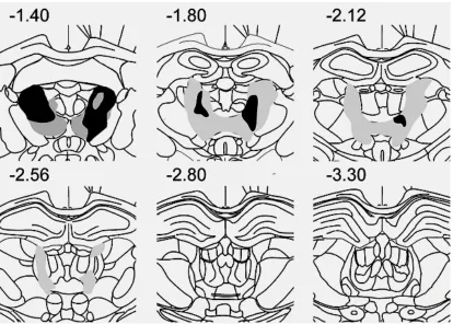
acceptable lesions in the AT group ... 25 Figure 3.2 Schematic coronal sections through the rat brain showing the largest and smallest
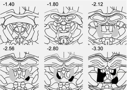
acceptable lesions in the LT group ... 26 Figure 3.3 Average latency difference for the AT, LT and control groups in the object-odour
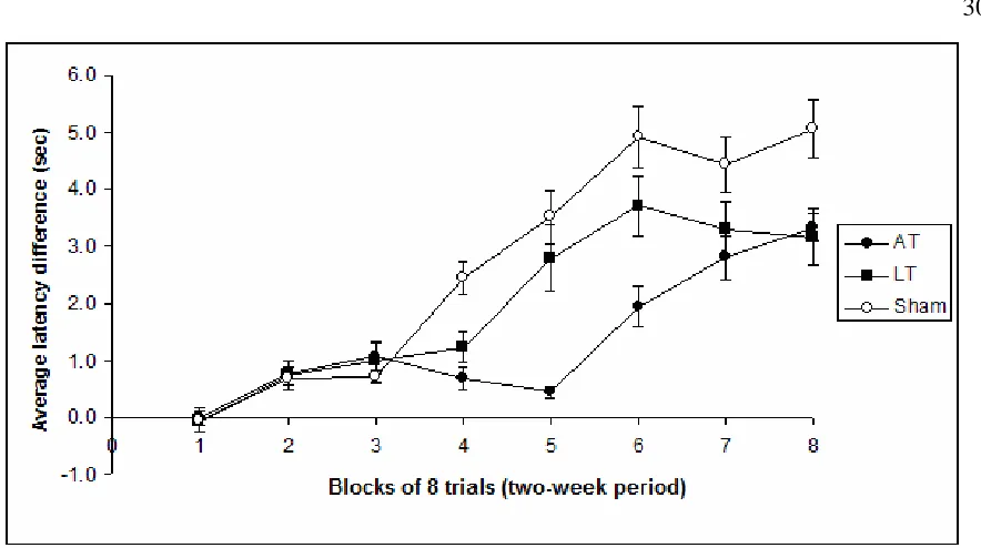
paired associate task ... 30 Figure 3.4 Average change in latency from Week 1 to Week 16 in the object-odour paired
associate task.. ... 32 Figure 3.5 Average latency difference for the AT, LT and control groups in the simple object
discrimination task... 33 Figure 3.6 Average latency difference for the AT, LT and control groups in the simple odour
discrimination task... 34 Figure 3.7 Scatterplot of performance during the last two weeks of training of AT, LT and control
groups against percent bilateral damage to the AT region. ... 35 Figure 3.8 Average latency difference between the AT rats that received the biggest and smallest
AT lesions... 36 Figure 3.9 Scatterplot of performance during the last two weeks of training of AT, LT and control
groups against percent bilateral damage to the LT region... 36 Figure 3.10 Average latency difference between the LT rats that received the biggest and smallest
List of Tables
Table 1.1 Summary of categorization of memory systems based on their function and connections with other parts of the brain... 7 Table 1.2 Summary of three different models of multiple neural memory systems based on kinds
of memory associated with key brain areas ... 8 Table 2.1 Lesion coordinates and related parameters for individual Bregma-Lambda distances
and corresponding AP coordinates for AT and LT lesions ... 20 Table 2.2 Pairings and mis-pairings of objects and odours in the object-odour paired associate
task ... 22 Table 3.1 Percent bilateral damage to selected areas for each of the rats in this study ... 28 Table 3.2 Fisher LSD scores showing the p values for AT, LT and Sham groups performance in
Acknowledgements
I would like to thank my supervisor Assoc. Prof. John Dalrymple-Alford and my co-supervisor Assoc. Prof. Larry Field for their help, support and expertise to help my write this thesis. Their patience, advice and hard work over the year are very much appreciated.
Thanks to Mathieu Wolff and Sheree Gibb for their advice, support and friendship right through the various stages of my experiment. Thanks to all the other postgraduate students in the Psychology animal lab for their help and support. Thanks to the Emilie Doat, technical staff at the animal lab, for caring for my animals.
Abstract
1. Introduction
1.1 General introduction
It has been widely shown that damage to the medial temporal lobe, particularly the
hippocampus, can result in severe amnesia (Gilbert & Kesner, 2002a, 2003; Squire &
Knowlton, 2000). More recently, however, increased attention has been paid to the
influence on learning and memory processes of the diencephalon, especially the medial
thalamus (Aggleton & Brown, 1999; Sziklas & Petrides, 1999). Damage to the
diencephalon essentially mimics aspects of the episodic memory deficit observed with
damage to the medial temporal lobe (Aggleton & Brown, 1999). The brain damage in
clinical cases of thalamic amnesia is problematic because it affects overlapping thalamic
regions. The various regions of interest that may be responsible for amnesia are in close
proximity to each other and the injury may damage fibres of passage, both of which make it
difficult to specify the nature or extent of the injury. The current study addresses this
problem of lesion specificity by focusing on the effects of selective lesions to the anterior
thalamic nuclei (AT) and the lateral thalamic nuclei (LT), using rat lesion models in which
injury is induced using microscopic quantities of a neurotoxin, N-methyl-D-aspartic acid
(NMDA) that spares fibres of passage. The AT and LT regions include thalamic nuclei
within the medial thalamus that have been implicated in diencephalic amnesia.
The medial thalamus is also known as the limbic thalamus because of its connections with
structures of the limbic system. It consists of the anterior and middle regions of the
thalamus. The two nuclei, AT and LT make up part of the medial (limbic) thalamus, with
each region providing different neural connections with other parts of the brain. By
identifying the effects of lesions to these two thalamic nuclei we hope to understand more
about memory systems in which they are involved and how the thalamus as a whole is
involved in memory processes.
The aim of this study was to extend the work done by Gibb et al. (2006) by examining the
effects of AT and LT lesions on a different paired-associate task to that used in their work.
In the current study, acquisition of an object-odour paired-associate task examined the rat’s
paired-associate memory impairment is one important characteristic in amnesia
(Eichenbaum et al, 2000; Aggleton & Pearce, 2001; Eacott et al, 2004), the current study
used a paired-associate memory task to examine the influence of these thalamic regions in
rats with AT and LT lesions. Simple object discrimination and simple odour discrimination
tasks were also examined to determine the effects of these lesions on non-paired associate
memory and served as a control.
1.2 Human studies of diencephalic amnesia
Amnesia or memory loss is a common feature of many human brain disorders. Amnesia
can result from injury to several parts of the brain and is the severe inability to display
normal levels of memory. Of particular interest to the present study is the inability to
acquire new memory - also known as anterograde amnesia - when brain injury occurs in the
diencephalon. Diencephalic amnesia can result from brain injury due to tumours,
infarction or from the alcoholic Korsakoff’s syndrome.
Findings collated from clinical studies of brain damage in humans have shown that
diencephalic amnesia can arise from damage to many structures. Anterograde amnesia has
been shown to result from damage to the AT, mediodorsal nuclei (MD) and mammillary
bodies and also from damage to the fibre pathways connecting cortical and sub-cortical
regions of the thalamus (Harding et al, 2000; Kapur et al, 1996; Mayes et al, 1988; Victor et
al, 1971). Previous case studies on Korsakoff syndrome suggest that damage to the MD
alone causes severe impairment but damage to the mammillary bodies alone does not cause
any major impairment in memory (Victor et al, 1971). This indicates that MD damage on
its own is sufficient to cause amnesia. A post mortem examination by Victor et al (1971)
revealed that 38 of 43 patients who suffered from the alcoholic Korsakoff syndrome had
damage to the MD region. The 5 other patients recovered from their amnesic condition and
were found not to have sustained much, if any, damage to the MD. Other studies have
shown amnesia arising from damage to the mammillary bodies and paratenial nucleus but
with no damage to the MD (Mair et al., 1979; Mayes et al., 1988). Conversely, Burk &
Mair (1998) showed that the intralaminar nuclei (ILn) are more critical for amnesia. They
showed that lesions to the ILn, but not the MDn, impaired performance of rats on a delay
that the ILn impaired learning on tasks that involved a delay component similar to the
amnesic condition observed in humans with damage to the ILn.
Other work supports the view that the AT region may be of particular significance in
amnesia. Graff-Radford et al (1990) examined four patients who suffered bilateral medial
thalamic damage, two of whom were found to be severely impaired. One of these impaired
patients had extensive damage to the mamillothalamic tract and ventroamygdalofugal
pathway (an amygdala related neural pathway) and the other had damage to the
mamillothalamic tract, the anterior thalamic nuclei and only the superior part of the
ventroamygdalofugal pathway. The remaining two patients were not as severely impaired
in the memory tasks and had damage to only one of the mentioned regions. Importantly,
the size of the damage did not necessarily influence the severity of amnesia, as in the case
of patient one who had smaller lesions than the other three patients but had the most severe
amnesic condition. Graff-Radford et al (1990) noted that amnesia occurred when damage
was in the anterior part of the thalamus. Several other authors came to the same
conclusions that the anterior thalamus may be responsible for amnesia. Gentilini et al
(1987) reported 8 cases of bilateral medial thalamic lesions (in three cases lesion locations
were uncertain). Three cases had the infarction located posteriorly and recovered from
their amnesia. Two cases were reported as having severe amnesia and had anterior lesions.
Similarly, Kritchevsky et al (1987) reported two patients with thalamic infarctions located
posteriorly without damage to mammillary bodies who showed no amnesia. Recent work
by Harding, Halliday, Caine and Kril (2000) has shown that while damage to the AT, MD
and mammillary bodies are all found in Korsakoff syndrome cases, damage to the AT may
be primarily responsible for the amnesic condition. They found that other alcoholic cases,
who only experienced the preliminary “Wernicke’s” phase, with neurological symptoms
but no permanent amnesic condition, were uniquely characterised by little or no AT
damage.
Nevertheless, human studies have key limitations since they cannot address the role of
individual regions within the diencephalon. For instance, current imaging tools pose
difficulty in specifying the locus of diencephalic injury in human subjects and the delay
between the injury and analysis during which the size and structure of lesions may change
fibre-tract connections to various and distant parts of the brain. In addition, the close
proximity and the complex structures of the diencephalon make it impossible to allow
accurate predictions of the role that these different regions play in memory.
Animal models on the other hand provide a means of overcoming some of the lesion
problems that arise in human studies. The location and size of the lesions and the time
delay between lesions and analysis can be controlled in animal models. Also, behavioural
analysis prior to lesions can be readily assessed and ensure that all subjects begin at the
same level of performance for the intended task. In addition, subjects’ age and sex can be
selected to remove any influence these parameters may have across subjects.
1.3 Animal studies
Animal models have used lesions made by various methods that include the use of
electrolytic or radiofrequency signals (Harrison & Mair, 1996; Koger & Mair, 1994;),
ablation, excitotoxins (Burk & Mair, 1998; Zhang, Burk, Glode, & Mair, 1998), or the
pyrithiamine induced thiamine deficiency method (Mumby et al 1995).
It has been suggested that no single nucleus within the thalamus is responsible for
diencephalic amnesia on its own (Aggleton & Brown 1999). Perhaps different regions play
a subtly different role in memory and these regions are interconnected to form memory
systems that work in parallel. For example, Aggleton and Brown (1999) proposed that
anterograde amnesia may be the result of damage to two different memory systems - one
system based on recall/ recollection (focused on AT and hippocampal connections) and the
second system based on familiarity-based recognition (focused on MD and perirhinal
cortex connections). They also presume that damage to any area within each system can
result in similar impairments.
Previous evidence has focused on three sites within the thalamus: the anterior thalamic
nuclei (AT) (Sziklas & Petrides 1999, Moran & Dalrymple-Alford 2003, Byatt &
Dalrymple-Alford 1996), the intralaminar nuclei (ILn) (Burk & Mair 1998) and the
damage to each of the cited nuclei can result in some aspects of amnesia, but with subtly
different characteristics.
The basis for suggesting involvement of the AT in diencephalic amnesia can be explained
by the fact that both the medial temporal lobe and diencephalon neural systems are
connected via the fornix, primarily, creating a hippocampus-anterior thalamic axis. In other
words, the AT is considered to be a part of an ‘extended hippocampal system’, since
hippocampal efferents to the medial thalamus (particularly the AT) are considered vital for
proper hippocampal functioning (Aggleton & Brown 1999). Several studies have shown
similar deficits in spatial memory processing in animals with AT and hippocampal lesions
(e.g. Gilbert & Kesner 2003, Mitchell & Dalrymple-Alford 2005, Gibb et al 2006). Sziklas
& Petrides (1999) investigated the effects of AT lesions in an object-place paired associate
task. The task consisted of allocentric and egocentric components. They found that AT
lesions impaired performance in allocentric spatial tasks (radial arm maze) but not in the
egocentric spatial tasks. It is important to note that in the egocentric task the AT rats
performed at a similar rate to the control rats, but in the probe trials the controls performed
at a rate higher than 50%. Sziklas & Petrides (1999) suggested that this could be because
the controls rats are able to use a combination of allocentric and egocentric cues to solve
the task.
The basis for suggesting the LT to be involved in diencephalic amnesia can be explained by
experiments conducted by Mair and his colleagues (Burk & Mair 1998). They compared
the effects of intralaminar (ILn), mediodorsal (MD) and lateral internal medullary lamina
(L-IML) lesions on a delayed match to sample task in an operant chamber. They found that
the MD group was only slightly impaired and had an intermediate effect on the task, but the
ILn rats were severely impaired in this task. In addition, the effects of ILn lesions were
independent of delays. Specific L-IML lesions produced a smaller more transient
impairment. In another study Mair and his colleagues (Burk, Mair & Porter 1998) found
ILn lesions to produce a general impairment in radial arm maze tasks. They proposed that
ILn lesions have a significantly more severe memory impairment that lesions to other
regions of the thalamus or the hippocampus. They draw many similarities between ILn
Lesion studies involving many brain regions have supported the idea of the multiple
memory systems in the brain. Consistent with Aggleton and Brown (1999), it is also
possible that different regions of the diencephalon have subtly different roles in memory.
This has prompted alternative suggestions that a number of different thalamic areas, each
underlying a different type of memory or memory related factor, are responsible for intact
performance in memory tasks (Aggleton & Brown, 1999; Mitchell & Dalrymple-Alford,
2005; Van der Werf, Jolles, Witter, & Uylings, 2003). Hence, the extent of damage to
specific thalamic structures would determine the characteristics or range of memory
impairment.
Mitchell & Dalrymple-Alford (2005, 2006) and Gibb et al (2006) conducted studies on rats
with AT, LT and MT regions (MT = medial and central MD), based on their anatomical
connections in the brain; their LT lesions included the ILn and the lateral MD nucleus.
Mitchell & Dalrymple-Alford’s studies concluded that different regions of the medial
thalamus participate in multiple memory systems, in that only AT lesions produced marked
reference and working memory or spatial memory deficits in the radial arm maze, and only
LT lesions produced working memory deficits for response memory, and only MT lesions
produced working memory deficits for reward magnitude. Memory systems are
traditionally classified in terms of the cognitive operations they perform and their
underlying cerebral structures (Table 1.1). For example, White and McDonald (2002)
proposed that memory systems function simultaneously and independently of each other.
They focus on three parallel independent memory systems, each with a central structure:
hippocampus, amygdala or dorsal-striatum. All three systems receive the same information
(stimuli, response and reinforcer) but their processing styles differ for this information.
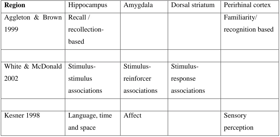
Table 1.2 shows the suggested different associations represented by each of their three
memory systems: the hippocampus deals with stimulus-stimulus associations, the amygdala
deals with reward associations and the dorsal-striatum deals with
stimulus-reinforcer associations. The idea is that lesions to any region within a system would result
in an impaired performance in a memory task whose attributes or elements are coherently
represented by the system (White & McDonald 2002).
By contrast, Kesner (1998) proposed multiple parallel memory systems that are primarily
language (in humans). He has three over-riding memory systems that process event-based,
knowledge-based and rule-based memory. Memory for new information depends on the
event-based system, in which processing of space, time, and language, is largely undertaken
by the hippocampus, processing of emotional / affect / reward information is undertaken by
the amygdala and event-based information about individual objects is processed by
perirhinal cortex.
Table 1.1: Standard categorization of memory systems (Squire & Knowlton, 2000; Squire
et al, 2004)
Declarative memory (hippocampal dependent)
• Concerns long-term factual or episodic memory that is recalled consciously
• The neural system involved in episodic/ declarative memory is comprised of the hippocampus and the anterior thalamic nuclei; traditionally, aspects of the medial
temporal lobes are also included as part of this system.
Non-declarative memory (non-hippocampal dependent)
• Concerns a range of different memory systems, such as procedural memory which in humans is characterised by evidence of memory that occurs without conscious
awareness
Working memory
• Short term memory that allows the brain to regularly update and is useful in trial unique or inconsistent (everyday) situations. This type of memory may represent
Table 1.2: Summary of three different models of multiple neural memory systems based on
the different kinds of memory associated with some key brain structures. The difference
between systems is presumed to reflect either their ‘information processing style’
(Aggleton & Brown, 1999; White & McDonald, 2002) or the specific kinds of information
that are processed (Kesner, 1998).
Region Hippocampus Amygdala Dorsal striatum Perirhinal cortex
Aggleton & Brown
1999 Recall / recollection-based Familiarity/ recognition based
White & McDonald
2002 Stimulus-stimulus associations Stimulus-reinforcer associations Stimulus-response associations
Kesner 1998 Language, time
and space
Affect Sensory
perception
The cognitive operations of the memory systems listed in Table 1.2 are based on lesion
studies on animals. Lesion data from animals have shown that damage to the medial
temporal lobe, particularly the hippocampus, can also result in amnesic conditions that are
similar to those that occur in humans (Gilbert & Kesner, 2002, 2003; Squire & Knowlton,
2000). The hippocampus is thought to play an important role in spatial tasks (Kesner 2002,
Aggleton & Brown 1999), associative memory tasks (Eichenbaum 1999) and other
conditioning tasks (Nadel et al 1997). Lesion experiments in monkeys and rats showed that
amygdaloid damage resulted in different memory deficits, particularly in memory tasks that
require associations between neutral and incentive stimuli and fear conditioning (Kesner,
1998; White and McDonald, 2002). Damage to the dorsal striatum has been shown to
result in memory deficits in tasks that involve a motor response in the presence of a single
cue (Kesner, 1998; White and McDonald, 2002). Thus far, the studies by Mitchell &
Dalrymple-Alford (2005, 2006) and Gibb et al (2006) provide some support to the idea that
different parts of the limbic thalamus are involved in different memory systems, at least
1.4 The thalamic nuclei used in this study
The location, size and specificity of lesions have varied across previous studies, making it
difficult to interpret the role of individual thalamic structures (see section 1.7). The
thalamic lesions targeted in this study are the AT (including the anterodorsal, anteromedial
and anteroventral nuclei) and LT (including the intralaminar nuclei and the lateral
mediodorsal nuclei). This selection was made because studies using lesions of the
traditional AT and ILn regions have produced the most consistent evidence of memory
impairments in rat models. Both the AT and the LT have different neural connections with
respect to the hippocampal system and the striatum, respectively (see Figures 1.1, 1.2), but
they also have partially overlapping connections with the prefrontal cortex. It is thought
that the confounding results (differential and coincidental effects of lesions) from lesion
studies that included AT or ILn regions are due to the fact that the lesions used in previous
studies may have overlapped the two regions, resulting in uncertainty as to their role in
memory (Mitchell & Dalrymple-Alford 2006). The specificities of the lesions used in this
study are based on previous findings from Mitchell & Dalrymple-Alford (2005, 2006) in
comparative studies as well as their afferent and efferent connections to other major regions
of the brain.
1.5 The Anterior thalamic nuclei
As stated above, disruption of AT in humans is associated with anterograde amnesia
(Harding et al 2000, Van der Werf et al 2000). The anterior thalamic nuclei have major
connections with the retrohippocampal region and are considered to be part of the
‘extended hippocampal system’ (Aggleton & Brown 1999, Vann & Aggleton 2004a)
(Figure 1.1). For example, Warburton et al (1999) showed that impairments caused by AT
lesions in rats were comparable to those caused by fimbria-fornix lesions. This and other
evidence mentioned earlier indicates that the AT has a close functional relationship with the
hippocampus and is involved in acquisition of spatial memory. Mair et al. (2002) showed
that AT and parahippocampal lesions have separate effects on spatial memory but together
they can disrupt hippocampal-dependent spatial memory. Dalrymple-Alford’s group has
or the MD, produces profound deficits on standard spatial memory tasks (Mitchell &
[image:17.595.86.322.147.300.2]Dalrymple-Alford 2005, 2006).
Figure 1.1: Conceptual diagram showing the connections of the Anterior Thalamic (AT)
region. Note particularly its connection to the hippocampus via the fornix. Reproduced
with permission from Dalrymple-Alford (2005) and adapted from Aggleton and Brown
(1999).
As the purpose of the current study was to examine paired-associative memory when object
and odour cues were paired, it is relevant to briefly first discuss prior research on simple
odour memory, simple object memory and related tasks or the effects of related (i.e.,
hippocampal) lesions.
1.5.1 The role of the AT region in odour memory
The Gibb et al (2006) study, in which odour-place association memory was markedly
impaired by AT lesions, found that acquisition of a simple odour discrimination was only
mildly impaired, although this may in part be due to the prior training on the
paired-association task. With regard to hippocampal lesions these do not impair performance in an
odour-odour (Alvarez et al 2002) nor an object-odour paired association task (Gilbert and
Kesner 2002a). However, hippocampal lesions do impair performance in an object-place
and odour-place association task, presumably because these tasks involved a spatial
component (Gilbert and Kesner 2002a). For this reason it is expected that the AT lesions
will not markedly impair acquisition of an object-odour task as there is no spatial
1.5.2 The role of the AT region in object memory
Studies with thalamic lesions in rats on spontaneous object recognition have shown no
evidence that AT lesions impair memory even if there is a delay component involved in the
task (Aggleton et al, 1995; Moran & Dalrymple-Alford 2001).
In terms of associative memory involving objects as one of the component attributes,
Sziklas and Petrides (1999) tested the effects of AT lesions in rats in an allocentric object
recognition task designed to study the ability of rats to choose between two objects
depending on their location in an enclosed arena (one object was correct on one side of the
field, whereas the other object was correct on the other side of the field). They found that
AT lesions severely impaired performance in this task. However, this may be solely due to
the spatial component involved in this task. As mentioned, Gibb et al (2006) reported that
AT lesions severely impaired performance on an object-place paired associate task.
However, AT lesions did not impair performance in the spontaneous object recognition or
simple object discrimination tasks (Gibb et al, 2006).
One study examined the ability of rat to use unique stimulus cues (complex objects, such as
multiple small blocks of wood, white wire mesh) in the context of a configural learning task
(Moran and Dalrymple-Alford 2001). The rats were required to learn the reward/
non-reward that either appeared alone or in combination along the runway arm of a radial maze.
For example, they were rewarded for responding when two complex cues, A and B
appeared alone, but they were not rewarded when the two types of cue appeared together
(A+, B+, AB-). AT lesions, which impaired spatial memory in a standard (no cues) radial
arm maze task, did not impair performance in this cue-based configural task. Acquisition of
the configural task, but not the standard spatial memory task, was impaired by perirhinal
cortex lesions.
1.6 The Lateral thalamic nuclei
The lateral thalamic nuclei of interest include the rostral intralaminar nuclei (ILn) and
lateral mediodorsal nuclei (MDn). Both these regions have strong connections with the
lower brain-stem structures including the substantia nigra. As a whole the LT has
reciprocal connections with overlapping regions of the striatum as well as the anterior
cingulate and precentral cortices of the PFC, which thereby form a
fronto-striatopallidal-medial thalamic neural circuit (Berendse and Groenewegen 1991; Van der Werf et al 2002).
As stated earlier, LT lesions produce only minor, if any, spatial memory impairments,
consistent with the idea that they alter normal functioning between the striatum and frontal
[image:19.595.88.296.249.407.2]cortex, not hippocampal system function (Mitchell & Dalrymple-Alford 2005).
Figure 1.2: Conceptual diagram showing the connections of the Lateral Thalamic (LT)
region. Reproduced with permission from Dalrymple-Alford (2005) and adapted from
Aggleton and Brown (1999).
1.6.1 The role of the LT region in various memory tasks
Relatively fewer studies have examined the effects of ILn lesions, often in the context of
lesions that include the lateral internal medullary laminae (L-IML). L-IML lesions impair
olfactory continuous DMS learning but odour discrimination was not impaired (Zhang et al
1998). LT lesions have been shown to produce impairments in the radial arm maze (Mair
et al 1998), but this may have been partly due to encroachment of the lesions to adjacent
regions (Mitchell & Dalrymple-Alford 2005). For example, localized LT lesions when
confined to the ILn and lateral MD produce only minor impairments in working memory in
the radial arm maze (Mitchell & Dalrymple-Alford 2005) and no impairments in the Morris
water maze (Wolff et al personal communication, June 2007). Mair’s group have also
instead of the LT being involved in odour memory it could be responsible for general
learning and memory.
Savage et al (1998) investigated the effects of L-IML and ILn lesions on an object pair
discrimination task in rats. Although this task tested the retention of information acquired
prior to surgery (retrograde amnesia) it still showed that the ILn had no effects on object
memory.
Gibb et al (2006) found that LT lesions severely impaired acquisition of the object-place
paired associate task but did not have any effect on the acquisition of simple object or
simple place discrimination. This indicates that damage to the LT is sufficient to cause a
serious learning deficit. However, since the LT did not impair learning on the simple
discrimination tasks or on the spontaneous object recognition tasks it is unclear whether its
role involves general learning and memory or a particular memory attribute such as relevant
cues, strategies and involve memory systems that the rats may use to solve the tasks.
1.7 Issues of location, method, specificity and analysis of lesions
As stated previously, the thalamus is made up of a very small and complex array of nuclei
that are interconnected to other parts of the brain. Due to the complexity of the thalamic
nuclei significant methodological issues arise with the location, size and specificity of
lesions produced. Small intrusions into other thalamic areas have probably compromised
the experimental results of many previous experimental studies using thalamic lesions.
One important issue in such studies is the method used to produce lesions in the thalamus.
Methods like electrolytic or radiofrequency (heat-produced) lesions (e.g., Byatt and
Dalrymple-Alford, 1996) destroy both cell bodies and fibres of passage, whereas the
excitotoxic method employed in this study only destroys cell bodies. The excitotoxic
method is used here because of the high density of fibres of passage in the AT nuclei of the
thalamus.
However, some unintentional damage to other structures is inevitable due to the close
example neurotoxic AT lesions can inadvertently damaged LT and MT tissue (Mair et al.
2003), and IL tissue (Warburton et al. 1999). MT lesions damaged tissue in the AT (Hunt
& Aggleton 1998), and L-IML tissue (Burk & Mair 1998). IL lesions often damage AT
tissue (Savage et al 1998) and MT tissue (Mair et al 1998). Hence there is a need for
quantative analyses on lesions, rather that qualitative alone. Our research group at the
University of Canterbury have made a detailed quantative analysis of lesions produced and
attempted more localised thalamic lesions (Gibb et al., 2006; Mitchell and
Dalrymple-Alford, 2005, 2006; Wolff et al, 2006).
The main purpose of the study was to compare the effects of the two thalamic lesions (AT
and LT) on an object-odour paired associate task. The task used here was the object-odour
paired associate task employed previously by Gilbert and Kesner (2002a) who found that
hippocampal lesions had no effect on acquisition of this task, unlike deficits found when
the task included a spatial component as part of the association. Their task used a go/ no-go
procedure. Rats were required to learn the arbitrary association between an object and an
odour to receive a food reward, where two specific pairs of odour and objects signal a
reward (“go”), but any mispairing signalled no reward (“no-go”). It is important to note
here that while the task involved an odour and an object attribute it did not involve a spatial
attribute.
The role of the hippocampus and AT in non-spatial memory is not clear other than memory
for the temporal order of events (Fortin et al, 2002; Kesner et al, 2002; Hopkins et al 1995)
but considerably evidence exists to show their involvement in spatial memory (Sziklas &
Petrides 1999; Gibb et al. 2006; see Kesner 1998 for a review). As the AT nuclei are
considered to be part of the ‘extended hippocampal system’ we do not expect AT lesions to
impair performance in this object-odour paired associate task since there is no spatial
component to this task.
1.8 Simple object discrimination and simple odour discrimination
Additional go/ no-go simple discrimination tasks followed the main task. In these
additional tasks, rats were required to make a simple discrimination between either two
performance in the main (paired-associate) task was impaired by an inability to
discriminate between the objects or the odours used and/or simply the inability to inhibit a
response. That is, the simple discrimination tasks did not involve a paired association and
thus served as a performance comparison for the main task.
1.9 Summary: Aims of the current study
This study aims to extend earlier work done at the University of Canterbury, on
comparative influence of selective AT and LT lesions by examining their effects on Gilbert
and Kesner’s (2002) object-odour paired-associate task. The formation of an episodic or
declarative memory is thought to require not only that a number of attributes of an event are
encoded but that they are bound together in a unique way (Aggleton & Pearce 2001).
The study of Gibb et al. (2006) was modelled on Gilbert and Kesner’s work but
investigated the effect of thalamic lesions, not hippocampal lesions, in an odour-place
paired association task. This task included major spatial attributes. They found that the AT
lesions severely impaired performance in this task, whereas the LT lesions tended to
produce less impairment.
The next logical step from Gibb’s (2006) work was to compare the lesions of the thalamic
nuclei on an object-odour paired association task. Since there is no spatial component in
this task it would be ideal to further investigate the role of the thalamus. It assesses the
ability to learn an arbitrary association between one of two objects and the presence of one
of two types of odorized sand in which the rat digs for a food reward, without any spatial
cues.
We have focused on two of the nuclei to build on these findings and to attempt to identify
what roles each region plays. The only previous study to comparatively test the effects of
these nuclei of the thalamus was that of Gibb et al. (2006) in an odour-place paired
associate task.
It was predicted that the AT would not be impaired in this object-odour paired associate
sensitive to hippocampal lesions and thus AT lesions). It was predicted that the LT lesions
would impair performance in this task because the LT is thought to play a general role in
memory and learning (that is, not limited to any specific attribute of the task).
The specific hypotheses were:
a) AT lesions should not affect this type of associate memory task (as this task is
unimpaired by hippocampal lesions) (Gilbert and Kesner 2002; Gilbert and Kesner 2003).
b) LT lesions should produce a deficit, on the basis that this region may have a general role
in higher order memory and learning tasks, thereby disrupting the acquisition of any
arbitrary association
c) None of these lesions should impair, or should only minimally impair, acquisition of
2. Methods
2.1 Subjects
Forty PVGc Hooded female rats were bred in the Psychology animal facility and were
housed in opaque plastic cages (50cm long, 30cm wide and 23cm high) in groups of four
per cage and maintained in a 12 hour light-dark cycle (8am-8pm). All testing was
conducted during the dark cycle. Rats received restricted food access to maintain their
body weights at 80-85% ad libitum throughout all training. The rats weighted between
150g and 200g at time of surgery and were allowed free food access during the first week
of the recovery period before returning to restricted food access. All procedures conformed
to the NIH Guide for the Care and Use of Laboratory Animals and were approved by the
University of Canterbury Animal Ethics Committee (refer Appendix A for ethics
committee approval).
2.2 Apparatus
The large circular wood board used in this task was 119 cm in diameter, 3.5cm thick and
was painted white with no wall around the perimeter. A start box (24cm long, 15cm wide
and 17cm high) painted black with a hinged lid and manually operated guillotine-like door
was placed at the perimeter of the board. A ceiling mounted camera recorded behavioural
data. The room also contained a chair, two desks and beige curtains surrounding the board.
Small terracotta pots (6cm wide at top, 6cm high) were painted black and filled with
sterilized sand to 1cm below rim. The pots were attached to a platform (25cm X 25cm) to
stabilize pot and minimize spillage of sand onto board. Rats were trained to dig for food in
these pots. To minimize the use of any food odour cues a wire mesh overlaid inaccessable
“Froot loops” cereal placed at the bottom of the pot. Only one of these pots was on the
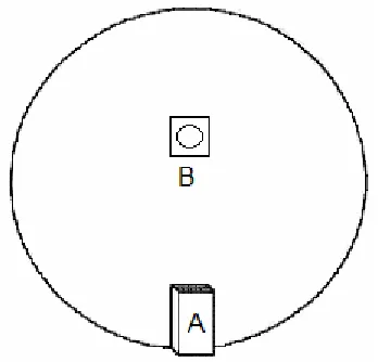
board at any time during training. They always appeared 67.5cm from back edge of start
box (Figure 2.1).
Two objects, a yellow plastic spade (22cm high and 4.5 cm diameter at base) and a green
one object was placed directly behind the pot on the platform and was visible to the rats
from the start box. The objects always appeared in the same place as the pots did. The
main object-odour paired-associate task as well as the simple odour and object
discrimination were conducted on the same board, with the same pots, start box and
objects.
2.3 Pre-training
Individual rats were placed in a large 1m square box, containing a pot filled with
non-odourised sterile sand in the experiment room and were shaped to retrieve “Froot Loops”
cereal buried successively deeper in the sand. Subsequently each cage of four rats was
placed on the board and allowed to eat “Froot Loops” scattered across the board (15 mins/
day, for 2 weeks). Then each rat was placed in the start box on the board and trained to
[image:25.595.219.393.396.563.2]retrieve “Froot loops” from the pot using 10 trials per day, 5 days a week for 3 weeks.
Figure 2.1: Schematic diagram showing the start box (A) shown at one edge of the board,
behind which the experimenter stood. The pot and object (B) was 67.5cm from back edge
of the start box. On any given trial for the object-odour paired associate task only one pot
with odourised sand and object was present on the board. During the simple odour
discrimination tasks only one pot containing the odourised sand was on the board (no object
present). During the simple object discrimination task only one pot containing
2.4 Surgical procedures
Rats were randomly assigned to receive either AT, LT or control (sham) lesions. Rats were
anaesthetized IP with sodium pentobarbitone (50mg/ ml, at a dose of 1.65ml/ kg) 25
minutes after Atropine IP (0.065mg/ml/kg) supplemented by Mepivacaine (5mg/ml/kg,
subcutaneously under the scalp) and Norocarp (5mg/ml/kg, subcutaneously at the nape of
the neck). Rats were then placed in the Kopf stereotaxic clamp with incisor bar 7.5mm
below interaural line to minimize damage to the fornix via passage of the 1ul Hamilton
syringe operated by a Stoelting motorized infusion pump.
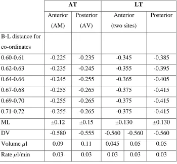
Excitotoxic lesions were made using 0.12M of NMDA dissolved in phosphate buffer (pH
7.20). Anterior-posterior coordinates were varied slightly among individual rats based on
the distance between lambda and bregma (Table 2.1). Stereotaxic co-ordinates for lesion
placements were based on those used previously by Mitchell & Dalrymple-Alford (2005),
after further verification and minor improvements in pilot work. Infusions were made by
lowering the Hamilton needle slowly to the site and allowing it to rest for 30 seconds prior
to infusion; neurotoxin was dispensed at a rate of 0.003ul/ min with 3 min post infusion for
diffusion at the site and then slowly retracting the needle. Control lesions (3AT and 2LT
sites) received the same surgery but with no neurotoxin in the infusion needle which was
lowered to a coordinate 0.3cm above that used for the respective lesion sites (to minimise
damage to thalamic structures).
2.4.1 Anterior thalamic lesions
The AT lesions consisted of bilateral infusions aimed at the anteroventral nucleus (AV)
using 0.11ul of NMDA, and the anteromedial nucleus (AM) using 0.09ul of NMDA. All
measurements were taken from bregma. To ensure accurate placing of lesions the
anterior-posterior (A-P) co-ordinates varied between each rat in line with the Bregma-Lambda
distance (which ranged from 0.60 to 0.72cm for each lesion type, for clarity see Table 2.1).
Hence the AP distances for AT lesions varied from -0.235 to -0.265cm, 0.15cm from the
midline and -0.555cm ventral from the dura at the AV site. At the AM site AP distances
varied from -0.225 to -0.255cm, 0.12cm from the midline and -0.580cm ventral from the
2.4.2 Lateral thalamic lesions
The LT lesions consisted of three bilateral sites (two anterior LT depths/sites and one
posterior LT site). At the anterior LT site AP distances varied from -0.345 to -0.375cm at a
distance of 0.130cm from the midline and at a depth of -0.560cm and -0.600cm from the
Dura using 0.045ul NMDA at 0.03ul/min at each site. At the posterior LT site, the AP
distances varied from -0.385 to -0.415cm, and 0.130cm from the midline and at -0.560cm
from the dura using 0.05ul NMDA at 0.03ul/min.
Table 2.1: Lesion coordinates and related parameters for individual Bregma-Lambda
distances and corresponding anterior-posterior coordinates for AT and LT lesions
AT LT
Anterior
(AM)
Posterior
(AV)
Anterior
(two sites)
Posterior
B-L distance for
co-ordinates
0.60-0.61 -0.225 -0.235 -0.345 -0.385
0.62-0.63 -0.235 -0.245 -0.355 -0.395
0.64-0.66 -0.245 -0.255 -0.365 -0.405
0.67-0.68 -0.255 -0.265 -0.375 -0.415
0.69-0.70 -0.255 -0.265 -0.375 -0.415
0.71-0.72 -0.255 -0.265 -0.375 -0.415
ML ±0.12 ±0.15 ±0.130 ±0.130
DV -0.580 -0.555 -0.560 -0.560 -0.560
Volume µl 0.09 0.11 0.045 0.05 0.05
[image:27.595.83.433.312.635.2]2.5 Object-odour paired associate task
2.5.1 Re-familiarisation
Rats were allowed to recover for three weeks post-surgery and were re-familiarized with
the board. Rats were placed in the start box and pots filled with non-odourised sand were
placed within 10cm from the start box during re-familiarisation and were allowed to dig in
the pot for cereal (12 trials per rat for 4 days).
2.5.2 Object-odour paired associate task
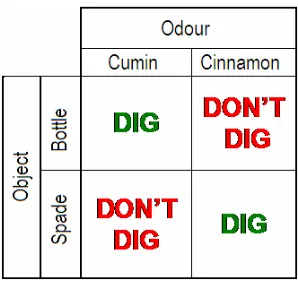
The rats were then required to retrieve “Froot loops” cereal buried 1cm under odourised
sand in a pot (1% cinnamon or 0.4% cumin w/w sand). Only one pot paired with only one
object (spade or bottle) was present on the board at any one time. When the rat approached
the pot it received a food reward if the correct object-odour pairing occurred on that trial
(go trial) but no reward for a mis-pairing of an object and odour (no-go trial) (Table 2.2).
The reward contingency was counterbalanced across rats within each lesion group and
across home cages. There were thus two correct and two incorrect pairings of object and
odour per rat and they received 12 trials (six correct pairings and six incorrect pairings) per
day. Rats were trained 5 days a week for 16 weeks (until they reached an asymptotical level
of performance).
The time taken from when the back feet of the rat exited the start box to when it started
digging in the pot was recorded. Rats were given a total of 10s within which to start
digging. If they did not dig within this 10s they were returned to the start box for the next
trial. Digging was defined as more than 2 consecutive strokes in the sand with one or both
paws. Daily average latencies (No-go minus go trials) were used as a dependent measure
of acquisition. Optimal acquisition of this object-odour paired associate task required rats
Table 2.2: Parings and mispairings of objects and odours in the object-odour paired
associate task
2.6 Simple discrimination tasks
After completion of the main paired-associate task the rats were required to make a simple
discrimination between the two objects or between the two odours. One half of the rats
were assigned to the simple odour discrimination and the other half to the simple object
discrimination. The subjects were balanced across lesion groups, performance and home
cages (time of testing during the day).
2.6.1 Object discrimination task
In this task rats were required to dig in a pot of non-odourised sand when the correct object
(go trial) was present and not when the incorrect object was present (no-go trial). The
‘correct’ allocation of objects was balanced across lesions and home cages. Again time was
measured when rats back feet exited box to when it began digging in sand. Rats received
12 trials per day for 5 days a week across a period of four weeks.
2.6.2 Odour discrimination task
In this task rats were required to dig in a pot of odourised sand when the correct odour (go
trial) was present and not when the incorrect odour was present (no-go trial). No objects
were present on the board during this task. The ‘correct’ allocation of odours was balanced
[image:29.595.306.455.145.287.2]when it began digging in sand. Rats received 12 trials per day for 5 days a week across a
period of four weeks.
2.7 Histology
After completion of the simple discrimination tasks the rats were injected with an overdose
of sodium pentobarbitone and perfused with 0.9% saline and 10% formalin solution. Their
brains were carefully removed and stored in 4% formalin before sectioning at 50um using a
vibratome. Each section was taken throughout the thalamus and mounted on glass slides.
The sections were stained using a standard procedure with cresyl violet (cell bodies,
including neurones and glial cells). One in every five sections was stained with NeuN (a
neurone-specific immuno-marker; Jongen-Relo et al 2002).
2.7.1 NeuN staining procedure
NeuN is a protein that is expressed in cell bodies of most neuronal cells in rodents, humans
and some other animals. The advantage of staining brain sections with NeuN is that this
protein is not expressed by glial cells and hence can be used to clearly label lesion sites
(Jongen-Relo et al 2002).
For NeuN staining, brain sections were treated with 0.5% H2O2 in 0.1M phosphate buffer
solution (PBS) for 30 minutes to suppress peroxidase activity. The sections were incubated
for an hour in PBS with horse serum and bovine albumin serum and Triton X-100 and then
in anti-NeuN serum, visualised using a biotinylated secondary antibody and DAB reaction
3. Results
3.1 Histology
A minimum of 40% damage to the target thalamic areas was used as the inclusion criterion
for an acceptable lesion in this study. Examples of lesions are shown in Figs. 3.1 and 3.2,
in which schematic diagrams indicate the largest and smallest acceptable lesions from the
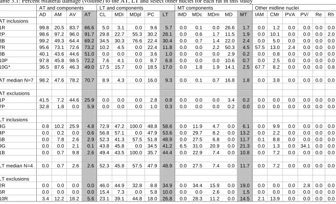
AT and LT lesion groups. A list of percent bilateral lesion damage for AT and LT lesion
rats is shown in Table 3.1. As shown in Table 3.1, seven rats sustained greater than 40%
damage to the AT region, with a median value of 70.7% (range, 49.0% to 91.7%),
including one rat that had been intended as an LT rat but in which the lesion was anterior
and thus in the AT region (the LT damage in this rat was only 17%). In the AT group with
acceptable lesions, minimal to relatively minor damage occurred in the adjacent LT region
(median, 9.3%; range, 1.0% to 30.4%), minimal damage to the MT region and no damage
to midline nuclei except in two cases which had extensive damage to the interanteromedial
nucleus. Two AT rats had insufficient AT damage and were excluded from the main
behavioural analyses. Five rats had acceptable LT lesions (median 48.9%; range, 41.2% to
58.6%) and three LT rats had insufficient damage and were discarded from behavioural
analysis (see Table 3.1). Damage to the AT region was minimal in rats with acceptable LT
lesions (median, 48.9%; range, 0.1% to 4.8%) and generally small or absent in other
Figure 3.1 Schematic coronal sections through the rat brain showing the locations of the
largest (grey) and smallest (black) acceptable lesions in the AT group. Numbers are
[image:32.595.89.503.83.379.2]Figure 3.2 Schematic coronal sections through the rat brain showing the locations of the
largest (grey) and smallest (black) acceptable lesions in the LT group. Numbers are
[image:33.595.88.502.84.377.2]Abbreviations for Table 3.1: AD= anterodorsal nucleus; AM= anteromedial nucleus; AT= anterior
thalamic aggregate; AT median= median percent damage for all included AT rats; AV=
anteroventral nucleus; CL= centrolateral nuclei; IAM= interanteromedial nucleus; LT= lateral
thalamic aggregate; LT median= median percent damage for all included LT rats; MDl= lateral
segment of the mediodorsal nucleus; MDpl= paralamellar segment of the mediodorsal nucleus; PC=
paracentral nucleus; PVA= anterior paraventricular nucleus; PV/PVP= paraventricular
nucleus/posterior paraventricular nucleus; Re= reunions nucleus; Rh= rhomboid nucleus. * 10G
intended to be LT lesion but met criterion for AT lesion. No damage was sustained by the LD
Table 3.1: Percent bilateral damage (volume) to the AT, LT and select other nuclei for each rat in this study
AT and components LT and components MT components Other midline nuclei
AD AM AV AT CL MDI MDpl PC LT IMD MDc MDm MD MT IAM CMr PVA PV/ Re Rh AT inclusions
1R 99.8 20.5 83.7 66.6 5.0 3.1 0.0 9.6 5.7 0.0 0.1 0.0 26.6 1.7 0.0 1.2 0.0 0.0 0.0 0.0 2P 98.6 97.2 96.0 91.7 29.8 22.7 55.3 30.2 28.1 0.0 0.6 1.7 11.5 1.9 0.0 10.1 0.0 0.0 0.0 2.0 3B 99.2 49.3 64.4 69.2 34.5 30.3 76.6 22.4 30.4 0.0 0.7 1.4 22.0 2.4 0.0 5.0 0.0 0.0 0.0 0.0 7R 95.6 73.1 72.6 73.2 10.2 4.5 0.0 22.4 11.8 0.0 0.0 2.2 50.3 4.5 57.5 13.0 2.4 0.0 0.0 0.0 8B 40.1 43.6 44.6 51.0 0.0 0.0 0.0 3.6 1.0 0.0 0.0 0.0 2.9 0.2 0.0 0.8 0.0 0.0 0.0 0.0 10P 97.8 45.8 98.5 72.2 7.6 4.1 0.0 8.7 6.8 0.0 0.0 0.0 10.6 0.7 0.0 2.5 0.0 0.0 0.0 0.0 10G* 36.5 87.6 46.3 49.0 17.5 15.7 0.0 18.5 17.0 0.0 1.8 1.9 14.1 2.5 67.7 8.2 0.0 0.0 0.0 0.8
AT median N=7 98.2 47.6 78.2 70.7 8.9 4.3 0.0 16.0 9.3 0.0 0.1 0.7 16.8 1.8 0.0 3.8 0.0 0.0 0.0 0.0
AT exclusions
1P 41.5 7.2 44.6 25.9 0.0 0.0 0.0 2.8 0.8 0.0 0.0 0.0 3.4 0.2 0.0 0.0 0.0 0.0 0.0 0.0 7P 32.8 1.8 0.0 5.9 0.0 0.0 0.0 1.0 0.3 0.0 0.0 0.0 0.2 0.0 0.0 0.0 0.0 0.0 0.0 0.0
LT inclusions
4G 0.8 10.2 25.9 4.8 72.9 47.2 100.0 48.8 58.6 0.0 11.9 4.7 0.0 6.1 0.0 9.9 0.0 0.0 0.0 0.0 4P 0.0 0.2 0.0 0.6 56.8 57.1 0.0 47.9 53.6 0.0 29.7 8.2 0.0 13.2 0.0 2.2 0.0 0.0 0.0 0.0 5B 0.0 7.8 2.6 2.9 52.3 41.3 57.5 51.8 48.9 0.0 27.5 6.8 0.0 11.7 0.1 8.8 0.0 0.0 0.0 0.0 9G 0.0 0.0 2.1 0.1 43.8 45.8 0.0 34.5 41.2 6.5 31.0 20.9 0.0 21.3 0.0 1.3 0.0 34.1 0.0 0.0 1B 0.0 0.7 9.8 2.6 49.4 43.5 100.0 35.7 44.4 0.0 22.9 7.4 0.0 10.8 0.0 7.2 0.0 0.0 0.0 0.0
LT median N=4 0.0 0.7 2.6 2.6 52.3 45.8 57.5 47.9 48.9 0.0 27.5 7.4 0.0 11.7 0.0 7.2 0.0 0.0 0.0 0.0
LT exclusions
3.2 Object-odour paired associate task
A “latency difference” score was used as the dependent variable. For this measure, the average latency to dig in the pot of odourised sand on rewarded trials for each rat on each day was subtracted from the average latency to dig in the pot of odourised sand on the non-reward trials for that day. These scores were then averaged over 2-week blocks of testing. A maximum of 10 seconds on the non-reward trials was allowed, so optimal performance occurred when the rat responded quickly on rewarded trials but withheld responding on non-rewarded trials. That is, higher average latency scores indicated that a rat was making the correct associations between an object and an odour.
Figure 3.3 shows the average latency difference for the three groups, AT, LT and sham lesion groups, over a period of 8 blocks of testing (each block = 2 weeks of testing). There was no difference in performance between the three groups until the fourth block of testing (week 7 and 8) when the control group started to acquire the task. For both the AT and LT lesion groups the task was eventually acquired (block 8) but with impaired performance compared to the control group.
Figure 3.3: Average latency difference (seconds) for the AT, LT and control groups in the object-odour paired associate task over a period of 16 weeks (8 two-week blocks). Vertical bars are ±SEM. The average latency difference scores represent the average difference between the latencies on the go trials and the no-go trials.
These observations were verified statistically in a 3 (lesion: AT, LT and sham) X 8 (block) repeated measures MANOVA (Statistica) analysis, which revealed highly significant effects for Lesion (F(2,19)=8.939, p<0.001), Week (F(7,133)=42.979, p<0.0001), and a Lesion by Week interaction (F(14,133)=4.954, p<0.0001). A Post-hoc comparison (Fisher LSD) performed on the average latency difference scores over the entire testing period, confirmed that both the AT (p<0.001) and LT (p<0.01) groups were impaired relative to the Control group. However, there was no significant difference overall between the AT and LT groups (p>0.40).
[image:37.595.84.526.103.350.2]significantly worse than control group on blocks 4, 6, 7 and 8. For block 5, the AT group showed poorer performance than the control and LT groups, but the LT group did not differ significantly from controls. The AT and LT groups did not significantly differ for each other on any other block other than block 5. Together with the mean values across blocks, these analyses support a description that the AT and LT groups both showed poorer acquisition (rate and final level of performance) than the sham group and that there was evidence that the rate of acquisition was poorer in AT than in the LT group. For controls, the mean performance did not improve across the last three blocks of testing (last 6 weeks); the same was true of the LT group, but the AT group’s mean performance improved from block 5 to block 8.
A 3(lesion) X 2(last 2 weeks of training) repeated measures MANOVA confirmed the significant Lesion effect at the end of training (F(2,19)=5.99,p<0.01). A Post-hoc comparison (Fisher LSD) again showed a significant difference between the AT (p<0.02) and LT (p<0.01) when compared to the Control group.
[image:38.595.82.534.664.734.2]To provide a clearer picture of the relative degree of learning that occurred over the 16 week training period in the three groups, the difference between the average latencies at Block 8 and Block 1 is shown in Figure 3.4. The mean superiority of the control group is readily apparent. The LT and AT groups also had a similar increase in difference scores over the training period. A one-way ANOVA showed no a marginally non-significant lesion effect (F(2,16)=2.97, p>0.07). However, a post hoc (Fisher LSD) comparison again confirmed that there was a significant difference between the Control group and LT (p<0.006), Control and the AT (p<0.005). However there was no significance between the AT and the LT groups (p>0.50).
Table 3.2: Fisher LSD scores showing the p values for AT, LT and Sham groups performance in the object-odour paired associate task. Those values in bold indicate a significant lesion effect (p<0.05).
Block 1 Block 2 Block 3 Block 4 Block 5 Block 6 Block 7 Block 8
AT vs. LT 0.74 0.69 0.47 0.42 0.03 0.23 0.79 0.48
AT vs. Sham 0.77 0.56 0.15 0.001 0.001 0.001 0.04 0.01
0.0 1.0 2.0 3.0 4.0 5.0 6.0 7.0
AT LT Sham
[image:39.595.88.444.120.348.2]A v e ra g e d if fe re n c e b lo c k 8 b lo c k 1
Figure 3.4: Difference in average latencies from Week 16 to Week 1 for AT, LT and control groups. Vertical bars are ±SEM.
3.3 Simple object discrimination task
The simple discrimination tasks were conducted after completion of the object-odour paired associate task. The rats were divided into two groups, balanced across lesion groups and level of performance at the end of training in the main task, and each subgroup was tested on a simple discrimination task. The simple discrimination tasks required them to make a simple discrimination between either two objects or two odours (i.e. no paired-association was required). One group completed the object discrimination task and the other completed the odour discrimination task.
means were higher than that shown by even the control group after 16 weeks of training in the object-odour association task, despite the fact that the same objects were used for both tasks. A repeated measures MANOVA showed a significant effect of Week (F(3,39)=114.8, p<0.001) with a highly significant difference between week 1 performance and week 4 performance (Fisher LSD, p<0.0001). However, there was no significant Lesion effect (F(3,13)=0.24, p>0.8) or Lesion by Week interaction (F(9,39)=1.05, p>0.4). This suggests that all three lesion groups learned the simple object discrimination task at a similar rate.
0.0 1.0 2.0 3.0 4.0 5.0 6.0 7.0 8.0
WK1 WK2 WK3 WK4
W e e k ly a v e ra g e s ( s e c ) AT LT Sham
Figure 3.5: Average latency difference (seconds) for the AT, LT and control groups in the simple object discrimination task. Vertical bars are ±SEM.
3.4 Simple odour discrimination task
[image:40.595.85.472.283.504.2]latency differences of the AT and control group even at week 1 was above 3 seconds for the simple odour discrimination and were 5 to 6 seconds in all three groups by week 2.
A repeated measure MANOVA showed a significant effect of Week (F(3,24)=74.94, p<0.0001). However, there was no significant Lesion effect (F(3,14)=1.71, p>0.2) or Lesion by Week interaction (F(9,42)=0.61, p>0.7) which confirms that the all three lesion groups learned the simple odour discrimination task to similar levels and at similar rates. A post hoc (Fisher LSD) comparison showed no significant difference between the three lesion groups (p>0.05). 0.0 1.0 2.0 3.0 4.0 5.0 6.0 7.0 8.0
WK1 WK2 WK3 WK4
[image:41.595.86.448.301.523.2]W e e k ly a v e ra g e s ( s e c ) AT LT Sham
Figure 3.6: Average latency difference (seconds) for AT, LT and control groups in the simple odour discrimination task. Vertical bars are ±SEM.
3.5 Lesion-behaviour correlations
3.5.1 Relationship between lesion damage and performance on the odour-place
paired-associate task
object-odour paired-associate task. This was done to try and distinguish a relationship between amount of damage to the AT and impaired performance. From Figure 3.7 we can see that the level of performance for control rats varies (along the solid line at 0). A few control rats performed at the highest level and achieved an average latency difference above 7 seconds. However, some controls performed at a level similar to the highest performing lesion rats, but the slowest control rat’s performance was still well above that of the poorer lesion rats. Figure 3.7 also reveals a rather stable level of performance for AT rats, to the right of the dashed line, independent of the size of the AT lesion. There also appears to be no relationship between the amount of AT damage and the performance for the rats in the LT group, where unintentional AT damage in LT subjects was at a minimum.
0.0 1.0 2.0 3.0 4.0 5.0 6.0 7.0 8.0
-10 0 10 20 30 40 50 60 70 80 90 100
Percent bilateral AT damage
[image:42.595.89.454.343.566.2]B lo c k 8 a v e ra g e l a te n c y d if fe re n c e ( s e c ) AT LT Sham
Figure 3.7: Percent bilateral damage to the AT and average latency difference in week 15 and 16 for AT, LT and sham groups. The dashed line indicates inclusion criteria for AT lesions. All data points to the right of the dashed line meet inclusion criteria.
-1.0 0.0 1.0 2.0 3.0 4.0 5.0 6.0 7.0
1 2 3 4 5 6 7 8
Blocks of testing period
A v e ra g e l a te n c y d if fe re n c e ( s e c )
Bigges t AT les ion
Sm alles t AT les ion
Figure 3.8: Average latency differences of two rats; one that received the biggest AT lesion and the other that received the smallest AT lesion.
0.0 1.0 2.0 3.0 4.0 5.0 6.0 7.0 8.0
-10 0 10 20 30 40 50 60 70 80 90 100
Percent bilateral LT damage
[image:43.595.87.446.118.349.2]B lo c k 8 a v e ra g e l a te n c y d if fe re n c e ( s e c ) AT LT Sham
[image:43.595.88.458.422.645.2]Inspection of Figure 3.9 reveals there is no clear relationship between performance and percent bilateral LT damage. The final level of performance observed for the LT rats may be due to the increased performance of just 3 of the 5 LT rats. Clearly, two LT rats and one AT rat showed very little or no acquisition of the object-odour task. Given the distributions shown in Figure 3.7 and Figure 3.9, Spearman rank correlations were low (r<0.05).
Figure 3.10 is a graph of the rate of learning of the rats that received the biggest and the smallest LT lesions. From this Figure we can see that the level of performance of the LT rats is independent of lesion size, with the biggest LT lesion rat performing at a higher rate than the smallest LT lesion rat.
-1.0 0.0 1.0 2.0 3.0 4.0 5.0 6.0 7.0
1 2 3 4 5 6 7 8
Blocks of testing period
A v e ra g e l a te n c y d if fe re n c e ( s e c )
Bigges t LT les ion
Sm alles t LT les ion
Figure 3.10: Average latency differences of two rats; one that received the biggest LT lesion and the other that received the smallest LT lesion.
[image:44.595.89.444.342.571.2]