Abstract— There have been many attempts to implement
traditional telemedicine across the world especially in the developing countries, but the efforts has been characterized with challenges such as high-cost of sustaining telemedicine solutions and non-availability of medical expertise. Cancerous Skin disease such as melanoma and nevi typically results from environmental factors (such as exposure to sunlight) among other causes. The necessary tools needed for early detection of these diseases are still not a reality in most African communities. In recent years, there have been high expectations for techniques such as Dermoscopy or Epiluminiscence Light Microscopy (ELM) in aiding diagnosis; however evaluation of pigmented skin lesions using ELM is not only non-affordable by most of African communities but also complex and highly subjective, thus motivating researches in diagnosis automation. This study would focus on designing and modeling a system that will collate past Pigmented Skin Lesion (PSL) image results, their analysis, corresponding observations and conclusions by medical experts using prototyping methodology. These wealth of information would be used as a library. A part of the system would use computational intelligence technique to analyze, process, and classify the
image library data based on texture and possibly
morphological features of the images. Trained medical personnel in a remote location can use mobile data acquisition devices (such as cell phone) to generate images of PSL, supply such images as input to the proposed system, which in turns should intelligently be able to specify the malignancy (life-threatening) or benign (non-(life-threatening) status of the imaged PSL.
Index Terms — automated diagnosis, computational
intelligence, medical imaging, remote health diagnosis, skin disease
I. INTRODUCTION
EDICAL Imaging provide techniques and processes in
creating images of human body or samples (such as blood or sputum) for clinical purposes - (medical procedures seeking to reveal, diagnose or examine disease), medical science - (study of normal anatomy and physiology) or for knowledge discovery. Digital image processing involves the screening of a region for processing and saving this region to a location (possibly a file) for processing using operations
Manuscript received June 30, 2013; revised August 1, 2013. This work was supported in part by Dermatology Society of South Africa (DSSA).
D. A. Okuboyejo is with Department of Software Engineering, Faculty of Information Communication and Technology, Tshwane University of Technology, South Africa (e-mail: [email protected]).
O. O. Olugbara is with Department of Information Technology, Faculty of Accounting and Informatics, Durban University of Technology, South Africa (e-mail: [email protected]).
S. A. Odunaike is with Department of Software Engineering, Faculty of Information Communication and Technology, Tshwane University of Technology, South Africa (e-mail: [email protected]).
such as fractal and texture analysis.
A Fractal is an object that technically displays self-similarity on all scales; such that even if the object does not exhibit exactly the same structure at all scales, it would have same type of structures appearing on all scales (Mathworld, 2012). Fractal Dimension is a ratio that provides a statistical index of complexity comparing how details in a fractal pattern changes with the scale at which it is measured, such as given by relationship between an object’s length (or area) and its diameter. In medical imaging, Fractal analysis typically determines the fractal dimension of an image using techniques such as global box-counting, fractal dimension, mean fractal dimension, and local connected fractal dimension for binary threshold used during analysis of an image. Texture analysis on the other hand uses methods such as co-occurrence matrices calculus; evaluation of texture features (such as energy, entropy, contrast and correlation during image processing).
The major aim of image analysis is to use image processing techniques to provide a machine interpretation of an image, typically in a format that could foster decision making process. In the past two decades, strong impulse has been given to developing automated systems capable of assisting physicians in medical imaging task (Dobrescu et al., 2010). However, the presence of noise, masking structures, variability of biological shapes and tissues, imaging system anisotropy etcetera making the automated analysis of medical images a very hard task (Rubegni et al., 2002; Stanganelli et al., 2005; Dobrescu et al., 2010). The skin’s surface is a detailed landscape, with complex geometry and local optical properties. Skin features depends on many variable such as body location (such as forehead or cheek), subject parameters (such as age or gender), and imaging parameters (such as lighting or camera, and also the direction from which it is viewed and illuminated). Bacterial and viral skin infections generally affect the skin by decolorizing and distorting pigmented skin areas, further making automation of medical image analysis difficult (Tushabe et al., 2011).
One of the best approaches to overcome aforementioned challenges in automating medical imaging diagnosis is to simplify the objective of the analysis and to exploit some kind of hypothetical information about the imaged structures. The information about the structures to be analyzed can be anatomical knowledge about their typical appearance (such as shape and grey levels) and position; or statistical knowledge of their properties (such as gray level of the tissues included in those structures). The images can then be classified using their morphological, color, fractal, and texture properties.
Laws, 1980 in his work transformed digital images to
Automating Skin Disease Diagnosis Using
Image Classification
Damilola A. Okuboyejo, Oludayo O. Olugbara, and Solomon A. Odunaike
identify regions of interest and provided an input dataset for segmentation and features detection operation. The author used operations such as thresholding, morphological analysis and texture detection to divide digital image into individual objects to perform a separate analysis of each region. The incidence of skin cancer is rapidly increasing throughout the world and it is gradually becoming one of the predominant forms of cancer especially in Caucasian population countries and among fair-skinned people (Snober et al., 2001; Han et al., 2006; Ali & Deserno, 2012). Skin cancer incidence is on the order of 10 to 12 in Europe, 18 to 20 in United States, and 30 to 40 in Australia per 100000 subjects (Schmid-Saugeona et al., 2003).
Over the years, it is been reported that Automatic Data Analysis used for Melanoma (a type of skin cancer) showed a higher diagnostic performance compared to physicians observation in terms of sensitivity (proportion of true positives), though lower in terms of specificity (proportion of true negatives). A common technique used for aforementioned automated data analysis is Dermoscopy or Epiluminiscence Light Microscopy (ELM), an in-vivo, non-invasive technique that in recent years has disclosed a new dimension of the clinical morphological features of Pigmented Skin Lesion (PSL) using different light magnification systems with oil immersion technique (Ali & Deserno, 2012). Dermoscopy or its synonymous Dermatoscopy provides dermatologist with a technique for in-vivo inspection of PSL and it renders higher accuracy for detecting suspicious cases than it is possible with popular practice of naked eye inspection (Kittler, 2004).
Interestingly, while merit of medical imaging is getting popular, World Health Organization reported in one of their findings that three quarter of entire world population is yet to have access to medical imaging which is an essential technique in new age telemedicine such as in automation of skin disease diagnosis (WHO, 2007). So far, medical imaging has contributed immensely towards advancing medical procedures. The fact that interpretation and analysis of medical imaging results are still heavily dependent on medical experts (whose availability are low or non-existence) is a serious concern for developing and underserved regions (especially rural settings). An approach is needed to minimize this dependency and also to limit probable bias of medical personnel in the analysis of a medical image result, hence this particular study.
II. PROBLEM STATEMENT
The doctors typically have assumed diagnosis opinion, which most likely begin by searching for further evidence that their assumption can be validated and in cases where it is not validated, they will have missed other potential diagnosis. Bias essentially influences analysis made by medical practitioners, just as with any human search that begins with keywords chosen by the user. Additionally, if a doctor begins searching by symptoms, while this may be accurate, the order or weight given to any of the symptoms would most likely give a bias towards related diagnosis when in fact, there may be a symptom that is not given any credit and thus not included in the search or considered in timely fashion.
The heavy dependencies on medical expert for medical image diagnosis analysis are a serious challenge for regions (especially Low and Medium Income Countries) where the expert might not be readily available, inadequate or non-responsive to an urgent medical need (such as dermatological-related). The aforementioned problems suggest that a better and manageable solution is needed urgently with the view to minimize these dependencies and human bias, thus leading to our research question.
III. RESEARCH FOCUS
Based on finding of Han et al., 2006, the incidence of skin cancer is rapidly increasing throughout the world and it is gradually becoming one of the predominant forms of cancer especially in Caucasian population countries and among fair-skinned people. Skin cancer incidence is on the order of 10 to 12 in Europe, 18 to 20 in United States, and 30 to 40 in Australia per 100000 subjects (Schmid-Saugeona et al., 2003; Stanganelli et al., 2005; Ali & Deserno, 2012). This research is motivated by the need for studies that can help provide ways or approaches that can be used by medical practitioners (such as Dermatologist) and at the same time not heavily dependent on opinions of the Dermatologist (which can often be subjective as reported by Rubegni et al., 2002; Stanganelli et al., 2005).
The primary research question is how can we minimize the heavy dependencies on medical experts for diagnosis procedure of Pigmented Skin Lesions (PSL) in patients residing in remote or underserved areas?
This question could be favorably answered if emphasis is placed on areas below:
i) Bickers et al., 2004 revealed that healthcare cost for diseases such as melanoma averaged at about $39.3 billion in 2004. To provide a portable inexpensive solution, what devices or tools are easily accessible or already owned by most people living in underserved areas that can be used as part of the diagnosis tool?
ii) What is the best approach to standardize image set to provide for library of past known diagnosis and corresponding results?
iii) For the proposed system to automate the diagnosis process, how can we classify medical images using the image properties (such as texture) as either malignant or benign?
iv) In a bid to improve the speed of diagnosis, how can bias made by medical experts, especially dermatologist be minimized?
IV. RESEARCH AIM AND OBJECTIVES
The purpose of the study is to design and model a system that uses medical imaging to reduce heavy dependencies on medical expert for diagnosis procedure of PSL (especially melanoma and nevi) in patients. The research would use pattern matching algorithms to analyze medical images against known values.
The research objectives are:
ii) To ease diagnosis and treatment of skin patient (by means of automation) and provide for cost effective way of treatment (by using devices or tools easily accessible or already possessed by most people living in remote and underserved areas).
iii) To improve the speed of diagnosing Pigmented Skin Lesion such as in melanoma and nevi (using ground truth knowledge of past diagnosis of PSL as medical image library).
V. LITERATURE REVIEW
During the last few years, telemedicine with remote image viewing and analysis has emerged as a highly valuable and versatile tool, particularly suited to places where local medical expertise is limited. Granot et al., 2008 worked on creating a medical imaging system consisting of physically separated components of medical imaging system in order to produce a robust and less expensive system that can be used by trained non-medical personnel. Adoption of simple method of microphotography which could significantly increase opportunities and quality diagnostics while lowering costs and considerably increasing connectivity between most isolated laboratories and distant reference center has been proposed by Aher & Kaore, 2010.
Dobrescu et al., 2010 described a method of an algorithm for automatic detection of malignancy of skin lesion which is based on both local fractal features (local fractal dimension) and texture features derived from medium co-occurrence matrices (such as contrast, energy, and homogeneity). Tushabe et al., 2011 proposed an image-based diagnosis method where images of skin disorder were used to classify skin diseases into broad category of either viral infected or bacterial infected.
Malignant melanoma currently accounts for a third of most frequent type of skin cancer and 79% of skin cancer death. The incidence of malignant melanoma in fair-skinned patients has increased histrionically in most parts of the world over the past few decades (Rubegni et al., 2002; Stanganelli et al., 2005; Dobrescu et al., 2010). In Europe, it’s been reported that malignant melanoma incidence is increasing by 5% every year and it is responsible for 91% of skin cancer death (Sboner et al., 2001; Ali & Deserno, 2012). In a bid to improving early detection, a number of diagnostic checklists and rules have been proposed such as Seven Point Checklist (Healsmith et al., 1994), and ABCDE: Asymmetry, Border, Color, Diameter, Evolution checklist (Fitzpatrick et al., 1998). These rules and checklists specify visual features associated with malignant lesion symptoms.
Stolz et al., 1994 in their work, developed a diagnosis scheme for dermoscopic images, accessing the Asymmetry (A), Border (B), Color (C), and Diameter (D) of different image structures. This ABCD rule became the standard in Dermoscopy for staging PSL into benign, suspicious, or malignant moles (melanoma). However, dermoscopic diagnosis is often complex and subjective, thus associated with poor reproducibility and low accuracy especially among inexperience dermatologist, as the accuracy of experts is 65-84% (Argenziano et al., 2003; Lee, 2001, Stanganelli et al., 2005). Also, visual interpretations of these features by dermatologist have so far proven to be a difficult
task. Lee 2001 in his study reported detection rate based on clinical visual investigation to be about 65%.
Melanoma is highly curable if diagnosed early and treated properly as survival rate varies between 15% and 65% from terminal to early stages respectively (Ali & Deserno, 2012). Depending on the observer’s experience, Dermoscopy improves the diagnostic accuracy for melanoma detection up to 50% as compared with traditional visual inspection (Kittler, 2004). In the last decade, usage of Dermoscopy or Epiluminiscence Light Microscopy (ELM) changed the dermatologist’s approach to suspicious PSL. However, the analysis made using ELM are extremely complex and subjective (Rubegni et al., 2002). To prevent aforementioned challenge of quantitative interpretation, methods based on Computer-Aided Diagnosis (CAD) have been introduced towards automating the diagnosis procedures, such as in Rubegni et al., 2002; Stanganelli et al., 2005; Mittra & Parekh, 2011.
Gilmore et al., 2009 used lacunarity (a measure of transitional invariance of an object used in quantifying aspects of patterns that exhibit scale-dependent changes in structure) to provide a promising method for automated assessment of melanocytic nevi and melanoma. The fuzzy-based histogram analysis technique used by Stanley et al., 2003 provided a possibility for automated skin lesion discrimination in dermatology clinical images. Rubegni et al, 2002 developed an automated process using artificial neural network methods based on mathematical analysis of pigmented skin lesions to avoid the problem of qualitative interpretation made by the use of ELM by Dermatologist. Kreutz et al., 2000 presented a combination of artificial neural network approach with texture analysis using digital image processing and mixture-of-experts to attempt automation of skin cancer diagnosis. Ganster et al., 2001 developed a system that provided for automated computerized analysis of images obtained from ELM to enhance the early recognition of malignant melanoma. Sheha et al. 2012 used Grey-Level Co-occurrence Matrix (GLCM) and Multilayer perceptron classifier (MLP) for automatic Detection of Melanoma Skin Cancer using Texture Analysis. One core challenge however with many of aforementioned approaches is their inability to integrate well with ubiquitous devices such as mobile telephony, now largely accessible to underserved areas (Mobithink, 2012).
VI. BENEFITS
Noticeable problems in current approach towards PSL diagnosis include the subjective nature in the use of dermoscopic diagnosis, its setup cost and its’ over dependency on medical expert (in this case, dermatologist) for analysis. This study tends to model a system that would help to improve remote patient diagnosis, screening and examination of skin problem at a reduced cost while reducing over dependencies on medical expert.
systems using digital technologies are increasingly been developed (Arani & Ghassemian, 2010; Baldi et al., 2009). So, it may be possible even with use of mobile device which are increasingly available in Low and Medium Income Countries (Mobithink, 2012) to diagnose a skin disease once the mobile phone uploads the captured image to a server (such as connected via Virtual Private Network) and in no time analysis would be done on the image and relevant diagnosis result would be automatically be published back to the phone.
The primary aim of this study is to build and model a system that will take advantage of all these developments can reduce previously mentioned challenges during analysis of PSL, which in effect would speed up treatment procedure for patients. No doubt, the death tolls as a result of lack or unavailability of dermatologist in underserved areas could be largely reduced. We believe the study would in bear future open opportunities in developing better and low-cost effective solutions to mitigate dermatological challenges.
VII. RESEARCH METHODOLOGY
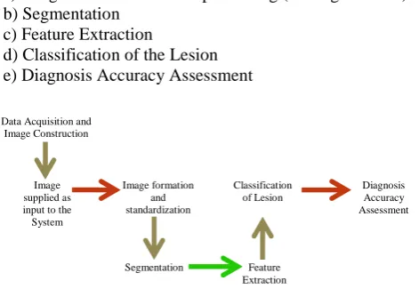
The methodology of this work is based on soft science design to realize a prototype system for the diagnosis of skin disease represented by a skin image. We intend using features based on texture analysis and classify the lesion using techniques such as thresholding and neural networks to develop and prototype a new algorithm for skin disease diagnosis. Figure 6.1 shows the overall systematic steps of the propose algorithm for skin disease diagnosis. Each important step of the algorithm is thereafter discussed. In a bid to build fast, better and manageable quality system, we intend using prototyping methodology. We would have an initial model that would include program modules, database, input and output unit that the system would use. We would continually work on the prototype by modifying, and adding to it to enable us eventually having a complete working system. The subsequent sub-section reveals the design and methods we intend using.
VIII. RESEARCH DESIGN AND METHODS
We are typically following a procedure that involves five main steps in processing the PSL:
a) Image Formation and Preprocessing (see Fig. I below) b) Segmentation
c) Feature Extraction
d) Classification of the Lesion e) Diagnosis Accuracy Assessment
After the acquisition and preprocessing of color images of the PSL, we intend performing segmentation of the lesion possibly from the surrounding skin (Zagrouba and
Barhoumi, 2004). The lesion areas and boundaries would be clearly identified, and various attributes of the lesion characteristics of the malignity or benign symptoms would be measured. Such characteristics features will then be the raw input to a recognition algorithm classifying the lesion as malignant melanoma, benign nevi or suspicious mole. In order to have successful classification system, section 6.1a – 6.1d identified above would be carried out with care. The accuracy assessment would help discover the sensitivity and specificity of the study result.
A. Image Formation and Preprocessing
PSL images to be used would be standardized into a chosen resolution. It is expected that
some of the images to be used might have features such as hairs and pigments which confuses analysis. These features are typically regarded as noise, and thus need to be filtered off in order to facilitate separation of the lesion area from the surrounding skin. A technique known as DullRazor would be implored for the median filtering of the noise as described by Lee et al., 1997 (see Fig 2 and Fig 3).
B. Segmentation Process
Majority of the techniques used in addressing segmentation of dermoscopic images
rely on color and grayscale thresholding which are often unable to define a clear criterion to separate with precision the pigmented lesion from the background healthy skin. We intend to follow an
approach that determines the boundary of the lesion by region growing (after a possible initial step based on fuzzy sets to enhance the lesion region of interest) to localize suspicious lesion region in the PSL images (Zagrouba & Barhoumi, 2004).
C. Feature Extraction and Classification
While the ABCD rule has become a standard used by many dermatologists, its diagnostic use by dermatologist based on visual and qualitative evaluation of such criteria are often subjective (Schmid-Saugeona et al., 2003). This study would however characterize the ABCD rule (which serves as the ground truth) into quantitative attributes measured by image analysis, and implore texture analysis technique (to describe the spatial arrangement of pixels which regional intensity or color alone might not sufficiently describe) with Gabor wavelet (to make scale, translation and rotation invariant) in order to automate the classification process. Multilayer Perceptron Classifier (MLP) can then be used to classify the lesion as being benign or malignant.
D. Diagnostic Accuracy Assessment
[image:4.595.45.281.571.738.2]We would use the degree of sensitivity and specificity of the result obtained by the classifier to assess the diagnostic accuracy of the system. Our approach would be to compare our implementation of ABCD rule (Stolz et al., 1994) with chosen machine learning alternatives classification such as logistic regression.
Fig. 1 Algorithm Method
Fig 2 Original Image
Fig 3 Processed Image
Fig 4 Segmentation
Data Acquisition and Image Construction
Image supplied as input to the System
Image formation and standardization
Segmentation
System
Feature Extraction
Diagnosis Accuracy Assessment Classification
Sensitivity is a statistical measure that defines the degree of true positive subjects with malignant status in the total group of subjects with the disease. Specificity measures the accuracy of true negative subjects without malignant status in the total group of subjects with the disease.
E. Data Collection Technique
Considerable large proportion of the PSL images is been collated from the Dermatology Society of South Africa (DSSA), and the rest is meant to be gotten via online web resource of known PSL diagnostic results and images.
F. Research Ethics
a) Protection from Harm: There is no foreseeable harm that the research team or possible participants would likely experience, be it physical, social, or emotional.
b) Right to Privacy: Privacy would be maintained by not disclosing identities of the subjects whose PSL image is being used for the study. Also, appropriate reference to relevant authors would be made in the course of the study.
c) Confidentiality: Should a need arise for the research team to communicate with any of the subject whose PSL image is being used, communications would be treated confidential.
d) Sample Data: Appropriate reference would be made to identify the sources of sample data used. The team would also ensure misrepresentation of sample data is avoided.
IX. CONCLUSION
Automatic diagnosis of skin cancer is feasible and achievable through the usage of well-defined segmentation and classification technique. While many success has been recorded in the current advances in automation of medical diagnosis, this study tends to maximize the large availability of ubiquitous devices and elicitation of past skin cancer diagnosis image set towards providing cost-effective, easier and faster diagnosis for underserved areas.
REFERENCES
[1] Aher, A., & Dr. Kaore, N. (2010), Application of Camera Phone
Technology in capturing microscopy images. Department of Microbiology, Peoples College of Medical Sciences & Research Centre Journal of recent advances in applied sciences (JRAAS) 25:05-07, 2010
[2] Ali, A., & Deserno, T.M. (2012), A systematic review of automated
melanoma detection in dermatoscopic images and its ground truth data. Proceedings of SPIE vol 8318, 831811
[3] Arani, M.N. & Ghassemian, H. (2010), A Hierarchical Content-Based
Image Retrieval Approach to Assisting Decision Support in Clinical Dermatology, Iranian Journal of Electrical and Computer Engineering, 9(1), pp. 23-33
[4] Argenziano, G., Soyer, H.P, Chimenti, S. (2003), Dermoscopy
pigmented skin lesions: Results of a consensus meeting via the internet. J Am Acad Derm 2003; 48(5):679-93
[5] Baldi, A., Murace, R., Dragonetti, E., Manganaro, M., Guerra, O.,
Bizzi, S., Galli, L. (2009), Definition of an automated Content-Based Image Retrieval (CBIR) system for the comparison of dermoscopic images of pigmented skin lesions, BioMedical Engineering Online , 8(18), pp. 1-10
[6] Bickers, M.R., Lim, H.W., Margolis, D., Weinstock, M.A., Goodman,
C., Faulkner, E., Gould, C., Gemmen, E., Dall, T. (2009), The burden of skin diseases: 2004 a joint project of the American Academy of Dermatology Association and Society for Investigative Dermatology.
[7] Dobrescu, R., Dobrescu, M., Mocanu, S., Popescu, D. (2010),
Medical images classification for skin cancer diagnosis based on combined texture and fractal analysis. Proceedings of WSEAS Transactions on Biology and Biomedicine, Isuue 3, Colume 7, July 2010.
[8] Fitzpatrick, T.B., Rhodes, A.J., Sober, A., Mihm, M. (1988), Primary
malignant melanoma of the skin: The call for action to identify persons at risk, to discover precursor lesion, to detect early melanoma. Pigm Cell Res 9(1): 110-7
[9] Ganster, H., Pinz, A., Rohrer, R., Wildling, E., Binder, M., Kittler, H.
(2001), Automated Melanoma Recognition. IEEE Transactions on Medical Imaging, Vol. 20, No. 3, March 2001
[10] Gilmore, S., Hofmann-Wellenhof, R., Mulr, J., Soyer, H.P., (2009),
Lacunarity Analysis: A Promising Method for the Automated Assessment of Melanocytic Naevi and melanoma. PLos ONE 4(10) e7449 doi: 10.1371/journal.pone.0007449
[11] Granot, Y., Ivorra, A., Rubinsky, B. (2008), A New Concept for
Medical Imaging Centered on Cellular Phone Technology PLoS ONE 3(4): e2075. doi:10.1371/journal.pone.0002075
[12] Han, J., Colditz, G., Hunter, D. (2006), Risk factors for skin cancer: A
nested case-control study within the Nurse’s health study. Proceedings of Int J Epidemiol 2006; 1514-21
[13] Healsmith, M.F., Bourke, J.E., Graham-Brown, R.C. (1994), An
evaluation of the revised seven-point checklist for the early diagnosis of cutaneous malignant melanoma. Brit J Dermat 130:48-50
[14] Kittler, H. (2004), Dermoscopy of pigmented skin lesions. Gital
Dermatol Venereol 2004; 139(6):541-6
[15] Kreutz, M., Anschutz, M., Gehlen, S., Grunendick, T., Hoffmann, K.
(2000), Automated Diagnosis of Skin Cancer Using Digital Image Processing and Mixture-of-Experts.
[16] Lee, K.T. (2001), Measuring border irregularity and shape of
cutaneous melanocytic lesions. PhD thesis, Simon Fraser University, Canada
[17] Lee, K.T., Galagher, R., Coldman, A., McLean, D. (1997), Dullrazor:
A software approach to heair removal from images. Comput Biol Med 27: 533-43
[18] Mathworld (2012), Definition of Fractal, Available at
http://mathworld.wolfram.com/Fractal.html, Last Accessed on
November 23rd, 2012
[19] Mittra, A.K., and Dr. Parekh, R. (2011), Automated detection of skin
disease using texture features. Proceedings of International Journal of Engineering Science and Technology vol 3 No 6 June 2011
[20] Mobithink, 2012;Active Global Mobile Subscriber, Available at
http://mobithinking.com/mobile-marketing-tools/latest-mobile-stats/a, Last Accessed on October 17th, 2012
[21] Rubegni, P., Cevenini, G., Burroni, M., Roberto, P., Eva, G.D.,
Sbano, P., Moracco, C., Luzi, P., Tosi, P., barbini, P., and Andreassi, L. (2002), Automated diagnosis of pigmented skin lesion. Int J Cancer: 101, 576-580
[22] Sboner, A., Blanzieri, E., Eccher, C., Baurer, P., Cristofolini, M.,
Zumiani, G., and Forti, S. (2001), A knowledge based system for early melanoma diagnosis support. Proceedings from 6th IDAMAP workshop – Intelligent Data Analysis in Medicine and Pharmacology, London, UK(2001)
[23] Schmid-Saugeona, P., Guillodb, J., Thirana, J.P. (2003), Towards a
computer-aided diagnosis system for pigmented skin lesions. Comput Med Imaging Graph 2003; 27(1):65-78
[24] Sheha, M.A., Mabrouk, M.S., Sharawy, A. (2012), Automatic
Detection of Melanoma Skin Cancer using Texture Analysis. International Journal of Computer Applications (0975 – 8887) Volume 42– No.20, March 2012
[25] Stanganelli, I., Brucale, A., Calori, L., Gori, R., Lovato, A., magi, S.,
Kopf, B., Bacchilega, R., Rapisarda, V., Testori, A., Ascierto, A., Simeone, E., and Ferri, M. (2005), Computer-based diagnosis of melanocytic lesions. Proceedings of Anticancer Research 25:4577-4582(2005)
[26] Stanley, R.J., Moss, R.H., Stoecker, W.V., Aggarwal, C. (2003), A
Fuzzy-Based histogram analysis technique for skin lesion discrimination in dermatology clinical images. Comput Med Imaging Graph 2003; 27(5): 387-396.
[27] Stolz, W., Riemann, A., Cognetta, A. (1994), ABCD rule of
dermoscopy: a new practical method for early recognition of malignant melanoma. Eur J Dermatol 1994;4(7)
[28] Tushabe, F., Mwebaze, E., Kiwanuka, F.N. (2011), An image-based
diagnosis of virus ad bacterial skin infections. Proceedings of ICCIR 2011.
[29] WHO Report: Essential Health technologies Strategy 2004-2007
(2007), Available at http://www.who.int/eht/en/EHT_strategy_2004-2007.pdf, Last Accessed on January 5th, 2012
[30] Zagrouba, E., & Barhoumi, W. (2004), A preliminary approach for