AN EVALUATION OF THE QUALITY
IMPROVEMENT POTENTIAL OF COMPUTER
ASSISTED SCREENING TECHNOLOGY WITHIN
A CERVICAL CANCER SCREENING
PROGRAMME
Histopathology
School of Medicine
A Thesis submitted to Trinity College Dublin for the degree of
Doctor of Philosophy (Ph.D.)
2018
1
Declaration
I declare that this thesis has not been submitted as an exercise for a degree at this or any other university and it is entirely my own work.
I agree to deposit this thesis in the University’s open access institutional repository or allow the library to do so on my behalf, subject to Irish Copyright Legislation and Trinity College Library conditions of use and acknowledgement.
David Samuel Nuttall Date
2
Abstract
Between 2006 and 2011, three studies - known as CAESAR (Computer Assisted Evaluation, Screening And Reporting) were carried out to evaluate Computer Assisted Screening (CAS) with manual primary screening to current procedures and protocols operated by the Welsh cervical screening programme – Cervical Screening Wales (CSW). A total of 45,317
SurePath™ liquid based cytology (LBC) cervical screening samples were submitted for CAS
within four Welsh Cytology laboratories as part of a multi-centre randomised controlled trial.
The CAS technology chosen was the Becton-Dickinson FocalPoint™ GS Slide Imaging System (FocalPoint™) technology and a comparative assessment was carried out between the slides
scanned and categorised using this technology (n=45,317) and those primary screened (n=137,806) over the same period and reported using established Cervical Screening Wales protocols, with a histological outcome where appropriate.
This thesis investigated several potential areas where this technology can be applied, in an effort to identify the overall benefits of the technology to the cervical screening programme.
These areas include:
• Rapid quality assurance screening
• Comparison of manual to automated primary screening • No further review (NFR) reporting category
• Evaluation of the automated detection of endocervical cells • Screener acceptance of the technology
• The relationship of the FocalPoint™ quintile ranking facility to sample Human Papillomavirus
(HrHPV) status.
3
• The timings for carrying out a rapid QA screen via FocalPoint™ are comparable to a manual rapid pre-screen/re-screen and the sensitivity of the technology was determined to be at least equivalent to the rapid QA screens currently employed in cervical cytology.
• FocalPoint™ as a primary screening tool is not as sensitive for high grade or all grades
dyskaryosis as manual primary screening as evidenced by not meeting the current minimum NHS Cervical Screening Programme (CSP) standards. Furthermore, the interval outcome rates for FocalPoint™ primary screened samples were greater than those for
manually screened samples, both at 2 and at 3-year intervals.
• The 3-year interval outcome rates for CIN 2+ and cervical pre-cancer are significantly lower for the FocalPoint™ NFR category than those for manual screening. Interval cancer
rates were similar, indicating that the FocalPoint NFR™ was demonstrably superior to
manual primary screening in terms of fewer false negative results.
• System calibration and operational monitoring of the FocalPoint™ technology is vital for
correct operation and optimised performance of the technology. This study highlighted on a hitherto unprecedented behaviour of the NFR reporting technology and brought about a significant revision of the manufacturer’s operating, calibration and
monitoring procedures. The revised protocol was communicated to the NHS CSP task and finish group producing the NFR guidance document and the updated LPCA
calibration procedure incorporated into the guidance (Denton et al., 2013). Therefore, as a direct result of this study, a major change in practice benefited a very large population of women (mainly outside the UK and Ireland) who received an improved screening outcome as a consequence.
4
• Screener perceptions and acceptance of the FocalPoint™ technology were positive with participating individuals mainly appreciative of the diversion offered by FocalPoint™ from manual primary screening. Respondent numbers were small, however, and further, more structured investigation and analysis should be undertaken to qualify these
findings.
The outcome of the economic analysis initially indicated that FocalPoint™ was expensive to implement and therefore offered little advantage to manual screening in a working
laboratory. Further analysis, considering insufficient staffing levels and the resultant requirement for backlog management indicated that FocalPoint™ NFR and rapid QA screening offered a viable alternative to overtime working at enhanced pay rates for staff and would be of use in situations where screening staff were difficult to recruit.
• Comparison of FocalPoint™ quintile ranking rates with HrHPV results on LBC samples
provided unexpected results, however, the samples compared were from a specific cohort of women that had received recent treatment for high grade CIN and this might account for the results of the comparison. Numbers of samples compared were low (n = 124) and this is a factor for consideration, indicating that further work is required to evaluate the FocalPoint™ technology in conjunction with HPV primary screening.
• In conclusion, the BD FocalPoint™ GS imaging system offers several advantages that are
worthy of consideration and implementation by laboratories offering a cervical screening service.
5 Contents
Page
Declaration………….……….………..1
Abstract ... ……2
List of Figures ... 9
List of Tables ... 11
List of Presentations and Publications ... 17
Acknowledgements ... 20
Chapter 1 ... 22
Introduction and Background ... 22
1.1 Cervical cancer – definition, development rates and time trends ... 22
1.2 The National Health Service cervical screening programme ... 27
1.2.1. Evidence of the effectiveness of cervical screening ... 27
1.2.2. Current manual screening practices in the UK ... 27
1.2.3. Screening intervals and coverage ... 28
1.2.4. Future NHS cervical screening programme considerations ... 28
1.3 The National Service Framework for the Cervical Screening Programme in Wales ... 29
1.3.1. Cervical Screening Wales ... 30
1.3.2. Cervical Screening Intervals ... 30
1.3.3. Burden of cervical cancer in Wales ... 31
1.4 The technologies used in cervical screening ... 31
1.4.1. The Papanicolaou smear test ... 31
1.4.2. Liquid Based Cytology ... 32
1.4.3. HPV DNA testing ... ………..33
1.4.4. Cervical screening and HPV vaccination ... 33
1.5 Development of CAS technologies ... 33
1.5.1. Current systems in production ... 36
1.5.2. Computer Assisted Screening in cytology – where are we? ... 37
1.5.3. The FocalPoint™ technology ... 47
1.5.3.1. Laboratory Preparation Calibration Assessment ... 47
1.5.3.2. The scanning process………48
Chapter 2 ... 58
Materials and Methods... 58
2.1 Study Design ……….……… ………..58
2.1.1 Study phases ... ………..59
2.1.2 Protocols for the studies ... 62
2.2Sample LBC Processing ... 66
2.3 Use of the Guided Screener Workstation ... 69
2.4 Staff Training ……….……….71
6
2.6 Further arrangements required prior to initiation of the CAESAR projects ... 73
2.7 Study Participants ……….………...74
2.8 Ethical Approval …… ………..75
2.9 Statistical Considerations ... 76
2.9.1 Power calculation ….………..76
2.9.2 Statistical analyses performed as part of this project ...78
2.10 Data collection ………..78
2.11 Methodology used to evaluate the research objectives of the study ... 79
2.11.1 Performance against Key Performance Indicators (KPIs) ...79
2.11.2 Rapid quality assurance screening ...81
2.11.3 Evaluation of automated screening compared to manual primary screening ...83
2.11.4 No Further Review (NFR) reporting category ...84
2.11.5 Automated detection of endocervical cells ...86
2.11.6 Economic Analysis ….……… ………87
2.11.7 Screener acceptance of the FocalPoint™ GS technology ...89
2.11.8 FocalPoint™ GS Imaging and HPV testing ...90
Chapter 3 ….………... 91
FocalPoint™ performance for Rapid Quality Assurance and Primary Screening ... 91
3.1 Project Overview ……… ……….91
3.2 Sample Processing Considerations ... 93
3.2.1 FocalPoint™ Process Review ...93
3.2.2 Re-Run Samples ... ….………..94
3.3 Rapid Quality Assurance Screening - ... 94
3.3.1 Comparison of manual to automated rapid quality assurance screening performance ..……… ...95
3.4 Comparison of manual to automated primary screening performance ... 97
3.5 Observations on the relationship between FocalPoint™ Quintile Ranking and cytology outcome….………..100
Chapter 4 ... 103
Report on the NFR technology and the automated detection of endocervical cells ... 103
4.1 No Further Review (NFR) reporting category ... 103
4.2 CIN 2+(HSIL+) disease outcome data for all CAESAR studies ... 103
4.3 Pre-cancer and Cancer outcome data for all CAESAR studies ... 104
4.4 Unpredicted behaviour of the NFR technology ... 105
4.4.1 Finding of high grade cases categorised as NFR ...105
4.4.2 Evaluation of the FocalPoint™ scanning workload and outcomes ...110
4.5 Evaluation of the automated detection of endocervical cells ... 113
4.5.1 Comparison of transformation zone reporting rates ...113
7
Chapter 5 ... 116
Economic Analysis, Screener Acceptance of FocalPoint™and comparison of FocalPoint™results with HPV test results ... 116
5.1 Economic Analysis….……… . 116
5.1.1 Processes exempt from the EA ... 116
5.1.2 Processes Included in the EA ... 117
5.1.3 Samples rejected by the FocalPoint™ GS Slide Imager ... 117
5.1.4 Workload data (for the year 2013-14, identified on an all-Wales basis ... 118
5.1.5 Modelling and calculation of the cost of manual primary screening ... 121
5.1.6 FocalPoint™ GS Imaging system potential for improvement in laboratory throughput….………. ... 123
5.1.7 Operational potential of using the FocalPoint™ technology in the cytology laboratory: ….……… ... 125
5.2 Screener acceptance of the FocalPoint™ GS technology ... 126
5.3 Observations on the relationship between FocalPoint™ Quintile Ranking and HrHPV test results….……… ... 129
Chapter 6 ... 131
Discussion ... 131
6.1 Introduction……… ... ……131
6.2 Technology Advances in Laboratory Cervical Screening ... 132
6.2.1 Liquid Based Cytology ... 132
6.2.2 Automation in cervical cytology ... 132
6.3 CAESAR’s contribution in developing CAS in Wales and the UK ... 133
6.4 Discussion of the results of this study... 134
6.4.1 FocalPoint™ performance for Rapid Quality Assurance and Primary Screening ... 134
6.4.1.1 Rapid Quality Assurance Screening ... 134
6.4.1.2 Comparison of manual to automated primary screening performance ... 135
6.4.2 Report on the NFR technology and the automated detection of endocervical cells ... 136
6.4.2.1 The performance of the FocalPoint™ NFR reporting category compared to manual primary screening……… ... 136
6.4.2.2Unpredicted behaviour of the NFR technology in respect of an unprecedented increase in high grade cases categorised as NFR ... 137
6.4.3 Evaluation of the automated detection of endocervical cells ... 141
6.4.4 Cost-minimisation analysis carried out on a modelled implementation of the FocalPoint™ technology……… ... 143
6.4.5 Screener acceptance of the FocalPointhnology ... 144
6.4.6 Observations on the relationship between FocalPoint™ Quintile Ranking and HrHPV test results……… ... 145
8
Chapter 7 ... 147
Future Directions ... 147
7.1 Introduction……… ... 147
7.2 SWOT analysis of the current situation in the laboratory cervical screening programme .……… ... 148
7.2.1 Strengths……… ...148
7.2.1.1Biochemical analysis……… ... 149
7.2.1.2 Immunochemical biomarkers ... 149
7.2.1.3 Computer Assisted Screening ... 150
7.2.2 Weaknesses……… ...150
7.2.3 Opportunities……… ...152
7.2.4 Threats……… ...152
Chapter 8 ... 153
Conclusion ... 153
Appendix 1 ... 155
Appendix 2 ... 169
Appendix 3 ... 172
Appendix 4 ... 175
Appendix 5 ... 177
Appendix 6 ... 181
Appendix 7 ... 185
Appendix 8 ... 204
Appendix 9 ... 215
Appendix 10 ... 219
Appendix 11 ... 226
Appendix 12 ... 230
Appendix 13 ... 233
9 List of Figures
Page
Figure 1.1: European Age-Standardised Incidence Rates of Cervical Cancer per 100,000
population. Source: CRUK data – accessed September 8th, 2017 ... 23
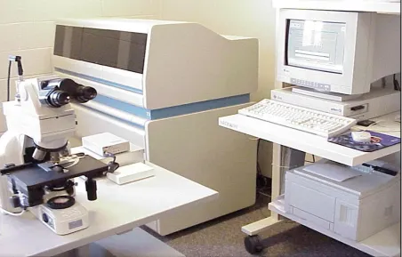
Figure 1.2: BD FocalPoint™ GS Imaging System, along with GSW microscope workstation and PC interface and printer ... 48
Figure 1.3: BD FocalPoint™ Low Resolution Scanning process ... 49
Figure 1.4: BD FocalPoint™ High Resolution Scanning process ... 50
Figure 1.5: Schematic diagram of the FocalPoint™ process for ranking slides and sorting them into quintiles according to probability of morphological abnormality ... 50
Figure 1.6: Labelling and Loading the FocalPoint™ GS Imaging System ... 51
Figure 1.7: FocalPoint™ Calibration Plate required to perform the System Integrity Test ……….……… ... 52
Figure 1.8: FocalPoint image and data transfer pathways for various laboratory service configurations ... 52
Figure 2.1: FocalPoint™ Guided Screener™ Workstation (GSW) system or “SlideWizard™” ………. ... 63
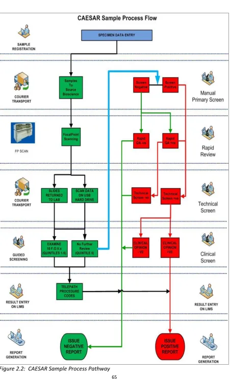
Figure 2.2: CAESAR sample process pathway ... 65
Figure 2.3: SurePath™ Liquid Based Cytology sample taking process: ... 66
Figure 2.4: Sample pre-treatment to remove excess inflammatory and blood elements using the PrepMate™ pipetting station ... 67
Figure 2.5: Slide preparation using the SurePath™ Autocyte Prep™ instrument ... 68
Figure 2.6: Stage calibration of the FocalPoint™ Guided Screener™ Workstation ... 69
Figure 2.7: Image presentation by the FocalPoint™ Guided Screener™ Workstation ... 70
Figure 2.8: GSW screen presented to the cytologist when reviewing FOVs ... 71
10
Figure.2.11: FocalPoint™ GS Imager primary cytology screening mode pathway ... 84
Figure 2.12: FocalPoint™ NFR and manual primary screening pathway ... 86
Figure 3.1: CAESAR study periods, total manually screened and scanned by FocalPoint™ ……….……… ... 91
Figure 3.2: FocalPoint Quintile Ranking by abnormal cytology result ... 102
Figure 4.1: NFR case finally reported as high grade dyskaryosis. Microbiopsy. X40 objective………... 107
Figure 4.2: NFR case finally reported as high grade dyskaryosis. X40 objective. Individual dyskaryotic squamous cells are featured ... 108
Figure 4.3: NFR case finally reported as high grade dyskaryosis. Microbiopsy. X40 objective………... 109
Figure 4.4: NFR case finally reported as high grade dyskaryosis. X40 objective. Single small dyskaryotic squamous cells are featured ... 110
Figure 4.5: Comparative high-grade cytology prevalence July 2009 to February 2010 ... 111
Figure 4.6: High grade cytology incidence in NFR cases/total number of NFR cases ... 113
Figure 5.1: Proposed laboratory screening pathway ... 119
11 List of Tables
Page
Table 1.1: Cervical age of first invitation and subsequent screening intervals ... 31
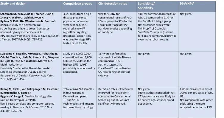
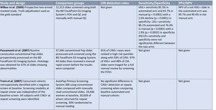
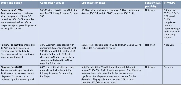
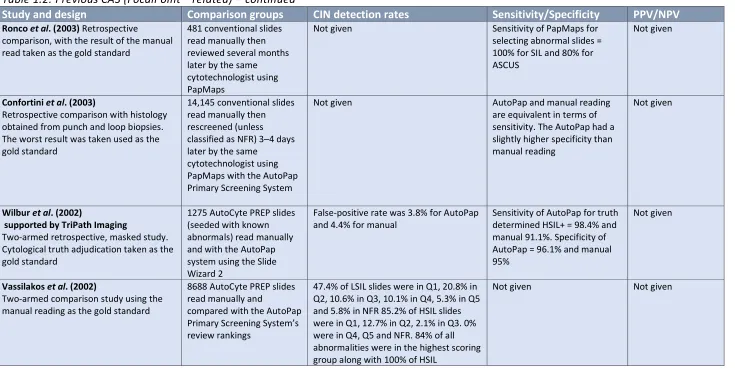
Table 1.2: Previous CAS (FocalPoint™ related) publications ... 39
Table 1.2: Previous CAS (FocalPoint™ related) publications – continued ... 39
Table 1.2: Previous CAS (FocalPoint™ related) publications – continued ... 41
Table 1.2: Previous CAS (FocalPoint™ related) publications – continued ... 43
Table 1.2: Previous CAS (FocalPoint™ related) – continued ... 44
Table 1.2: Previous CAS (FocalPoint™ related) – continued ... 45
Table 2.1: CAESAR studies – dates of sampling ... 61
Table 2.2: Total numbers of cases scanned by FocalPoint, categorised as NFR and manually screened during the CAESAR studies ... 85
Table 3.1: Total samples processed, by participating laboratory ... 92
Table 3.2: Total samples processed by cytological outcome ... 92
Table 3.3: Total samples scanned by FocalPoint™, by participating laboratory ... 93
Table 3.4: Total samples scanned by FocalPoint™, by cytological outcome of manual screening ……… ... 93
Table 3.5: Process Review rates reported during the CAESAR 1 study ... 94
Table 3.6: Average time to examine 10 FOV (CAESAR 1) ... 95
Table 3.7: Average time to manually Rapid QC a slide (CAESAR 1) ... 95
Table 3.8: FocalPoint™ LGS Rapid QA data against manual cytology final report ... 95
Table 3.9: Manual rapid preview screen data against manual cytology final report ... 96
Table 3.10: Summary of published results for sensitivity of rapid QA screening (for HG dyskaryosis+ (HSIL+) unless otherwise stated) compared to CAESAR manual and automated results……… ... 97
Table 3.11: Comparison of the sensitivity of 10 FOV presented to cytotechnologists by FocalPoint™ compared to NHS CSP national minimum standards ... 98
Table 3.12: Comparison of the sensitivity of 10 FOV presented to cytotechnologists by FocalPoint™ to that of manual screening in the participant laboratories during the CAESAR studies……… ... 98
12
Table 3.14: Cancers and Pre-cancers at 2 and 3 years for FocalPoint™ LGS 10 FOV vs.
manually screened negative samples ... 99
Table 3.15: The distribution of final cytology test results within the FocalPoint™ Quintiles ……… ... 101
Table 3.16: NFR cases by laboratory and final cytology result ... 101
Table 4.1: CIN 2+(HSIL+) cases at 2 and 3 years for NFR vs. manually screened negative samples……….……… ... …….104
Table 4.2: Cancers and pre-cancers at 2 and 3 years for NFR vs. manually screened negative samples……….……… ... 104
Table 4.3: Samples assigned to the FocalPoint™ NFR category by laboratory and final cytology outcome ... 105
Table 4.4: Abnormal samples assigned to the FocalPoint™ NFR category by laboratory and final cytology and histology outcomes ... 106
Table 4.5: Total slides scanned by FocalPoint™ from July 2009 to February 2010 . ... 111
Table 4.6: High grade cytology prevalence during the period of July 2009 to February 2010 . ……… ... 111
Table 4.7: High grade cytology incidence in NFR cases/total number of NFR cases . ... 112
Table 4.8: Manual TZ reporting versus FocalPoint endocervical reporting rates ... 114
Table 4.9: All-Wales laboratory TZ detection rates ... 114
Table 4.10: Level of agreement between FocalPoint™ endocervical component and manual detection of endocervical cells ... 115
Table 5.1: Non-medical staff working within Cytology in Wales, 2013-14 . ... 120
Table 5.2: Annual FocalPoint™ costs . ... 120
Table 5.3: Annual staffing establishment required to screen Welsh workload in 2013-14 and costs……… ... 121
Table 5.4: Annual staffing establishment capacity in 2013-14 and costs ... 122
Table 5.5: Staffing model and costs for samples screened via overtime working. ... 123
Table 5.6: Productivity gain and associated cost saving realised using the NFR feature in the proposed cytology screening process pathway at normal time rates ... 124
Table 5.7: Productivity gain and associated cost saving realised using the NFR feature in the proposed cytology screening process pathway at overtime rates . ... 124
13
Table 5.9: Labour costs associated with manual Rapid QC screening in the 2013-14
14
List of Abbreviations
A4C Agenda for Change
Adeno Ca Adenocarcinoma
ASCUS Atypical Squamous Cells of Uncertain Significance ASM Annual Scientific Meeting
BD Becton Dickinson
BMS Biomedical Scientist
CAESAR Computer Assisted Evaluation, Screening and Reporting
Ca Carcinoma
CAS Computer Assisted Screening CEA Cost Evaluation Analysis
CI Confidence Interval
CIN Cervical Intraepithelial Neoplasia
CIN1 Cervical Intraepithelial Neoplasia Grade I
CIN1- Any Lesion of CIN Grade I or Less – Cases Not Requiring Treatment
CIN2 Cervical Intraepithelial Neoplasia Grade II CIN2+ Any Lesion of CIN2 or worse
CIN2– Any Lesion of CIN Grade II or Less
CIN3 Cervical Intraepithelial Neoplasia Grade III CIN3+ Any Lesion of CIN3 or Worse
CONSORT Consolidated Standards of Reporting Trials
CSP Cervical Screening Programme
CSW Cervical Screening Wales
DNA Deoxyribonucleic Acid
EASR European Age Standardised Incidence Rate FDA United States Food and Drug Administration
FEG FocalPoint™ Executive Group
FOG FocalPoint™ Operational Group
FOV Fields of View
FP Becton-Dickinson FocalPoint™ GS Imaging System
GLP Good Laboratory Practice
15
GUM Genito Urinary Medicine
HB Health Board
HC2 Qiagen™ High-risk HPV Hybrid Capture 2 HG High Grade (as in High Grade Dyskaryosis) HSIL High Grade Squamous Intraepithelial Lesion
HSIL+ High Grade Squamous Intraepithelial Lesion or worse
HPV Human papillomavirus
HrHPV High Risk Human Papillomavirus
HTA Health Technology Assessment
IARC International Agency for Research on Cancer
IBM™ International Business Machines
IQC Internal Quality Control
KPI Key Performance Indicator
LBC Liquid Based Cytology
LLDNO Llandudno General Hospital
LSIL Low Grade Squamous Intraepithelial Lesion
LSIL+ LSIL or worse
LIMS Laboratory Information Management System LPCA Laboratory Process Calibration Assessment LREC Local Research Ethics Committee
MAVARIC Manual Assessment versus Automated Reading in Cytology
MDM Multi-disciplinary meeting MLA medical laboratory assistant
mRNA Messenger Ribonucleic Acid
MSC Managed Service Contract
MSD Most Significant Dot
NFR No Further Review
NHS National Health Service
NHSCSP NHS Cervical Screening Programme
NICE National Institute for Health and Clinical Excellence NPP Negative Predictive Potential
NPV Negative Predictive Value
16
Pap Papanicolaou
PC Personal Computer
PCR Polymerase Chain Reaction
PCT Primary Care Trust
PHE Public Health England
PHW Public Health Wales
PII Patient Identifiable Information
PPV Positive Predictive Value
QC Quality Control
RGH Royal Gwent Hospital
RNA Ribonucleic Acid
ROU Resolution of Uncertainty
RQA Rapid Quality Assurance
SBS Source BioScience
SCJ Squamo-columnar Junction
SD Standard Deviation
SIL Squamous Intraepithelial Lesion
SOP Standard Operating Procedure
TIS ThinPrep™ Imaging System
TOC Test of cure
TPR True Positive Rate
TZ Transformation Zone
UK United Kingdom
UKAS United Kingdom Assessment Service
VDU Visual Display Unit
VPN Virtual Private Network
WG Welsh Government
WHO World Health Organisation
WTE Whole Time Equivalent
WXM Wrexham
Y2K Year 2000 bug (Year coded in computer software as “YY” and not “YYYY”)
17
List of Presentations and Publications
Published papers, and professional guidance documents
Cuscheri K, Denton K, Nuttall D, Sargent A. Laboratory quality control and assurance for human papillomavirus testing. January 2017. Public Health England. NHS Cervical Screening Programme. www.gov.uk – accessed June 1, 2017.
Hibbitts S, Tristram A, Beer H, McRea J, Rose B, Hauke A, Nuttall D, Dallimore N, Newcombe RG, Fiander A. UK population based study to predict impact of HPV vaccination. 2014. Journal of Clinical Virology. Feb;59(2):109-114.
Denton K, Nuttall D, Cropper A, Desai M. Implementation of ‘No Further Review’ (NFR) using the BD FocalPointTM Slide Profiler. 2013. NHS Cervical Screening Programme: Good Practice Guide No. 4.
Submitted for publication – awaiting decision:
Nuttall DS, Fox R, Hillier S, Dallimore N, Clayton H, Martin C, O’Leary JJ, Sloan
S, Savage A. A Retrospective Validation of the Becton Dickinson Focal Point GS Slide Profiler NFR Technology by Analysis of Interval Disease Outcomes Compared to Manual Cytology Screening.
In preparation:
18 Presentations
2016:
European Congress of Cytology, 40th Conference. Liverpool.
Oral Presentation: Improved Detection of CIN 2+ Lesions by the Becton Dickinson Focal Point™ GS Slide Profiler No Further Review Technology Compared to Routine Manual Slide Reading: An analysis of interval outcomes.”
United States and Canadian Association of Pathology (USCAP) Annual Scientific Meeting. Seattle, USA.
Oral Presentation: Improved Detection of CIN 2+ Lesions by the Becton Dickinson Focal Point™ GS Slide Profiler No Further Review Technology Compared to Routine Manual Slide Reading: An analysis of interval outcomes.
2014:
Cervical Screening Wales 15th Anniversary Conference – Cardiff.
Oral Presentation: Cervical Screening Laboratory Services – the next 15 years!
2013:
Update course for Consultant Biomedical Scientists East Pennine Cytology Training School.
Oral Presentation: Cervical Screening Wales –A Different Perspective”.
Cervical Screening Wales Colposcopy Conference – Llandrindod Wells. Oral Presentation: Introduction and Clinical Management of HPV Testing.
2012:
British Association for Cytopathology. Annual Scientific Conference.
19 2011:
Conference of the Cytology Society of Belgium - Antwerp.
Oral Presentation: The Role of Biomarker Assisted Morphology in the Cervical Cytology Laboratory – The Welsh SuPerLy Project.
European Congress of Cytology – Istanbul.
Poster Presentation: “An Evaluation of the BD Focal Point™ Imaging System – The
Significance of the No Further Review Slide Scan Result Category for Routine Use within the Cervical Screening Programme in Wales.
Nuttall DS, Rose B.
Meeting of the Cytology Society for the South of England and South Wales.
Oral Presentation: The Role of Biomarker Assisted Morphology in the Cervical Cytology Laboratory – The Welsh SuPerLy Project.
2010:
National Association of Cytology. Keele University.
20
Acknowledgements
During the course of carrying out this research study, I have been very fortunate to receive a great deal of help, and I wish to gratefully acknowledge those that have provided that support.
Firstly, I am indebted to Professor John O’Leary and I wish to acknowledge his great support and unstinting advice and guidance. I have had the good fortune to collaborate with Prof. O’Leary on several
occasions and his enthusiasm for his field of endeavour, his teaching and his mentorship of the students fortunate enough to study under his supervision, is truly inspirational. I clearly remember that day in 2009 when I was introduced to Prof. O’Leary and his subsequent invitation to study under his
mentorship at TCD. It has been a roller coaster of personal highs and lows, mostly highs and I shall be eternally grateful for the opportunity.
I wish to express my sincerest thanks to Prof. Cara Martin, who has provided immeasurable support, help and advice in writing up this thesis as well as helping me fulfil the requirements of TCD of its students. Without Prof. Martin, I do not think that I would be in the fortunate position of writing up this acknowledgement!
Sincerest thanks are also due to Dr James O’Mahony for his help and advice with the cost evaluation
assessment and with proof reading the thesis.
I would also like to acknowledge the Screening Division, Public Health Wales for partly financing my candidature. I would like to thank the following individuals for their help and support:
Mr. Bryan Rose, former Head of Programme – Cervical Screening Wales for his enthusiasm, his willingness to help and advice and for being a friend.
Mrs Gwyneth Carey, for all the help with the formatting of this thesis, which she carried out with consummate ease. Gwyn, thanks for all you’ve done over the years!
Drs. Hilary Fielder and Rosemary Fox, former Directors of Screening Division for their support and I am also grateful to Dr Sharon Hillier, currently Acting Director of Screening Division.
21
To the staff of the Welsh laboratories that have contributed to this study, in particular to Amanda Savage, Sonia Sloan, Virginia Seager and Steve Court. Your contributions have been invaluable.
22
Chapter 1
Introduction and Background
1.1 Cervical cancer – definition, development rates and time trends
Cervical cancer is a malignant neoplasm of the cervix uteri. In 2012, 528,000 new cases of cervical cancer were diagnosed worldwide, and 266,000 women died of the disease - almost 90% of these women were from low to middle income countries. Cervical cancer is the leading cause of cancer deaths in Eastern and Central Africa. Without urgent attention, deaths due to cervical cancer are projected to rise by up to 25% over the next 10 years (WHO; 2014)
The incidence of cervical cancer and mortality rates in most countries has decreased significantly in the last 30 years (WHO, 2014). In the UK, mortality from cervical cancer has been declining and in 2012, was at a low of <5 deaths per 100,000 women. However, there were 3,224 new cases of cervical cancer reported in the UK in 2014 and the peak rate of these cases were in the 25-29 year age group. Cervical cancer European Age Standardised (EASR) incidence rates have decreased in the UK since the early 1990s, however, in the last decade EASR incidence rates have increased by 5%. This increase reflects the death by cervical cancer of a young celebrity (Lancucki et al., 2012).
1.1.1. Epidemiology of cervical cancer
The incidence rates of cervical cancer show strong birth cohort effects (Sasieni, Adams, 2000). This means that women born at one time might be at relatively high risk of cervical cancer in their 20s and 30s and remain at relatively high risk through their 40s, 50s, 60s and 70s. My understanding of this effect is that there is an underlying characteristic of increasing rate of disease with age, but the level is determined by environmental exposure (to a
23
Figure 1.1: European Age-Standardised Incidence Rates of Cervical Cancer per 100,000 population. Source: CRUK data – accessed September 8th, 2017
1.1.2. Incidence of cervical cancer
From a public health perspective, it is important to note that:
• women born in the 1960s are three to four times greater risk of cervical cancer than women born in the 1930s.
• after adjusting for cohort effects, the incidence of cervical cancer increases between ages 20 and 55 years, most rapidly between ages 30 and 40, and decreases steadily after age 55.
Therefore, the cumulative risk of cervical cancer in women born during the 1960s is likely to be around 4-5%, thus emphasising the importance of cervical screening.
1.1.3. Risk factors
24
factors usually quoted include number of sexual partners and age at first sexual intercourse (Brinton et al. 1992). The behaviour of men is also a factor, as shown by increasing risk in women with just one partner, depending on the number of partners that their husband has had (Buckley et al., 1981). More recently, the sexually transmitted agent has been identified as certain types of the human papillomavirus (HPV), now known as high-risk human
papillomaviruses (Hr-HPV).
Human papillomavirus (HPV) is a common, sexually transmitted infection. In rare cases, infection with high-risk forms of this virus can cause a woman to develop cervical cancer. There is consistent evidence from across the world that high-risk HPV infection is a necessary cause of cervical cancer, and optimal testing systems have identified the virus in all invasive specimens (NHS Cancer Screening Programmes, 2008). HPV is implicated in both squamous cell carcinoma (SCC) and adenocarcinoma (Adeno Ca), as well as in over 95% cases of the cancerous precursor, cervical intraepithelial neoplasia, grade 3 (CIN3).
Co-factors that appear to increase the risk of developing cervical cancer in HPV-infected women include the use of oral contraceptives, smoking, high parity, unidentified genetic factors possibly related to immunity, and previous exposure to other sexually transmitted diseases, such as chlamydia trachomatis and herpes virus type 2. Women exposed to human immunodeficiency virus (HIV) are at high risk of HPV infection, HPV persistence, and cervical cancer. Immunosuppression certainly increases the risk of cervical cancer, as evidenced by studies on renal transplant patients receiving immunosuppressive drugs (NHS Cervical Screening Programme Publication No. 20; 3rd edition; 2016), and smokers are thought to be at increased risk due to the immunosuppressive effects of tobacco smoke inhalation. Diet may play a role in the immune response to HPV, but studies on diet and cervical cancer are inconclusive to date (Garcia-Closas et al. 2005).
More recently, genetic factors that modify the risk of cervical cancer have been identified, but the understanding of the factors that determine why some women develop cervical cancer after infection with oncogenic HPVs, whilst the majority do not, is incomplete.
25
had a substantial impact on cervical cancer incidence in many countries (NHS Cancer Screening Programmes, 2008).
1.1.4. Squamous cell carcinoma and adenocarcinoma
There are two main types of cervical cancer. The most common is squamous cell carcinoma. It used to be said that this accounted for around 90% of all cervical cancer. However, more recent data shows that adenocarcinoma (including adeno-squamous) is accounting for a growing proportion of diagnoses particularly in younger women (Stockton et al., 1997). Squamous cell carcinoma now only accounts for around 75% of all cervical cancer. The reason for the increasing proportion of adenocarcinoma seems to be three-fold:
• Adenocarcinoma really is becoming more common having been a very rare disease because of the greater awareness of adenocarcinoma.
• It is being reported more often on pathology reports - previously the cell type may not have been reported and so was assumed to be squamous.
• Cytological screening is better able to detect cancerous squamous lesions than pre-cancerous glandular (adeno) lesions and thus the relative incidence of the two types of cancer has changed.
There is some suggestion that adenocarcinoma is associated with HPV type 18, whereas squamous cell carcinoma is associated with the more common type 16 (International Agency for Research on Cancer). HPV types 31 and 33, although less common in the UK, are also associated with cervical cancer (IARC, 1995).
1.1.5. Natural history
26
1995) and viral load can be used as a surrogate for persistence (Cuzick, 1997). It is now generally accepted that one of the key steps in the development of cancer is integration of the viral DNA in the host genome (Cullen et al., 1991).
Cervical neoplasia appears to constitute a disease continuum (Kiviat et al., 1992) ranging from cervical intraepithelial neoplasia (CIN) grades 1 to 3, to micro-invasive and finally fully invasive cancer. Follow-up studies of women with CIN have found that about 60% of CIN 1 regresses compared to about 33% of CIN 3; whilst 11% and 22% of CIN 1 and 2, respectively, progressed to CIN3 (Ostor et al., 1993). Although the details of progression and regression are largely speculative, at most about a third of high grade CIN will progress to cancer over about 15 years and that the majority of CIN 1 will regress.
CIN 3 is very rare in women under the age of 20 (Evans et al. 1997). The rates rise rapidly peaking at about age 30 and fall again rather more slowly to about half their peak by age 40 and just 10-20% by age 50.
1.1.6. Treatment and survival
Five-year survival after diagnosis of cervical cancer is strongly related to the stage of the tumour at diagnosis. Studies show over 90% survival for discreet tumours in women under 50 at diagnosis, 50% for cancers with local involvement and just 11% in women over 50 years with distant metastases. Survival (5-year) after diagnosis of micro-invasive cancer (stage 1a1/1a2) is around 94-98% (Quinn et al., 2006).
Micro-invasive cancers may be treated by cone biopsy alone, but hysterectomy may be the preferred treatment, particularly if the patient has completed her family. Surgery
(Wertheim's hysterectomy) is the usual treatment for invasive cancer that has not spread beyond the pelvic area. It may be followed by radiotherapy if the cancer recurs.
Radiotherapy alone is the standard treatment for more advanced cancer, chemotherapy is also used.
27
CIN (CIN 2 and 3) will develop invasive cancer within 10 years. Ablative treatments used to be popular but are now becoming less so. Excision can be done by various means. LLETZ (large loop excision of the transformation zone) is widely used, but laser, cold-knife cone biopsy, cold coagulation and cryotherapy are still used on occasion.
1.2 The National Health Service cervical screening programme
1.2.1. Evidence of the effectiveness of cervical screening
The NHS Cervical Screening Programme (NHSCSP) implemented a managed, structured programme of call and recall of screening participants in 1988 and this programme is estimated to save as many as 5000 lives per year in the UK Peto et al. 2004). It is now recognised as one of the leading cervical cancer prevention programmes worldwide.
The use of new technology to improve service quality and efficiency is a key strategy of the NHSCSP. Within screening cytology, improving sensitivity and specificity and reducing human workload are key desirables and the number of screening tests has dropped in recent years because of service improvement. For example, the implementation of liquid-based cytology (LBC) in 2004-2008, saw the number of inadequate samples and subsequent repeat testing drop from 9% in 2004–5 to 2.9% in 2007–8 (Kitchener et al., 2011). The introduction of human papillomavirus (HPV) triage and test of cure (TOC) in the UK has reduced the number of repeat tests taken by triaging the treatment and management of women based on their HPV results. Women attending for routine tests who are found to have a low-grade
abnormality and a positive HPV result are referred directly to colposcopy without repeat cytology testing, and those who are HPV negative are returned to routine recall without cytological follow-up. (HPV Sentinel Sites implementation project, 2008).
1.2.2. Current manual screening practices in the UK
28 least every 2 hours.
Rapid QA screening is carried out by screening staff performing a rapid internal quality control (IQC) review of the whole slide in around 90 seconds. Current screening techniques are labour intensive requiring a large and committed laboratory workforce. The initial
training and ongoing competency assessment of staff is managed on a national scale and the external quality assessment schemes (EQA) for participating staff and technical slide
preparation are a seriously resource intensive undertaking.
1.2.3. Screening intervals and coverage
In England, currently, women aged 25–49 years are invited every 3 years, and women aged 50–64 years are invited every 5 years (NHS Cancer Screening Programme Annual Review, 2008). Of the 3.6 million women aged 25–64 years who were screened in 2008–9, around 6.7% received an abnormal result. In the same period, there were 134,000 referrals to colposcopy prompted by an abnormal screening result, 28.9% of which were for results of moderate dyskaryosis or worse,the remainder resulting from low-grade cytological abnormalities (The Health and Social Care Information Centre. Cervical Screening Programme, England 2008–09).
1.2.4. Future NHS cervical screening programme considerations
Following the publication of the MAVARIC report (Kitchener et al., 2011), computer assisted screening (CAS) was approved for use in the UK within the NHS cervical screening
programme (NHS CSP). This approval concerned the BD (Becton Dickinson - BD, Franklin lakes, NJ, USA) FocalPoint™ GS Imaging System “No Further Review” (NFR) reporting
technology and the guidance for implementation is set out in NHS CSP Guidance Document No. 4 (Denton et al.; 2013 “Implementation of ‘No Further Review’ (NFR) using the BD FocalPointTM Slide Profiler”).
As well as the implementation of CAS, several other organisational challenges faced the NHSCSP at that time. In 2007 the Department of Health (DoH) published the Cancer Reform Strategy, (Department of Health; 2007). This document recommended that to achieve the Government’s target of a 14-day turnaround time (from cervical sample being taken to the
29
reconfigured to make them larger and more efficient. Some laboratories currently operate as “hub and spoke” with larger central laboratories processing the LBC samples and
returning them to the smaller laboratories for screening. Amalgamation of smaller laboratories will see further changes to this service configuration. In the NHS in Wales, pathology cervical screening services were reviewed and restructured in 2009-10 in anticipation of the implementation of CAS and HPV testing. This resulted in two “hub and
spoke” networks implemented in Wales – one in North Wales linking the laboratories in
Llandudno, Bodelwyddan and Wrexham via the A55 trunk road and the other in South Wales, linking laboratories in West Wales (Carmarthen and Haverfordwest), Swansea, Cardiff and Newport via the M4 motorway corridor. In 2010-11, this structure was replaced by a single hub laboratory (Magden Park, Llantrisant, Cardiff) servicing a single North Wales screening laboratory at Glan Clwyd Hospital along with two other laboratories in South Wales, at Newport and Swansea. Following the recommendations and subsequent validation of CAS (Appendix 13), arising from the CAESAR studies, and presented to the all-Wales Management Group of Cervical Screening Wales (CSW) the FocalPoint™ NFR technology was
approved for use in Wales in 2012.
The HPV vaccination programme will also impact on the cervical screening programme. Implemented in September 2008, young girls were vaccinated at ages 12–13 years followed by a 3-year catch-up campaign to vaccinate older girls aged 14–17 years. Once the evidence regarding screening in a vaccinated population becomes clearer, screening intervals and follow-up protocols will need to be reviewed to reflect these findings. The importance of following up the screening outcomes of recently vaccinated girls was stressed by the
Advisory Committee on Cervical Screening (ACCS) in 2009 when reviewing current screening policy in women aged 20–24 years. Following recommendations from the ACCS, the DoH decided against making any changes to current policy regarding screening in women aged 20–24 years. Instead, further education of general practice staff will ensure that
symptomatic women aged < 25 years are assessed appropriately.
1.3 The National Service Framework for the Cervical Screening Programme in Wales
30
published standards were in place and that all eligible women receive the same standard of service and quality of care for the same level of need. The decision to develop an NSF
followed concern about failures of the UK programme, particularly in the Kent & Canterbury NHS Trust, and about the fact that previous reports and recommendations in Wales had not been fully implemented. The Health Minister at that time, Win Griffiths, invited Velindre NHS Trust, to work alongside the Welsh Office to develop and implement the National Service Framework.
1.3.1 Cervical Screening Wales
Cervical screening began in Britain in the mid-1960s. By the mid-1980s, although many women were having regular cytology tests, there was concern that those at greatest risk were not being tested, and that those who had positive results were not being followed up and treated effectively. The NHS Cervical Screening programme was set-up in 1988 when the Department of Health instructed all health authorities to introduce computerised call-recall systems and to meet certain quality standards. Cervical Screening Wales (CSW) is
responsible for the Cervical Screening Programme (CSP) in Wales. CSW was launched in 1999 to provide women with equal access to a uniform and high quality cervical
screening service across Wales. The programme aims to reduce the incidence of and mortality from invasive cervical cancer. This is accomplished by regularly screening all women at risk so that conditions which might otherwise develop into invasive cancer can be identified and treated. At the time of this study, Cervical Screening Wales invited women aged 20-64 for a cytology test every three years. Cervical screening is free for all eligible women. Screening is not offered to women who have had a total hysterectomy.
1.3.2 Cervical Screening Intervals
At the time of this study the cervical screening invitation and screening intervals were as presented in Table 1.1. The NHS call-and-recall system invites women who are registered with a GP. It also keeps track of any follow-up investigation, and, if all is well, recalls the woman for screening after three years.
31
Table 1.1: Cervical age of first invitation and subsequent screening intervals
Age group (years) Frequency of screening
20 First invitation
20-64 3 yearly
65+ Screen those who have recent abnormal tests until these have been resolved
The programme screens on average around 223,500 women in Wales each year. For clinical reasons, some women have more than one test during a year thus around 236,800 samples are examined by pathology laboratories annually. Whilst no cervical screening intervention can be 100 percent effective, cervical screening programmes have been shown to
dramatically reduce the incidence of cancer in a population of women.
Since the introduction of the NHSCSP in the UK in 1988, the number of diagnoses has halved, from 16.5 per 100,000 women in 1988 to 8.5 per 100,000 women in 2008 – despite
increased rates of underlying disease.
1.3.3 Burden of cervical cancer in Wales
Between 1999 and 2009, 1863 cases of cervical cancer were registered in Wales with an average European age-standardised incidence rate (EASR) of 10.3 per 100,000 women. Mortality is substantially lower than incidence of cervical cancer with 735 cases reported between 1999 and 2009 (average EASR mortality rate 4.1 per 100,000).
1.4 The technologies used in cervical screening
1.4.1 The Papanicolaou smear test
The Papanicolaou or “Pap” smear was first described in 1943, (Papanicolaou GN, Traut H,
1943) but was not implemented in the UK until 1964 (Appendix 1). The screening of a cervical cytology sample is performed manually under a cytologist’s microscope. Apart from
great improvements in quality assurance and structured training and assessment of the cytologists working in the laboratories that provided a cervical cytology service, little changed in the production of the “Pap” smear in the UK until the advent of Liquid Based
32
This important milestone changed cervical cytology radically (see Section 1.4) and provided a springboard for the amalgamation of laboratories into large high-throughput units that have the required critical mass of work needed to implement expensive technologies such as Computer Assisted Screening technologies most effectively.
1.4.2 Liquid Based Cytology
The conventional method of producing cervical cells on a glass slide involved a sample being obtained from the cervix using a spatula which was smeared onto a glass slide and then fixed. Fifty years on, this method is still widely used worldwide. The quality of the slide material is variable, with the cells often unevenly spread along with excess blood cells and mucus capable of obscuring the cervical cells. This leads to a large number of slides being designated as ‘inadequate’ for reporting, resulting in the woman requiring a repeat sample
to complete the test.
With LBC, the cervical sample is dissipated in a fluid medium which contains a cell fixative. The liquid sample is then subjected to either a process which filters the cells onto a slide (ThinPrep™ LBC, Hologic, Bedford, MA, USA) or cell enrichment [Becton Dickinson (BD)
SurePath™ LBC, BD, Franklin lakes, NJ, USA] producing a cleaner, more homogeneous
preparation which facilitates improved examination of the cervical cells. From 2001–3 an NHSCSP pilot study was performed in England in order to evaluate LBC in comparison with conventional cytology in a historical population. The findings were that inadequate samples were reduced from around 7%–8% to around 1%, and that LBC was certainly not less
sensitive than conventional cytology and possibly more so, that laboratory throughput was more efficient, and that laboratory staff preferred LBC. LBC was determined to be cost-effective and meant that far fewer women were recalled because of an ‘inadequate’ smear.
The National Institute for Health and Clinical Excellence (NICE) recommended its adoption (National Institute of Clinical Excellence, 2003) and between 2003 and 2008 LBC was rolled out across the entire UK. The new technology (SurePath™ version) was introduced across
Wales between 2004-2005.
33
HPV testing and the potential to move to automated technology if the requirement arose.
1.4.3 HPV DNA testing
There are over 100 types of Human Papillomavirus (HPV) and most do not cause significant disease in humans. However, around 15 HPV types have been implicated in cervical cancer, notably types 16 and 18 which give rise to about 70% of all cervical cancers. Research has shown that women with no evidence of high risk HPV infection are extremely unlikely to have concurrent cervical pre-cancerous disease or to develop such disease or cervical cancer over the next 6 years.
HPV testing has been evaluated in various settings:
▪ To triage women with low grade dyskaryosis including borderline changes on cytology
▪ As a “test of cure” to reduce the duration of surveillance following treatment for CIN
▪ To replace cytology as the primary screening test
1.4.4 Cervical screening and HPV vaccination
Two prophylactic HPV vaccines have been shown to be highly effective at preventing persistent HPV infection and the high-grade disease (CIN3) caused by infection. In
September 2008, a national programme was introduced to vaccinate girls against HPV 16 and 18. This programme was aimed at girls aged 12-13, however a catch-up programme for those born during 1990-1995 was also initiated.
The NHS cervical screening programme continues to screen women who have not been vaccinated and the role of cervical screening for vaccinated women remains to be clarified. This role will depend on the age at which the woman was vaccinated, the cross-protection given by the vaccine for other HPV types and the duration of protection provided. The impact of HPV vaccination will require monitoring alongside the cervical screening programme. In the interim, continued work is needed to determine the most effective means of monitoring the impact of both vaccination and cervical screening.
1.5 Development of CAS technologies
34
and consistency (Desai, 2009). Detecting these rare occurrences with the high level of accuracy required has been a continual challenge to the early pioneers developing automated screening technologies.
Initial experimentation with CAS took place in Europe following attempts to automate scanning of samples stained with the Feulgen stain for DNA/RNA (Desai, 2009).
Developments then shifted to the USA with the development of the Cytoanalyser by the Airborne Instruments Laboratories, Inc. of Mineola, New York, as described by Tolles (1955). This company was exploring ways to utilise its war-time technologies for other uses,
including medical applications. The instrument was designed to compare cell size as well as nuclear size and density.
Early researchers soon discovered that the complexities of cellular morphological analysis and recognition were very challenging. The problems encountered included:
• Similarities between benign and abnormal cells were greater than the differences • The computing resources at that time were inadequate and unable to process the
amount of morphological data generated by several hundred thousand cells on a Pap slide.
• Thick, 3D clusters of cells compounded these problems • Detecting nuclear: cytoplasmic boundaries proved difficult.
Since these early beginnings, computers have become faster, the introduction of LBC has revolutionised cytology with the production of thinner slide preparations and the
advancement of technology in general means that CAS technology can now compete with manual cytology screening in terms of throughput and accuracy.
The US Food and Drug Administration (FDA) has since approved two automated machines that were developed in the 1990s, the AutoPap™ 300 QC (NeoPath™, Redmond, WA, USA) and the PapNet™ (Neuromedical Systems Inc., Suffern, NY, USA - NSI). Both these systems
were designed to work with conventional cytology slides.
35
AutoCyte-Prep slides (now BD SurePath LBC). AutoPap was a computerised image processor that used high-speed video microscopy, image analysis software and FOV computers to classify cell images of conventional Pap smears by means of special algorithms designed to classify cells and slides. A proportion of the ranked slides were then selected for manual screening or review by a cytologist.
The PapNet™ system used computer imaging technology and neural network software to
provide location-guided primary screening of conventional Pap smears. The system selected and presented up to 128 images of potentially abnormal cells to the cytologist.
These machines are no longer available. The AutoCyte™ was not year 2000 (Y2K) compliant
and the PapNet™ company went into receivership in 1999. The story of NSI’s PAPNET® System in the USA is an interesting one. In 1995, both NeoPath and NSI were approved by the FDA for the over-screening (secondary screen for quality assurance purposes) of negative conventional smears and they both then proceeded to market their products commercially. While NeoPath marketed its instrument to large laboratories by leasing the scanner for use on site, NSI aggressively marketed PAPNET® to clinicians and even directly to patients in the lay press, arguing that the added cost of PAPNET® QC screening ($40 a screen which was not covered by most health insurance companies) was worth it to ensure “peace
of mind”. The implication was that human screening was so unreliable and this undermined
conventional human screening to the point where it was perceived that its use alone was almost dangerous. Some laboratories used PAPNET® as a means of establishing cytological “truth”, even reprimanding cytologists whose negative cases turned positive on PAPNET® QC
screening. The impact on the morale of the laboratory community, already under siege by the lay press, was considerable. Adding to the company’s image problem were articles by a
representative of the company intimating that failure to offer the superior PAPNET® QC screening might result in medicolegal liability.
NSI had difficulty in getting consistent reimbursement by health insurers and several published studies suggested only a minimal improvement in detection rate over manual screening with poor cost effectiveness added to pressures on the company. In October 1999, and depleted of funds, NSI was declared bankrupt. NeoPath™ bought NSI’s intellectual
36
in 1999 to form TriPath™ Imaging Inc. (Burlington, NC, USA). TriPath™ discontinued both the
AutoCyte™ and the AutoPap 300 QC, replacing the systems with the AutoPap™ Primary
Screening System, which is now known as the BD FocalPoint™ GS Imaging System (BD
Diagnostics, Franklin Lakes, NJ, USA).
1.5.1 Current systems in production
Currently, two commercially available FDA-approved automated screening systems – the BD FocalPoint™ GS Imaging System and the ThinPrep™ Imaging System (Hologic™, Bedford, MA,
USA). The BD FocalPoint™ Slide Profiler scans the slides and assigns each one a score which
ranks them according to the likelihood of abnormal cells being present. The slides are assigned to quintiles, with quintile 1 containing the highest-ranking slides. The machine also categorises slides into one of four of categories: review (comprising quintiles 1–5), no further review (NFR; up to 25% of slides), process review (indicating a technical problem) and quality control review (requiring a full screen). The NFR category contains the slides least likely to contain an abnormality which could be reported as negative and archived without a full manual primary screen. The NFR technology is approved for use in the UK with each slide so designated being subjected to a rapid QA screen (rapid review or preview). Slides that are designated for review by the FocalPoint™ are examined by cytology screening
staff using the BD FocalPoint Guided Screener Workstation (GSW, previously known as the TriPath™ Slide Wizard™). This technology comprises of a standard screening microscope
fitted with an electronic stage linked to a desktop computer. The GSW directs screening staff towards 10 electronically marked fields of view on the slide. If abnormal cells are seen in any of the FOVs the entire slide is screened, and appropriate action taken in line with laboratory protocols. The BD FocalPoint Guided Screener (GS) Imaging System has received FDA
approval to scan both conventional and BD SurePath LBC slides.
In contrast, the ThinPrep™ Imaging System is designed to work with ThinPrep™ LBC slides (stained with the Hologic™ Imager stain) alone. The ThinPrep™ (TIS) Imaging System scans all
of the slides and presents 22 FOVs to screening staff on the review scope. The review scope is a Hologic™ automated screening microscope with a motorised stage to guide screeners to each of the 22 FOVs. If an abnormality is suspected in any of the 22 FOVs then a full screen of the slide is undertaken. Unlike the FocalPoint™, the TIS does not score and rank slides and
37
1.5.2 Computer Assisted Screening in cytology – where are we?
The use of Computer Assisted Screening (CAS) in cervical screening is well established in the USA (Wilbur et al., 2009) and Europe (Passamonti et al., 2007; Troni et al., 2007) and has undergone a major trial in the UK (MAVARIC – Kitchener et al., 2011) which was funded by the United Kingdom Health Technology Assessment (HTA) programme. Several other studies have been conducted in the UK and the Republic of Ireland, including the Cervical Screening Wales CAESAR Studies (described in this thesis and cited by Kitchener et al. 2011) and the study conducted by Cropper et al. 2010, using the BD FocalPoint™ GS Imaging System. Bolger et al. (2006) and the Scottish Cervical Cytology Review Group (Feasibility Sub Group - 2009) have reported on the Hologic ThinPrep™ Imager.
Four published systematic reviews were conducted on the potential of CAS (Broadstock, 2000; Willis et al., 2005; Kitchener et al., 2011; Della Palma et al., 2012). Willis et al, in a review commissioned by the Health Technology Assessment (HTA) programme and published in 2005 concluded that there was a need for rigorous, unbiased public-sector research into the effectiveness of automated screening technologies. In the earlier review by Broadstock in 2000 for the New Zealand HTA programme reached a similar conclusion and recommended large-scale trials to be conducted under normal laboratory conditions against reliable gold standards for diagnostic verification. Both these reviews focused on early technologies that are no longer commercially available. The study by Kitchener et al.,
“Manual Assessment Versus Automated Reading In Cytology” (MAVARIC) appraised both the
FocalPoint™ and TIS CAS systems but did not recommend either for implementation.
However, the study concluded that the NFR reporting technology of the FocalPoint™ system
presented promising results and recommended further research into its potential.
38 Efficacy:
1. Is there sufficient evidence that automatic screening is as least as accurate as the manual process?
2. If two methods are essentially equivalent in terms of accuracy, can it be stated that this also means that they are essentially equivalent in terms of efficacy in the prevention of cervical cancer?
Cost Effectiveness:
1. Is there evidence that automatic reading increases productivity per reader? 2. If so, by how much?
3. How is productivity affected by the screening of liquid-based cytology (LBC) slides?
Results: For conventional Pap-smear samples the break-even point was at about 49,000 cases per annum. For liquid based cytology samples, it was at the maximum capacity of the CAS technology – about 70,000 cases per annum. Therefore, efficiency increased with the volume of slides scanned – with screening time decreasing by two-thirds for conventional slides and by less than one half for LBC slides. It was also reported that acceptance of the technology by users was good.
39 Table 1.2: Previous CAS (FocalPoint™ related) publications
Study and design Comparison groups CIN detection rates Sensitivity/ Specificity
PPV/NPV
Schiffman M, Yu K, Zuna R, Terence Dunn S, Zhang H, Walker J, Gold M, Hyun N,
Rydzak G, Katki HA, Wentzensen N. Proof-of-principle study of a novel cervical
screening and triage strategy: Computer-analysed cytology to decide which
HPV-positive women are likely to have ≥CIN2. Int J Cancer. 2017 Feb;140(3):718-725.
3026 cases from a high disease prevalence population of women were scanned. This required a new FP algorithm targeting precancer/cancer. This was used to triage HPV tested cases for CIN
94% for ≥CIN2 for
conventional results of ASC-US compared to 91% for the FocalPoint triage of HPV positive samples depending on sub-type.
94% for conventional results of ASC-US compared to 91% for the FocalPoint triage group. Note: scanned slides were ThinPrep™ LBC samples, SurePath ™ samples (optimal for FocalPoint™) should provide even more robust results.
Not given
Sugiyama Y, Sasaki H, Komatsu K, Yabushita R, Oda M, Yanoh K, Ueda M, Itamochi H, Okugawa K, Fujita H, Tase T, Nakatani E, Moriya T. A Multi-Institutional
Feasibility Study on the Use of Automated Screening Systems for Quality Control Rescreening of Cervical Cytology. Acta Cytol. 2016;60(5):451-457.
Study of 12,000; 9,000 conventional and 3,000 LBC slides. Slides in the highest 15% (1,496) probability of abnormality rescreened.
117 were confirmed as abnormal of which 40 were confirmed as HGSIL. Authors suggest that FocalPoint™ is effective for QC rescreening of cervical cytology
Not given Not given
Rebolj M, Rask J, van Ballegooijen M, Kirschner B, Rozemeijer K, Bonde J,
Rygaard C, Lynge E. Cervical histology after routine ThinPrep or SurePath
liquid-based cytology and computer-assisted reading in Denmark. Br J Cancer. 2015 Nov 3;113(9):1259-74.
Total of 674,248 samples in four regions in Denmark compared TP and SP LBC
technologies and Imaging to conventional cytology.
Detection rates (≥CIN2) were improved for FocalPoint™ compared to Conventional Screening but TIS was not significantly improved.
Not given
(Note: authors concluded that CAS performance was likely to be patient age/scanner brand dependent.
Calculated as frequency of ≥CIN2 per 100 cases of ASC-US.
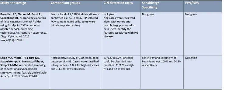
40 Table 1.2: Previous CAS (FocalPoint™ related) publications – continued
Study and design Comparison groups CIN detection rates Sensitivity/ Specificity
PPV/NPV
Bowditch RC, Clarke JM, Baird PJ, Greenberg ML. Morphologic analysis of false negative SurePath® slides using Focalpoint™ GS computer-assisted cervical screening
technology: An Australian experience. Diagn Cytopathol. 2015
Nov;43(11):870-8.
From a total of 2,198 SP slides, 47 were confirmed as HG. In all 47, FP selected FOV containing HG cells. Some were initially reported as Neg.
Not given.
Neg cases were reviewed along with others and morphology presented to help users identify the features associated with HG disease.
Not given Not given
Saieg MA, Motta TH, Fodra ME, Scapulatempo C, Longatto-Filho A, Stiepcich MM. Automated screening of conventional gynecological
cytology smears: feasible and reliable. Acta Cytol. 2014;58(4):378-82.
Retrospective study of 120 cases, aged between 18 – 85. Cases were classified into quintiles – 1 & 2 for high risk cases and 3,4,5 for low risk cases.
83/120 (69.2%) of cases could be classified into quintiles. 31/120 as high risk and 52 as low risk.
Sensitivity and specificity of FocalPoint was 100% and 70.3% respectively.
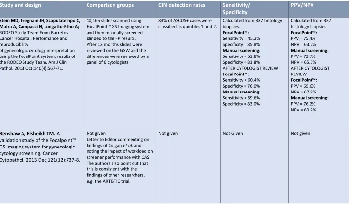
41 Table 1.2: Previous CAS (FocalPoint™ related) publications – continued
Study and design Comparison groups CIN detection rates Sensitivity/ Specificity
PPV/NPV
Stein MD, Fregnani JH, Scapulatempo C, Mafra A, Campacci N, Longatto-Filho A; RODEO Study Team From Barretos Cancer Hospital. Performance and reproducibility
of gynecologic cytology interpretation using the FocalPoint system: results of the RODEO Study Team. Am J Clin Pathol. 2013 Oct;140(4):567-71.
10,165 slides scanned using FocalPoint™ GS Imaging system and then manually screened blinded to the FP results. After 12 months slides were reviewed on the GSW and the differences were reviewed by a panel of 6 cytologists
83% of ASCUS+ cases were classified as quintiles 1 and 2.
Calculated from 337 histology biopsies.
FocalPoint™: Sensitivity = 45.3% Specificity = 85.8% Manual screening: Sensitivity = 52.8% Specificity = 81.8%
AFTER CYTOLOGIST REVIEW
FocalPoint™: Sensitivity = 60.4% Specificity = 76.0% Manual screening: Sensitivity = 59.6% Specificity = 83.0%
Calculated from 337 histology biopsies. FocalPoint™:
PPV = 75.4% NPV = 63.2% Manual screening: PPV = 72.7% NPV = 65.5% AFTER CYTOLOGIST REVIEW
FocalPoint™:
PPV = 69.6% NPV = 67.9% Manual screening: PPV = 76.2% NPV = 69.2%
Renshaw A, Elsheikh TM. A
validation study of the Focalpoint™ GS imaging system for gynecologic cytology screening. Cancer
Cytopathol. 2013 Dec;121(12):737-8.
Not given
Letter to Editor commenting on findings of Colgan et al. and noting the impact of workload on screener performance with CAS. The authors also point out that this is consistent with the findings of other researchers, e.g. the ARTISTIC trial.
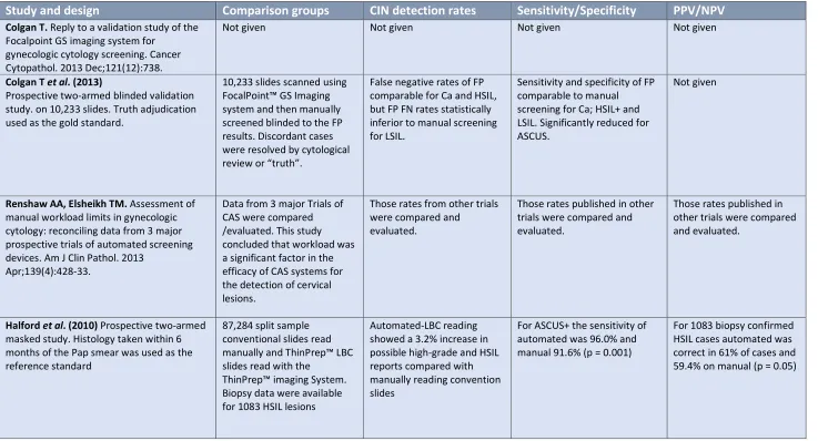
42 Table 1.2: Previous CAS (FocalPoint™ related) publications – continued
Study and design Comparison groups CIN detection rates Sensitivity/Specificity PPV/NPV
Colgan T. Reply to a validation study of the Focalpoint GS imaging system for
gynecologic cytology screening. Cancer Cytopathol. 2013 Dec;121(12):738.
Not given Not given Not given Not given
Colgan T et al. (2013)
Prospective two-armed blinded validation study. on 10,233 slides. Truth adjudication used as the gold standard.
10,233 slides scanned using FocalPoint™ GS Imaging system and then manually screened blinded to the FP results. Discordant cases were resolved by cytological review or “truth”.
False negative rates of FP comparable for Ca and HSIL, but FP FN rates statistically inferior to manual screening for LSIL.
Sensitivity and specificity of FP comparable to manual screening for Ca; HSIL+ and LSIL. Significantly reduced for ASCUS.
Not given
Renshaw AA, Elsheikh TM. Assessment of manual workload limits in gynecologic cytology: reconciling data from 3 major prospective trials of automated screening devices. Am J Clin Pathol. 2013
Apr;139(4):428-33.
Data from 3 major Trials of CAS were compared /evaluated. This study concluded that workload was a significant factor in the efficacy of CAS systems for the detection of cervical lesions.
Those rates from other trials were compared and
evaluated.
Those rates published in other trials were compared and evaluated.
Those rates publish