J. exp. Biol. 123, 27-41 (1986) 2 7 Printed in Great Britain © The Company ofBiobgists Limited 1986
EFFECTIVE AND MORPHOMETRIC OXYGEN-DIFFUSING
CAPACITY OF THE GILLS OF THE ELASMOBRANCH
SCYLIORHINUS STELLARIS
BY JOHANNES PIIPER1, PETER SCHEID2, STEVEN F. PERRY1* AND GEORGE M. HUGHES3
l
Abteilung Physiologie, Max-Planck-Institut fur experimentelle Medizin, Gottingen, FRG, zInstitut fur Physiologie, Ruhr-Universitdt, Bochum, FRG and ^Research
Unit for Comparative Animal Respiration, Bristol University, Bristol, UK Accepted 23 January 1986
SUMMARY
Calculations of the effective O2 conductance (diffusing capacity or transfer factor, Dcff) of fish gills, obtained from experimental data on gill O2 exchange, were
compared with the predicted CVexchange properties of gill models based on morphometric measurements of the elasmobranch, Scyliorhinus stellaris. Deff was
calculated from O2 uptake and PQ2 in gill water and blood, using a modified Bohr
integration technique. In the morphometric gill model, O2 conductance was con-sidered for both the water-blood tissue barrier (Dm) and the interlamellar water
(Dw). Dm was calculated from the total secondary lamellar surface area, the
har-monic mean water—blood barrier thickness, and an assumed Krogh 02-diffusion constant for gill tissue. Dw was estimated from the dimensions of the interlamellar
spaces, the mean respiratory water flow velocity, and the diffusion coefficient of O2 in water.
The ratio Dm/ Dw was 1-84 in quiescently resting, 1-68 in resting alert, and 1-47
in swimming fish, showing that diffusion across interlamellar water was somewhat more important than that across the water-blood barrier in limiting the diffusive O2 transfer between water and blood. The total morphometric diffusing capacity, Dmorphi estimated by the combined membrane-and-water diffusing capacity, Dm + W,
which is defined as l / Dm + w= l/Dm-l-l/Dw, was similar to Deff, the ratio Dm + W/
Dcff being 1-64 for quiescently resting, 1-02 for resting alert, and 0-92 for swimming
fish. The good agreement between the effective and morphometric D estimates validates the approach, and leaves, at least for the alert and swimming fish, little space for functional inhomogeneities, which are expected to reduce Deff as compared
t o Dm + w.
INTRODUCTION
There is a distinct discrepancy in fish between the effective conductance (diffusing capacity or transfer factor) for gill O2 exchange as determined by physiological methods and morphometric measurements of gill secondary lamellae (cf. Hughes,
#
28 J. PDPER, P. SCHEID, S. F. PERRY AND G. M. HUGHES
1972). One reason for the physiological estimates being lower than the morphometric has been claimed to be the diffusion resistance offered by the water passing through the interlamellar space (Scheid & Piiper, 1971; Hills & Hughes, 1970). The first attempt at a comparison of the effective, physiological conductance for O2 (Deff)
with morphometric measurements that accounted for resistance in water showed a reasonable agreement between Deff and preliminary morphometric data for the gills
in the elasmobranch Scyliorhinus stellaris (Scheid & Piiper, 1976).
Recently, the morphometric gill data of the same species have been reanalysed and completed, with particular attention paid to corrections for shrinkage and for optical artefacts (Hughes, Perry & Piiper, 1986). The aim of this study is to compare diffusing capacity for O2 derived from this newer set of morphometric data with Deff calculated from physiological measurements in the same species both at rest
and during swimming activity (Baumgarten-Schumann & Piiper, 1968; Piiper & Baumgarten-Schumann, 19686; Piiper, Meyer, Worth & Willmer, 1977). This comparison is based on the approach used in the previous study (Scheid & Piiper,
1976).
MATERIALS AND METHODS
A hypothetical Po2 profile across a secondary lamella and the adjacent interlamellar
water space is schematically shown in Fig. 1, which is based on the morphometry of Hughes et al. (1986). The PQ2 profile results from the resistances to O2 diffusion, in
interlamellar water and in the tissue barrier, and to the O2 uptake resistance offered by the blood. Since the diffusivity of O2 in water is about twice that in tissue (in terms of both Krogh's diffusion constant, K, and diffusion coefficient, d = K/ar; where a is the solubility of O2) but the maximum diffusion pathway in water, equal to one-half the interlamellar distance (b), is about five times the thickness of the water-blood tissue barrier (s), an appreciable part of the total O2 pressure drop is expected to reside within the interlamellar water.
An attempt will be made to estimate the relative magnitudes of the resistances to O2 diffusion in interlamellar water and across the water-blood barrier and to compare their sum with in vivo measurements of branchial O2 transfer. In accordance with customary usage, the reciprocal of O2 diffusion resistance, i.e. the
O2 conductance or CVdiffusing capacity, will be used as the characteristic par-ameter. In particular, we intend to compare the 'membrane' 02-diffusing capacity (Dm) with that of interlamellar water (Dw) and both of these with the effective
dif-fusing capacity (Deff), which includes both components, tissue barrier ('membrane')
and water.
RESULTS
Measurements
Physiology
Calculations are based on measurements of ventilation and gas exchange in
Baumgarten-O2 diffusing capacity of gills
29
Secondary lamellaTissue barrier Blood
O,
50 100 150
(/an)
Fig. 1. Schematic cross-section through a secondary lamella and an interlamellar space. The Po2 profile and the diffusion flux of O2 are indicated. The scale refers to dimensions
in 2-5-kg Scyliorfrinus stellaris.
Schumann & Piiper (1968), the animals were quiescently resting; i.e. although awake and unanaesthetized, their metabolic rate was probably close to basal. In the more recent experiments of Piiper et al. (1977) the same species was investigated in conditions of spontaneous periodic swimming and resting periods between swimming bouts. These resting periods can be regarded as a state of alertness, the metabolic rate being above basal. Table 1 shows ventilation and O2 uptake for these
series.
Morphometry
30 J. PHPER, P. SCHEID, S. F. PERRY AND G. M. HUGHES
The morphometric values required for this study, corrected for shrinkage as well as for the slant and Holmes effects (both due to non-perpendicular sectioning) are presented in Table 2, which also lists the magnitudes of the corrections.
Calculations
Effective diffusing capacity (Deg)
The effective O2-diffusion conductance of any gas exchange system can be obtained as the ratio of O2 uptake and mean PQ2 difference between medium, e.g.
water, and blood (cf. Piiper & Scheid, 1975). In Table 1 the effective diffusing capacity (= transfer factor) for O2 (Deff) was calculated from experimental data of O2
uptake (MQ2) and of Po2 in inspired water (Pi), expired water (PE), mixed venous
blood (Pv) and arterial blood (Pa) using three different methods.
(1) MQ2 divided by the arithmetic mean water —blood Po2 difference [i.e.
(Pi+PE-Pa-Pv)/2] (Randall, Holeton & Stevens, 1967).
(2) According to the theory of the counter-current model, assuming all resist-ance to O2 diffusion to reside in a membrane separating blood and water, and the blood O2 dissociation curve to be linear (Scheid & Piiper, 1976).
(3) The same as method 2, but using the blood (^-dissociation curve and a graphical Bohr integration technique adjusted to the counter-current model (Piiper & Baumgarten-Schumann, 19686; cf. Piiper & Scheid, 1984).
Since method 3 is the most accurate in theory, the Deff values based on this method
are used in the present study. The method is shown diagramatically in Fig. 2. Deff is
calculated as
M V ^N AC
D
X
L <*>
[image:4.451.48.414.495.586.2]where M is O2 uptake, Ca and Cv are O2 concentrations in arterial and mixed venous blood, N is the number of (not necessarily constant) blood O2 concentration
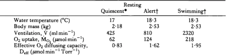
Table 1. Physiological measurements in Scyliorhinus stellaris
Water temperature (°C) Body mass (kg)
Ventilation, V (mlmin"1) O2 uptake, M Q (/tfnol min"1) Effective O2 diffusing capacity,
Dcff (/anol min"1 Torr"1)
• Baumgarten-Schumann & Piiper (1968). Deff calculated from data of these authors by Piiper &
Baumgarten-Schumann (19686).
f Piiper, Meyer, Worth & Willmer (1977). V and MQJ from their table 1, Deff calculated from
data of their table 4, using a mass of 2-53 kg which is the average body mass of their entire series (their table 1).
0
2diffusing capacity of gills
31
Table 2. Morphometric measurements of gill structures in Scyliorhinus stellaris of
2-18 and 2-53 kg body mass, used for calculations of morphometric O2 diffusing
capacity
Body mass (kg) 2-18 2-53
Ratio corrected to unconnected Total secondary lamellar surface area, A (cm2)
Harmonic mean thickness of water-blood barrier, s (/xm) Interlamellar distance, 2b (mm)
Base length of a secondary lamella, / (mm)* Height of secondary lamella, h (mm) Total number of secondary lamellae, N
Total cross-sectional area of interlamellar spaces, F (cm2) Base-to-top taper index, A
O2 diffusing capacity of membrane, Dm
(/flnol min"1 Torr"1)
Correction factors for shrinkage and distortion of tissue after Hughes, Perry & Piiper (1986). •Value adjusted to fit the A, N and h values using a base-to-top taper index, A= 0-75.
3218 9.4 0-102 1-8 0-45 226000 103-7
0-75 3-87
3614 9.4 0-112 2-0 0-45 234000 117-9
0-75 4-42
1-30 1-13 112 1-09 1-10 1-00 1-23
50 100 150
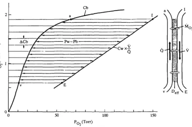
Fig. 2. Right-hand side: counter-current model for O2 exchange in fish gills. V, water flow; Q, blood flow; Mo2, O2 uptake. Left-hand side: Bohr integration technique for
determination of effective C>2-diffusing capacity (Deff). Cb is the effective blood O2
dissociation curve (Piiper & Baumgarten-Schumann, 1968a). The straight line is its water counterpart (Cw) standardized to the same total O2 concentration change (by multiplication by V/Q). The subdivision of the O2 content change in blood (ACb) and water into 10 elements is shown by the thin lines. The double-headed arrows indicate the O2 pressure difference effective for O2 uptake (Pw—Pb). Note that equal AC values do not correspond to equal Dc(f elements (due to variation of Pw—Pb). I, inspired;
32 J. PHPER, P. SCHEID, S. F. PERRY AND G. M. HUGHES
increments (AC) in the interval Ca—Cv, Pw and Pb are the PQ values of water and blood, respectively; for the integration, the limiting values of Pw—Pb are PE—Pv and Pi-Pa.
Evidently equation 1 defines a mean Pw—Pb (= M/Deff) as a harmonic mean.
The same applies to method 2, whereas method 1 uses an arithmetic mean. The mean Def{ values thus obtained are presented in Table 1.
Diffusing capacity of the water-blood barrier
According to Fick's diffusion equation, the diffusive conductance or diffusing capacity of a (tissue) sheet depends on the following physical and geometrical properties: d, diffusion coefficient; a, solubility; K, Krogh's diffusion constant; A, surface area; s, thickness:
Dm = d x a x A/s = K x A/s. (2)
The values for secondary lamellar surface area (A) and harmonic mean thickness of water-blood (tissue) barrier, s, can be taken from Table 2.
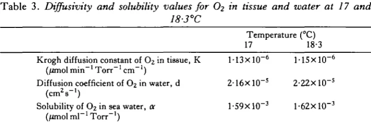
Unfortunately, no experimental data exist for d, a or K of secondary lamellar tissue for O2. We adopted the KQ2 value for human lung tissue (Grote, 1967)
extrapolated to 17 and 18-3°C, the average water temperature in the experiments (Table 1). These values are listed in Table 3.
The values for Dm thus calculated from equation 2 are listed in Table 2.
Diffusing capacity of interlamellar water (Dw)
Scheid & Piiper (1971) have analysed the resistance to O2 diffusion offered by the
interlamellar water, using simple geometric models of secondary lamellae. In these models they calculated the Po profiles in the interlamellar water which entered the
[image:6.451.40.412.493.618.2]interlamellar space at a partial pressure, Pi, the Po at the secondary lamellar membrane being kept constant at Po. Using the PQ in mixed water leaving the gill
Table 3. Diffusivity and solubility values for O2 in tissue and water at 17 and
18-3°C
Temperature (°C) 17 18-3 Krogh diffusion constant of O2 in tissue, K M 3 x l O ~6 M 5 x l O ~6
(/imol min~' Torr~' cm"')
Diffusion coefficient of O2 in water, d 2- 16x 10~ 5
2-22X 10~s ( c m V )
Solubility of O2 in sea water, a 1-59X10"3 1-62X10"3
(/unolmT'Torr"1)
O2 diffusing capacity of gills 33
model, PE, they defined the equilibration inefficiency, e, to quantify the equili-bration deficit due to the diffusion resistance in interlamellar water:
P E - P O
<3>
They showed that the magnitude of e for given secondary lamellar geometry and given velocity profile in the secondary lamellar water can be described as a function of the dimensionless equilibration resistance index, cp:
b2Xv
where b is one-half the interlamellar distance; v is the mean water velocity; / is the length of secondary lamella at the base of the lamella and d is the diffusion coefficient of O2 in water. A large value of cp indicates poor conditions for O2 equilibration.
Fig. 3 illustrates the relationship between e and cp according to model B of Scheid & Piiper (1971) in which water flow is laminar in the interlamellar space (parabolic velocity distribution across the secondary lamellar space). These two curves rep-resent limiting cases of lamellar shape as expressed by the base-to-top taper index, A, i.e. the ratio of lamellar length at the top to that at the base. For A = 1-0 (rectangular secondary lamella) the water velocity is independent of the height, whereas there is a hyperbolic flow distribution for A = 0-5, accounting for the smaller resistance to water flow at the shorter top compared with the bottom.
The value of <pcan be calculated from the data presented in Tables 1—3. The mean velocity, v, is calculated from the measured ventilation, V, and the total cross-sectional area of the interlamellar spaces, F:
v = V / F . (5)
F is given by the individual cross-sectional area of pores (width, 2b, multiplied by height, h) multiplied by their total number (N):
F = 2 b X h x N . (6)
Values for the mean water flow velocity (v) obtained from V (Table 1) and F (Table 2) are presented in Table 4 which also contains the resulting values for the equilibration resistance index cp for rest and swimming activity.
Using these values for cp, and a mean taper index of A = 0-75 (Table 4), the corresponding values for the equilibration inefficiency, e, can be obtained from Fig. 3. They are listed in Table 4.
The inefficiency parameter, e, has the meaning of a fractional effective water shunt: it defines what fraction of the respiratory water may be considered as shunted (because the Po value is unchanged) when the remainder is assumed to equilibrate completely with the secondary lamellae. Scheid & Piiper (1971) have used e to calculate the effective diffusing capacity of interlamellar water, Dw. The
34 J. PIIPER, P. SCHEID, S. F. PERRY AND G. M. HUGHES
1-01—
0-8
0-6
0-4
0-2
01—
i i i i i i n i
0 1
0-450
0-135
0-045
30
0-45 0-81 2-32
<P
Fig. 3. Plot of 'equilibration inefficiency', e (equation 3), against 'equilibration resistance index', cp (equation 4). Abscissa (<p), logarithmic; ordinate (e), linear. The two curves are for a rectangular lamella (A = 1-0) and for a trapezoidal lamella, of same base length, but tapering to one-half length at the top edge (A = 0-5). The experimental points (open circle, quiescently resting; half-closed circle, resting; filled circle, 9wimming) are in the middle, corresponding to A = 0-75.
distributed water velocity of the laminar flow model is replaced by a model with a stagnant water layer lining the secondary lamellar surface and a central core of mixed flow. In this model the central core equilibrates with the wall according to the equation:
£ = exp [-DW/(V X a)}, (7)
where V is ventilation (water flow) and a the solubility of O2 in water. Trans-formation yields:
Dw = V x tfXln(l/£). (8)
With values for V (Table 1), a (Table 3) and e (Table 4), one obtains the Dw values
O2 diffusing capacity of gills 35
The thickness of the equivalent stagnant layer, s8t, can be calculated from the Fick
diffusion equation:
s,t = d x a x A / Dw. (9)
The values for s^ and for the ratio s^/b are presented in Table 4.
Combination and comparison
The values of Deff (Table 1), Dm (Table 2) and Dw (Table 4) are compiled and
compared in Table 5.
The ratio D j D , , is higher than unity, implying that the limitation to O2 diffusion
is greater in interlamellar water than in the water-blood tissue barrier.
In order to compare the results of model calculations with Deff, a 'total
membrane-and-water' diffusing capacity, Dm + W, is approximated by the addition of the
re-ciprocal 'component' D:
1 / Dm + W= l / Dm+ 1 / Dw. (10)
The Dm+W/Deff ratio for quiescent fish, 1-64, is significantly above unity, but the
ratios for resting alert and swimming fish (1-02 and 0-92, respectively) are close to unity, signifying a remarkably good agreement between gas exchange measurements, physical properties of tissue and water, and morphometric values.
DISCUSSION
Physiological conditions
In two previous studies on resting fish (Baumgarten-Schumann & Piiper, 1968; Piiper et al. 1977), there were important differences in the conditions under which relevant measurements (e.g. of ventilation) were made. In the former study, the fish were in a prolonged state of inactivity, whereas the animals in the latter study were alert, the relatively short resting periods (averaging about 30min) being interrupted by spontaneous swimming periods. This is evident from the marked differences in both ventilation and O2 uptake. Such an increase in Deff may in part be due to
increased water velocity in the interlamellar space, with an associated increase in water diffusing capacity, Dw (see below). On the other hand, it is conceivable that in
the resting quiescent condition the full capacity of the gill apparatus is not used,
Table 4. Values used in calculating the interlamellar water O2 diffusing capacity (Dw), derived from data of Tables 1-3 according to the text
Resting
Quiescent Alert Swimming Mean water flow velocity, v (mm s"1) 0-68 1-15 3-28 Equilibration resistance index, <p 0-45 0-81 2-32 Equilibration inefficiency, e 0045 0-135 0-450 O2 diffusing capacity of interlamellar water, Dw 2-10 2-63 3-00
(jtfnol min"1 Torr"1)
Thickness of stagnant layer, s,, (fan) 31-6 29-7 26-0 Ratio of stagnant layer thickness to half inter- 0-62 0-53 0-46
36 J. PIIPER, P. SCHEID, S. F. PERRY AND G. M. HUGHES
A B
LAMINAR FLOW MODEL EQUIVALENT STAGNANT LAYER MODEL
A
- Transversally mixed central flow
_ Stagnant layer
Fig. 4. The laminar flow model (A) and the equivalent stagnant layer model (B). Top: section perpendicular to secondary lamellae, parallel to the filament. Water flow direction is indicated by open arrows. Numbers refer to positions: 1, inflow end; 2, middle; 3, outflow end; 4, respired water after leaving the interlamellar space. In B, equivalent cross-sectional mixing within the flowing water is indicated by transverse arrows; the equivalent stagnant water layer is separated from flowing water by a broken line. Middle: water velocity (v) profiles: parabolic in A, step-like in B, with v = 0 in the stagnant layers; v is constant in the central flow. Bottom: cross-sectional O2 pressure (Po2) profiles at
positions 1-4 of the model. Continuous profiles in A. In B, linear POj drop in the stagnant
layer, no Po2 gradient within the flowing water.
because there is ample functional shunting. This hypothesis is supported by the observations of rapidly changing arterial Po2 in some fish, apparently reflecting
changing functional inhomogeneity (Piiper & Schumann, 1967).
Diffusion limitation in interlamellar water
0
2diffusing capacity of gills 37
important role in limiting branchial O2 transfer, both at rest and during swimming activity.
The relative roles of diffusion in interlamellar water and across the water—blood barrier depend on the diffusion distances (Fig. 1) and the diffusion properties of the media. For a first approximation, the average path length for lateral diffusion of O2 molecules in the interlamellar space may be taken as b/2, and that across the water-blood barrier as s. For Scyliorhinus stellaris the b/2: s ratio is between 2-7 and 3 (Table 2). The ratio of the assumed Krogh diffusion constant for tissue-water (Table 3) is between 0-55 and 0-53. Thus the estimated water/tissue diffusion resistance ratio, corresponding to the Dm/ Dw ratio, is expected to be between 1-5
and 1-6, which is in reasonable agreement with the calculated results of Table 5. The mean diffusion path length decreases with increasing water velocity, because gas exchange becomes restricted to layers close to the secondary lamellar surface. This is why Dw increases and the equivalent stagnant water layer decreases with
increasing water flow (Table 5). The exact quantitative relationships are influenced by the flow velocity profile (Scheid & Piiper, 1971).
Comparison of morphometric and physiological diffusing capacities
For the quiescently resting fish, the total morphometric diffusing capacity (Dmorph), estimated by the combined membrane-and-water diffusing capacity
(Dm+W), is considerably above the effective, physiological diffusing capacity, Deff.
This result, which is in qualitative agreement with the earlier analysis of Scheid & Piiper (1976), is not unexpected since in most reported cases Dmorph has been found
to be considerably higher, even by an order of magnitude, than the Deff. This has
been repeatedly documented for mammalian lungs (reviewed by Weibel, 1973), but also for reptilian lungs (Perry, 1978) and for avian lungs (Abdalla et al. 1982). Only for the skin of a lungless plethodontid salamander (Piiper, Gatz & Crawford, 1976) and for the pleural membrane of dog lungs (Magnussen, Perry, Willmer &c Piiper, 1974) has a reasonable agreement been found. But also in these cases, Dmorph was
slightly higher than Deff.
The conventional explanation for Dmorph/Deff > 1 is that the numerous parallel
units in the gas exchange organ are inhomogeneous with respect to ventilation,
Table 5. Comparison of diffusing capacities for O2 (D)
Physiological, D,.[f Membrane, Dm
Water, Dw
Membrane-and-water, Dm + W
Dm/ Dw
Dm + W/ De f f
D is in /imolmin"1 Torr"1.
38 J. PIIPER, P. SCHEID, S. F. PERRY AND G. M. HUGHES
diffusion and perfusion, which, unless properly accounted for, leads to an under-estimation of Deff. For example, in mammalian lungs, D for O2 is determined from
alveolar-arterial Po differences; since these are also generated by shunt and unequal distribution of ventilation to perfusion, D for O2 is underestimated if no appropriate corrections are applied (see Piiper & Scheid, 1980).
For fish gills, there is ample possibility of ventilation-perfusion inhomogeneity due to both morphological and functional factors. Extreme cases are blood shunting (e.g. due to perfusion of intrafilamentary afferent-efferent arterial connections or to perfusion of unventilated lamellae) and water shunting (due to passage of water between rows of secondary lamellae or between tips of filaments). Moreover, part of the O2 uptake resistance may reside in the blood (diffusion limitation; reaction limitation due to slow oxygenation of haemoglobin).
Whereas the finding that Dm + W/ De f f> l for the quiescently resting fish thus
appears to be readily explained, it was unexpected to find a close agreement between
Dm + W and Deff for resting and swimming fish. This would call for a critical
exam-ination of all the methods, including morphometric techniques, physical properties, models and physiological measurements, for directional errors potentially leading to an overestimation of De{! or to an underestimation of Dm o r p h.
Shrinkage
One of the basic problems in morphometry is deformation, due in great part to shrinkage and to the finite sectioning thickness. Hughes et al. (1986) have presented a detailed account of the procedures for morphometry and the determination of factors for corrections for deformation (see Table 2).
Dm is proportional to A/s, thus the correction factor is l-30/l-13= 1-15. This
means that without correction, Dm is underestimated by (1 — l/l-15)Xl00 = 13%.
The effects of shrinkage on Dw are more complex. According to equation 4 the
anatomical dimensions determining qp are the interlamellar distance (2b) and the length of the secondary lamellae (/), <p being proportional to b2//. With the cor-rection factors from Table 2 this yields a combined corcor-rection factor for <p of 1-15. Thus shrinkage appears to lead to underestimation of <p and e, and thus to over-estimation of Dw.
This analysis assumes constancy of the interlamellar water velocity, v. Changes (errors) in the anatomical dimensions, however, influence v if a given (constant) V is considered. Combination of equations 4, 5 and 6 yields:
The combined correction factor of b/(hx/) is 0-934 (Table 2). In this case shrinkage, if not accounted for, leads to an overestimate of <p and e, and to an underestimation of Dw. But the error does not exceed 10% during either rest or
O2 diffusing capacity of gills 39
It is evident from equation 11 that not only the extent, but even more the anisotropy of the shrinkage, i.e. different functional shrinkage of b, h and /, play an important role in influencing the conditions for O2 diffusion in terms of Dw.
Physical diffusion properties
There are only few reports in the literature on measurements of the O2 diffusion coefficient, d, or of the Krogh diffusion constant for O2, K (=dXar), in tissues (see Bartels, 1971). Most authors have used the values of Krogh (1918/19) and of Thews & Grote (see Grote, 1967).
There are no measurements on fish gill tissue. We used the values obtained by Grote (1967) on rat lung tissue mainly because they appear to be derived from the most reliable determinations. Not only may the true value for fish gills be different, but also in calculations of Dm for lungs it must be considered that the measurements
were performed on slices of degassed whole lung tissue, only a small fraction of which constitutes the gas—blood barrier. A promising approach is determination of Dm
from oxygenation and deoxygenation kinetics of red cells in isolated secondary lamellae of fish gills (Hills, Hughes & Koyama, 1982). At present, nothing can be predicted concerning the direction or extent of errors due to the uncertainty about O2 diffusivity.
For this reason, i.e. lack of reliable data on physical diffusion properties, it appears to be generally preferable to express the results of morphometric studies on medium-blood barrier for gas exchange in terms of the surface area/mean harmonic thickness ratio (A/s; dimension: length) — the 'anatomical diffusion factor' of Perry (1978). The value can then be used for functional estimates in conjunction with the appropriate K value, of which more accurate determinations will be available in the future. In addition, the previously reported values for O2 diffusivity in water are rather unreliable (see Bartels, 1971).
Models
The flat sheet model for calculation of Dm is rather straightforward. But problems
arise from making appropriate allowance for the pillar cells, which support the secondary lamellae and reduce the surface area available for gas exchange.
More critical are the assumptions for calculation of Dw. Unfortunately, the
required morphometric measurements in large Scyliorhinus stellaris are very limited (Hughes et al. 1986). Moreover, the parabolic flow velocity profile, assumed in the model analysis, may not be fully developed in the interlamellar space. A square-front flow would be more efficient for gas exchange and would therefore yield higher Dw
values (Scheid & Piiper, 1971).
40 J. PUPER, P. SCHEID, S. F . PERRY AND G. M. HUGHES
The simplified model for isolated quantification of diffusion resistance in inter-lamellar water (Scheid & Piiper, 1971) does not take into account the change of Po in secondary lamellar blood, which in reality increases from the mixed venous to the arterial value. Instead, the lamellar Po is assumed to be constant throughout (Po in
equation 3 and Fig. 4). Therefore, the simple additive combination of Dw with Dm,
yielding Dm + W (equation 10), and comparison with D derived from a model that
accounts for changing Po in intralamellar blood is clearly incorrect because the
models are not consistent. However, a recent theoretical study shows that the error produced by this apparent incompatibility is relatively minor (Scheid, Hook & Piiper, 1986).
We conclude that resistance to O2 diffusion in the interlamellar water (l/Dw)
exceeds that of the secondary lamellar tissue membrane (l/Dm) and that this is
particularly pronounced at rest. When comparing morphometric with physiological estimates of the diffusing capacity, the good agreement between the Deff and Dm + W
values may be interpreted to show that the methods and models used are appropriate. On the other hand, local variations of physiological quantities like ventilation, dif-fusing capacity and blood flow, in part resulting from morphometric inhomogeneity, are expected to reduce gas exchange efficiency, i.e. to decrease Dcff. Possibly some
inhomogeneity effects were compensated by physiological control mechanisms. In any case, there seemed to be little space for mechanisms reducing O2 transfer efficiency, such as blood or water shunts.
REFERENCES
A B D A L L A . M . A., MAINA, J. N . , KING, A. S., KING, D . Z. & HENRY, J. (1982). Morphometrics of
the avian lung. I. The domestic fowl (Gallus gallus variant domesticus). Respir. Physiol. 47, 267-278.
BARTELS, H. (1971). Diffusion coefficients and Krogh's diffusion constants. Diffusion coefficients of gases in water. In Respiration and Circulation (ed. P. L. Altman & D. S. Dittmer), pp. 21-24. Bethesda MD: FASEB.
BAUMGARTEN-SCHUMANN, D. & PIIPER, J. (1968). Gas exchange in the gills of resting unanes-thetized dogfish (Scyliorhinus stellaris). Respir. Physiol. 5, 317-325.
GROTE, J. (1967). Die Sauerstoffdiffusionskonstanten im Lungengewebe und Wasser und ihre Temperaturabhangigkeit. Pflugers Arch. ges. Physiol. 295, 245-254.
HILLS, B. A. & HUGHES, G. M. (1970). A dimensional analysis of oxygen transfer in the fish gill.
Respir. Physiol. 9, 126-140.
HILLS, B. A., HUGHES, G. M. & KOYAMA, T . (1982). Oxygenation and deoxygenation kinetics of red cells in isolated lamellae of fish gills. J . exp. Biol. 98, 269-275.
HUGHES, G. M. (1972). Morphometrics of fish gills. Respir. Physiol. 14, 1-25.
HUGHES, G. M., PERRY, S. F. & PHPER, J. (1986). Morphometry of the gills of the elasmobranch
Scyliorhinus stellaris of varied body size.J. exp. Biol. 121, 27-42.
KROGH, A. (1918/19). The rate of diffusion of gases through animal tissues, with some remarks on the coefficient of invasion. J . Physiol., Land. 52, 391-408.
MAGNUSSEN, H., PERRY, S. F., WILLMER, H. & PIIPER, J. (1974). Transpleural diffusion of inert
gases in excised lung lobes of the dog. Respir. Physiol. 20, 1-5.
PERRY, S. F. (1978). Quantitative anatomy of the lungs of the red-eared turtle, Pseudemys scripta
elegans. Respir. Physiol. 35, 245-262.
PIIPEK, J. & BAUMGARTEN-SCHUMANN, D. (1968a). Transport of O2 and CO2 by water in gas
0
2diffusing capacity of gills 41
PliPER, J. & BAUMGARTEN-SCHUMANN, D. (19686). Effectiveness of 02 and C 02 exchange in the
gills of the dogfish (Scyliorhinus stellaris). Respir. Physiol. 5, 338-349.
PIIPER, J., GATZ, R. N. & CRAWFORD, E. C. JR (1976). Gas transport characteristics in an
exclusively skin-breathing salamander, Desmognathus fuscus (Plethodontidae). In Respiration
in Amphibious Vertebrates (ed. G. M. Hughes), pp. 339-356. London, New York, San
Francisco: Academic Press.
PUPER, J., MEYER, M., WORTH, H. & WILLMER, H. (1977). Respiration and circulation during
swimming activity in the dogfish Scyliorhinus stellaris. Respir. Physiol. 30, 221-239.
PnPER, J. & SCHEID, P. (1975). Transport efficacy of gills, lungs and skin: theory and experimental data. Respir. Physiol. 23, 209-221.
PlIPER, J. & SCHEID, P. (1980). Blood-gas equilibration in lungs. In Pulmonary Gas Exchange, vol. I, Ventilation, Blood Flow, and Diffusion (ed. J. B. West), pp. 131-171. New York, London, Toronto, Sydney, San Francisco: Academic Press.
PTIPER, J. & SCHEID, P. (1984). Model analysis of gas transfer in fish gills. In Fish Physiology, vol. XA (ed. W. S. Hoar & D. J. Randall), pp. 229-262. New York, London: Academic Press. PIIPER, J. & SCHUMANN, D. (1967). Efficiency of O2 exchange in the gills of the dogfish,
Scyliorhinus stellaris. Respir. Physiol. 2, 135-148.
RANDALL, D. J., HOLETON, G. F. & STEVENS, E. D. (1967). The exchange of oxygen and carbon
dioxide across the gill of rainbow trout. J. exp. Biol. 46, 339-348.
SCHEID, P., HOOK, C. & PIIPER, J. (1986). Model for analysis of counter-current gas transfer in fish gills. Respir. Physiol. (in press).
SCHEID, P. & PnPER, J. (1971). Theoretical analysis of respiratory gas equilibration in water passing through fish gills. Respir. Physiol. 13, 305-318.
SCHEID, P. & PIIPER, J. (1976). Quantitative functional analysis of branchial gas transfer: theory and application to Scyliorhinus stellaris (Elasmobranchii). In Respiration of Amphibious
Vertebrates (ed. G. M. Hughes), pp. 17-38. London, New York: Academic Press.
WEIBEL, E. R. (1973). Morphological basis of alveolar-capillary gas exchange. Physiol. Rev. 53,