R E S E A R C H A R T I C L E
Open Access
Stabilizing lateral ankle instability by suture
tape
–
a cadaver study
Heinz Lohrer
1,2,3*, Giuseppe Bonsignore
4, Nadja Dorn-Lange
4, Lu Li
3, Albert Gollhofer
3and Dominic Gehring
3Abstract
Background:Suture tape is a recent development to augment a Brostrom repair at least during the healing phase of the native tissues used for stabilization of the lateral ankle ligaments. The purpose of this study was to evaluate whether suture tape is an effective mechanical stabilizer against anterior talar drawer in a cadaver experiment when tested with a validated arthrometer.
Methods:Different stability conditions were created in 14 cadaveric foot and leg specimens. Following anterior talofibular ligament (ATFL) dissection, isolated suture tape ATFL reconstruction was compared to the unaltered specimens, to the condition with ATFL cut, to the ATFL plus calcaneofibular ligament (CFL) cut conditions, and to the ATFL, CFL, and posterior talofibular ligament transected specimens. Three-dimensional bone-to-bone
movement between fibula and calcaneus were simultaneously recorded using bone pin markers. Anterior translation was analysed between 20 and 40 N anterior talar drawer load, applied by an ankle arthrometer. Test conditions were compared using non-parametric statistics.
Results:Dissection of ATFL increased anterior talar drawer in arthrometer and bone pin marker analyses (p= 0.003 and 0.004, respectively). When the CFL was additionally cut, no further increase of the anterior instability could statistically be documented (p= 0.810 and 0.626, respectively). Following suture tape reconstruction of the ATFL, stability was not different from the unaltered ankle (p= 0.173).
Conclusions:Suture tape augmentation of the ATFL effectively protects the unstable anterolateral ankle in the sagittal plane. The CFL does not seem to stabilize against the anterior talar drawer load.
Keywords:Ankle, Lateral ankle instability, Suture anchor, Suture tape, Augmentation, Cadaver study
Background
Ankle sprain is the most common injury in the physically active population and development to chronic ankle in-stability (CAI) is frequent [1]. About one out of five osteo-arthritic ankles results from lateral ankle sprain [1]. The role of the mechanical component in CAI is a matter of
ongoing debate [2]. Most studies originating from a
kinesiology or sport scientific perspective do not report“a definitive association of ankle laxity with CAI”[3]. In the orthopaedic literature, however, CAI and mechanical ankle instability (MAI) are often used interchangeably [4–
8]. When MAI exceeds a certain amount and functional
deficits cannot adequately be restored by conservative
approaches, operative interventions have to be taken into consideration. In principle, tenodeses, anatomic recon-structions, and combined procedures are used to stabilize against ligamentous lateral ankle instability [9,10]. Repair using local tissue has been shown to effectively stabilize the lateral ankle joint [8,11]. Suture tape is a recent devel-opment to augment a Brostrom repair at least during the healing phase of the native tissues used for stabilization of the lateral ankle ligaments [12, 13]. Although there are concerns for progressive elongation of suture-tape, un-clear long-term stability (longevity of mechanical stability), and unexpected complications such as foreign body reac-tion, this procedure is increasingly used for various liga-ment reconstructions.
A clinical study examined the effectiveness of suture tape to stabilize the ankle against a manually performed anterior drawer test. Results led to the conclusion that using an
© The Author(s). 2019Open AccessThis article is distributed under the terms of the Creative Commons Attribution 4.0 International License (http://creativecommons.org/licenses/by/4.0/), which permits unrestricted use, distribution, and reproduction in any medium, provided you give appropriate credit to the original author(s) and the source, provide a link to the Creative Commons license, and indicate if changes were made. The Creative Commons Public Domain Dedication waiver (http://creativecommons.org/publicdomain/zero/1.0/) applies to the data made available in this article, unless otherwise stated. * Correspondence:[email protected]
1ESN–European Sportscare Network, Borsigstraße 2, 65205 Wiesbaden,
Germany
2Lilium Klinik, Borsigstraße 2, 65205 Wiesbaden, Germany
additional suture tape might be favoured compared to an isolated modified Brostrom repair [14]. Until now, only three cadaver experiments were published to demonstrate the stability of anterior talofibular ligament (ATFL) suture tape augmentation [13, 15, 16]. These experiments either investigated load to failure of the isolated ATFL [13], or ro-tation of the tibia, respective to the calcaneus [15, 16]. These experiments were designed to only load the ATFL (or the respective reconstructions), and specimens were therefore rigidly fixed to the testing apparatuses. In con-trast, our ankle arthrometer applied only low load to the unconstrained heel in an anterior direction. Due to the complex anatomy of the hindfoot, this load is transferred into a complex motion of the calcaneus and the talus with respect to the fixed leg. In contrast to previous cadaver ex-periments, we therefore aimed to more functionally test the whole lateral ankle ligament complex in a clinically relevant situation (anterior talar drawer), its contribution to stability, and the effect of suture tape ATFL augmenta-tion (Table 1). In previous experimental approaches, the loading of the ankle joint complex was quantified exter-nally with specific measurement devices, which can be ap-plied only to cadavers. Until now, no study compared the specific motion between the interacting bones (fibula and calcaneus) and its functional representation in an ankle arthrometer, which is validated for experimental and clin-ical use in a native condition, following incremental lateral ligament dissection, and following suture tape application.
The purpose of this study was to evaluate the effect of ATFL suture tape augmentation using a previously vali-dated anterior talar drawer arthrometer in a cadaver experi-ment. Additionally, the spatial bone-to-bone movement was examined by motion analyses using intraosseous markers.
Methods
This investigation is part of a larger research project, which also aims to experimentally evaluate and validate
the ankle arthrometer for assessing ankle laxity (anterior talar drawer).
The local Ethics Commission approved the study.
Specimens
Fourteen fresh-frozen cadaveric above-knee amputated foot and leg specimens, obtained from four female and
four male donors (median age = 78.5, range = 66–91
years), were thawed. We repeatedly irrigated the speci-mens with saline during dissection and testing to pre-vent desiccation. An experienced anatomist dissected all specimens down to the lateral ankle ligaments and cap-sule creating a square (5 × 5 cm) skin and subcutaneous window centered over the tip of the lateral malleolus. Lateral ankle ligaments were then identified, inspected for completeness, and were manually tested for anterior talar drawer and talar tilt. All ankles were stable and had no relevant ankle or hindfoot pathology. Following the experiments, the specimens were dissected to the bone to ensure that there was not any bony impingement or bone abnormality that could skew the data.
Test procedure
Initially, the unaltered specimen was placed on the ankle arthrometer and was tested. Five repeated trials were registered and the mean was calculated and used for fur-ther analyses. Then, the ATFL was cut and the testing procedure was repeated. Thereafter, the ATFL was
re-constructed by suture tape (InternalBraceTM, Arthrex,
Naples, FL) and the measurements were repeated. Then, the CFL was cut while ATFL tape augmentation was un-altered and measurements were repeated. In the next step, tape augmentation was cut and the measurements were repeated. Finally, the posterior talofibular ligament (PTFL) was cut and measurements were completed to test for maximum translation.
This procedure enabled registration of stability/in-stability data relative to sequentially increasing lateral ankle ligament instability and to the effect of ATFL
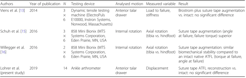
Table 1Overview of the literature presenting experimental suture tape testing
Authors Year of publication N Testing device Analysed motion Measured variable Result
Viens et al. [13] 2014 3
× 6
Dynamic tensile testing machine (ElectroPuls E10000, Instron Systems, Norwood, Massachusetts)
Anterior talar drawer
Load to failure, stiffness
Brostrom plus suture tape augmentation vs. intact: no significant difference
Schuh et al. [15] 2016 3
× 6
858 Mini Bionix (MTS Systems Corporation, Eden Prairie, MN, USA
Internal rotation Axial rotation (tibia vs. hindfoot)
Suture tape augmentation (angle at failure, failure torque) superior
Willegger et al. [16]
2016 2
× 6
858 Mini Bionix (MTS Systems Corporation, Eden Prairie, MN, USA
Internal rotation Axial rotation (tibia vs. hindfoot)
Suture tape augmentation: similar biomechanical stability compared to an intact native ATFL (torque at failure, angle at failure)
Lohrer et al. (present study)
2019 14 Ankle arthrometer Anterior talar
drawer
suture tape augmentation relative to isolated ATFL and to combined lateral ligament transections.
Surgical approach
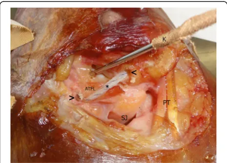
In our setup, the implantation of the suture tape was performed as an isolated ATFL reconstruction. The transected ATFL was not repaired. An experienced orthopaedic surgeon performed the procedure according to the manufacturer’s guidelines (fibula to talus) [12] with the manufacturer’s single-use instruments and im-plants (InternalBraceTM, Arthrex, Naples, FL). A 2.7-mm drill hole was created at the anterior margin of the fibula 2–3 mm lateral to the middle of the visible ATFL origin. After tapping, a 3.5-mm knotless anchor, preloaded with suture tape, was inserted (Fig.1). The talar ATFL inser-tion was then identified and drilled in a 45° posterome-dial direction with a 3.4-mm drill. After tapping, a 4.75-mm knotless anchor was preloaded with the free ends of the suture tape that was already attached to the fibula. According to implantation guidelines, it was inserted creating moderate tension to the tape [12].
Ankle arthrometer
Non-invasively, the anterior drawer was induced and mea-sured with an arthrometer. The technical principle of the construction and the previous validation process has been described in detail elsewhere [2, 17–20]. In short, the ankle arthrometer is featured by a sliding plantar plate with heel pad which induces anterior translation of the foot relative to the leg, which is fixed to an anterior shin pad (Fig.2).
Following a pilot study, the ankle arthrometer was modi-fied for the current study. The drawer plate containing the
foot of the arthrometer was additionally equipped with a ball bearing plate to reduce friction between the specimen’s sole and the drawer plate.
The ankle arthrometer induces an anterior drawer with a velocity of 8 mm/s. The arthrometer applied load
from 0–80 N and displacement was simultaneously
re-corded from the ankle arthrometer and kinematic-ally (bone pin coordinate system). The displacement between 20 and 40 N was calculated from the load-displacement curves. This interval was selected following results from previous work, indicating that ankle in-stability can best be differentiated in that low load re-gion, representing the linear slope of the force-displacement curve [2, 18–20]. A possible slack length of the suture tape should be compensated for within the first 20 N of load application and therefore might not affect the measurements.
Biomechanical testing
Three-dimensional bone-to-bone displacement during the anterior drawer within the ankle arthrometer was measured simultaneously and directly for the different test conditions. The methodology is described in detail in a further manu-script [21]. In short, using a motion analysis system (Vicon Motion Systems, Oxford, UK) with 11 cameras, the bony movement of the fibula, talus, and calcaneus were tracked at 200 Hz. For this purpose, 1.5 mm K-wires were drilled into the distal anterolateral fibula, the talar neck, and the lateral calcanear facet. Each K-wire was equipped with a frame, containing four 6 mm spherical reflective markers which defined a 3D Cartesian coordinate system (Fig. 2). These technical coordinate systems tracked the three-dimensional motion of each bone when performing the arthrometer testing. The relative motions between the fib-ula, talus, and calcaneus were calculated following the
rec-ommendations of the International Society of
Biomechanics for the ankle joint using a joint coordinate system approach [22]. This approach allows for calculating three-dimensional rotations and translations of bone seg-ments and had previously demonstrated its potential in de-tecting anterior translation instability of the ankle [23].
Statistical analyses
Statistical analyses were performed using the SPSS statis-tical package 22.0 for macOS (IBM Inc.) and MATLAB-Software (MathWorks Inc.). As evaluation with the Kolmogorov-Smirnov test indicated that most of the vari-ables were not normally distributed, dependent non-parametric comparisons were made for further analyses. Therefore, dependent descriptive non-parametric com-parison (median and mean deviation) was made. For post hoc testing between two separate conditions, the Wil-coxon sign-rank test was applied and adjusted according to the Bonferroni-Holm correction procedure. For all
analyses, the overall level of significance was defined atp < 0.05. Descriptive results are reported as median ± mean deviation around the median. Pearson correlations were calculated between all arthrometer data for all conditions (ligaments intact, ATFL cut, ATFL and CFL cut, ATFL cut and internal brace, ATFL and CFL cut and internal brace, ATFL and CFL and PTFL cut) and the 3D kinematic data (calcaneus vs. fibula).
Results
Macroscopically, there was no failure of the suture tapes or the anchor fixations during the testing procedures.
Ankle arthrometry (Fig. 3): Measured with the ankle
arthrometer, the median anterior translation of the intact specimens was 4.0 ± 1.1 mm. Following the dissection of the ATFL median displacement increased to 6.1 ± 2.9
mm (p= 0.003) and to 7.6 ± 3.6 mm when the CFL was
Fig. 2Photograph, demonstrating the test setup. The leg is fixed to the shin pad of the arthrometer with straps. The footplate induces anterior translation load via the heel pad. Fibula, talus, and calcaneus are equipped with K-wires, which carry 3D Cartesian coordinate systems with spherical reflective markers
additionally cut (p = 0.001). With suture tape recon-struction, anterior translation was 5.0 ± 1.2 mm and 5.0 ± 2.1 mm, when solely the ATFL or both, ATFL and CFL were cut, respectively. Both reconstructed condi-tions were not different from the intact condition (p = 0.173 and 0.078, respectively).
Bone pin measurements (Table 2): Compared to the
intact condition (0.2 ± 0.6 mm), anterior translation in-creased following the dissection of the ATFL (2.4 ± 4.9
mm;p= 0.004) and additionally the CFL (2.9 ± 6.5 mm;
p< 0.001). Adding the suture tape reduced the anterior translation to 0.9 ± 1.0 mm when solely the ATFL was
cut (p = 0.173) and to 0.7 ± 1.3 mm when additionally
the CFL was cut (p= 0.007).
The correlation for the comparison between the exact motion of interacting bones (fibula vs. calcaneus) and its functional representation in the ankle arthrometer for the 20–40 N anterior talar drawer load was r= 0.851 (p < 0.001).
Discussion
This study clearly demonstrates that an experimentally created anterolateral ankle instability (ATFL dissection) can effectively be reduced to anterior talar drawer base-line values by suture tape implantation. When compared to the intact condition and for the tested load (20–40 N), the CFL does not seem to play a relevant role for stabilizing against the anterior talar drawer, if the ATFL is reconstructed by suture tape. This behaviour is inter-esting, because our experimental setup tested for anter-ior translation, but the CFL is thought to protect mainly against ankle inversion [24, 25]. Because a large portion of patients with chronic ankle instability have insuffi-ciency of both ligaments, further study regarding the
suture tape ATFL and CFL reconstruction can be im-portant and interesting.
Anatomic repair/reconstruction is currently the main-stay for operative treatment of MAI [10, 14, 26, 27]. In the last decade, anchor systems have been introduced to secure transplants [28] or as a knotless fixation for
su-tures [8]. Suture anchors are most important for the
evolving arthroscopic Brostrom techniques [29,30]. The stability of anchor systems has been tested in cadaver studies, but the strength of these repairs was inferior to native ATFL [31–34]. Recently, suture tape techniques
for augmentation of the ATFL were described [12, 35]
and are proposed at least for patients with generalized ligamentous laxity [26], athletes, or patients with poor local tissue quality, e.g., following failed previous repair or reconstruction [13,14].
An advantage of these techniques is the additional mechanical stability, provided by the suture tape which is securely fixed to cover the ATFL from its anatomic fibular origin to its talar insertion [13, 15, 16]. So, safer and faster rehabilitation is thought to be possible [12,
14, 27]. Compared with tenodesis, no “donor site mor-bidity”can occur [35].
In a clinical study, young females with MAI underwent an isolated minimally invasive suture tape augmentation of the ATFL and CFL. After a minimum follow-up of 2 years, “91.2% achieved satisfactory functional results” [35]. In another study, patients“were able to quickly re-turn to activity and sports”[14].
A disadvantage of the suture tape procedure is the “possibility of progressive elongation over time”[35]. But otherwise, this could turn into an advantage by“allowing the natural tissues to progressively strengthen” [12]. An
uncontrolled study described “favourable” short-term
Table 2Median distance as simultaneously measured with the ankle arthrometer and with bone pins between 20 and 40 N anterior talar drawer for the sequential ligament dissection and reconstruction compared with the intact ankle. The ankle arthrometer measures the anterior translation of the calcaneus with its surrounding soft tissue against the leg. The bone pin measures represent the translation between fibula and talus
Ankle arthrometer Bone pin measures (calcaneus vs. fibula)
Median (mean deviation) [mm]
Δto intact [mm]
Δto intact [%]
Pvalue vs. intact
Bonferroni-Holm-corrected threshold
Median (mean deviation) [mm] Δ
to intact
[mm] Δ
to intact [%]
Pvalue vs. intact
Bonferroni- Holm-corrected threshold
Intact 4.0 (1.1) 0.2 (0.6)
ATFL cut 6.1 (2.9) 2.1 53 0.003 0.012 2.4 (4.9) 2.17 1033 0.004 0.012
ATFL cut + suture tape
5.0 (1.2) 1.0 25 0.173 0.405 0.9 (1.0) 0.64 304 0.173 0.306
ATFL cut + suture tape + CFL cut
5.0 (2.1) 1.0 25 0.078 0.312 0.7 (1.3) 0.50 238 0.007 0.035
ATFL cut, CFL cut, suture tape cut
7.6 (3.6) 3.6 90 0.001 0.006 2.9 (6.5) 2.66 1266 < 0.001 0.001
ATFL cut, CFL cut, PTFL cut, suture tape cut
7.3 (3.1) 3.3 82 0.001 0.006 6.1 (6.0) 5.93 2823 < 0.001 0.001
outcome [27]. The most critical point for the suture tape implementation until now is that no long-term follow-up is available to determine effects and side effects.
There are few reports to demonstrate the stability of the suture tape augmentation in cadaver experiments. Previous cadaver experiments tested load to failure of suture tape augmentation (Table 1). In these experimental setups, the talofibular joints were extensively dissected and isolated. The specimens were rigidly fixed in the test apparatuses while internal rotation load was applied to the calcaneus relative to tibia/fibula [13, 15, 16, 31, 32]. That highly standardized procedure tries to apply the load exactly in the direction of the suture tape. Contrasting to this, our in-vestigation was performed following only minimal dissec-tion of the specimens to mimic the specific modissec-tion of the involved bones during anterior talar drawer. The measure-ments were performed between 20 and 40 N anterior talar drawer load with our arthrometer which was developed, validated, and used for testing in the clinical situation [2,
17–20]. However, our arthrometer does not evaluate the coronal plane motion (varus/valgus) of the ankle, but this motion has been analysed in a different approach within the same project [25].
Discussion is open whether additional CFL repair or aug-mentation is necessary. The effect of suture tape ATFL augmentation on talar tilt has not been addressed in cadaver studies (Table1). However, a cadaver study demon-strated no difference in initial varus instability between isolated ATFL and combined CFL and ATFL repair with a Brostrom-Gould procedure [36]. This conclusion is also supported by an uncontrolled clinical study in an athletic population using a modified Brostrom procedure without
CFL reconstruction [37]. When compared to the intact
condition and for the tested load (20–40 N), the CFL does not seem to play a relevant role for stabilizing against the anterior talar drawer load, if the ATFL is stabilized by suture tape (Table2). Recently, a minimally invasive tech-nique was presented to anatomically augment both ATFL and CFL [35]. To further elucidate the role of the CFL, we addressed specifically the frontal plane instability in another approach [25].
Limitations to this study could be the higher age of our donors resulting in reduced bone and ligament qual-ity. However, due to intraindividual comparisons of the different testing conditions, these differences were not likely to influence the results. Ankle degeneration could also influence the mobility during our tests, but all our specimens were free from degenerative findings. To get information about the isolated suture tape effect, we per-formed no Brostrom repair and therefore the individual quality of the lateral ankle ligaments does not play a role for the comparisons. However, this procedure is different from the standard clinical setting. We do not suggest that this study should be interpreted to promote isolated
suture tape reconstruction to replace the modified Bros-trom repair. It can be expected that an additional repair of the local tissue by a Brostrom procedure would lead to even more stability if the balance of load between the suture tape and the repair were properly set. Interest-ingly, isolated suture tape augmentation without ad-dressing the ligaments has been recently shown to be effective in a clinical MAI study [35]. An additional limi-tation is that the cadaver study design only determines the response to the tape at the time of implantation. The stability measures would likely change over time due to wear out of the tape. Furthermore, this study evaluates the lateral ankle instability executed by an anteriorly ap-plied load to the posterior calcaneus. The lateral ankle ligaments, however, stabilize the hindfoot in more com-plex and three-dimensional ways. That behaviour should be subjected to further analyses.
In principle, differences between ankle arthrometer and bone pin measurements are to be expected. The arthrometer provides an external instrumented meas-ure, while the bone pins approach directly assesses the relative bone-to-bone movement between calca-neus and fibula. However, both measures demon-strated a strong relationship (r = 0.851). Our previous validation studies for the ankle arthrometer were
done in a 2D radiographic approach in cadavers [19]
or by in vivo comparison with a manual stress testing
[2, 18]. As we were interested in having the most
possible accuracy for the spatial bone-to-bone inter-action, and to further evaluate the functional repre-sentation of the bone-specific movements, we decided to also investigate the ankle arthrometer against mea-surements obtained from a very precise biomechanical measuring tool. Consequently, in further studies, intraosseous markers can therefore be omitted.
In summary, the presented data demonstrate the effectiveness of ATFL suture tape augmentation in a cadaver experiment and the validity of the eter to measure anterior talar drawer. This arthrom-eter therefore is recommended at least for quality management in the treatment of MAI patients and for preventive experimental and epidemiologic evalu-ations to further study CAI.
Conclusions
Suture tape augmentation of the ATFL effectively protects the unstable anterolateral ankle in the sagittal plane. For additional CFL lesions, the stabilizing effect in the sagittal plane was reduced.
Abbreviations
Acknowledgements
We are grateful to Dr. Peter Scholz-Kreisel, Institute for Medical Biostatistics, Epidemiology and Informatics (IMBEI), University of Mainz Medical Depart-ment, Germany, for his statistical support.
Authors’contributions
HL, DG, ND-L, and AG conceived the study. All authors participated in its de-sign. GB, LL, and DG performed the data acquisition. All authors interpreted the data. HL drafted the manuscript. DG helped to draft the manuscript. All authors read and approved the final manuscript. All authors have agreed both to be personally accountable for their own contributions and ensure that questions related to the accuracy or integrity of any part of the work, even ones in which the author was not personally involved, are appropriately investigated, resolved, and the resolution documented in the literature
Funding
Cadaver specimens were provided by the Institut für Funktionelle und Klinische Anatomie, Universitätsmedizin der Johannes Gutenberg-Universität Mainz, Germany.
The biomechanical setup was provided by the Institut für Sport und Sportwissenschaft, Albert-Ludwigs-Universität, Schwarzwaldstraße 175, 79117 Freiburg, Germany.
Arthrex provided surgical equipment and implants (InternalBrace) for this study.
ELMAKO (Iffezheim, Germany) manufactured and provided the ankle arthrometer.
The article processing charge was funded by the German Research Foundation (DFG) and the Albert Ludwigs University Freiburg in the funding programme Open Access Publishing.
All funding bodies did not influence the design of the study and collection, analysis, and interpretation of data and writing the manuscript.
Availability of data and materials
The datasets used and/or analysed during the current study are available from the corresponding author on reasonable request.
Ethics approval and consent to participate
The Ethics Commission of the Albert-Ludwigs-University, Freiburg, Germany, approved the study (Antrag-Nr. EK-Freiburg 10006 /18).
Consent for publication
Not applicable.
Competing interests
HL received fees for speaking from Arthrex. GB, ND-L, LL, AG, and DG declare that they have no competing interests.
Author details
1ESN–European Sportscare Network, Borsigstraße 2, 65205 Wiesbaden,
Germany.2Lilium Klinik, Borsigstraße 2, 65205 Wiesbaden, Germany.3Institut
für Sport und Sportwissenschaft, Albert-Ludwigs-Universität Freiburg, Schwarzwaldstraße 175, 79117 Freiburg, Germany.4Institut für funktionelle
und klinische Anatomie, Johannes Gutenberg-Universität Mainz, Johann-Joachim-Becher-Weg 13, 55128 Mainz, Germany.
Received: 28 January 2019 Accepted: 29 May 2019
References
1. Gribble PA, Bleakley CM, Caulfield BM, et al. Evidence review for the 2016 International Ankle Consortium consensus statement on the prevalence, impact and long-term consequences of lateral ankle sprains. Br J Sports Med. 2016;50:1496–505.
2. Lohrer H, Nauck T, Gehring D, et al. Differences between mechanically stable and unstable chronic ankle instability subgroups when examined by arthrometer and FAAM-G. J Orthop Surg Res. 2015;10:32.
3. Gribble PA, Delahunt E, Bleakley C, et al. Selection criteria for patients with chronic ankle instability in controlled research: a position statement of the International Ankle Consortium. J Orthop Sports Phys Ther. 2013;43:585–91.
4. Ahn HW, Lee KB. Comparison of the modified Brostrom procedure for chronic lateral ankle instability with and without subfibular ossicle. Am J Sports Med. 2016;44:3158–64.
5. Giannini S, Ruffilli A, Pagliazzi G, et al. Treatment algorithm for chronic lateral ankle instability. Muscles Ligaments Tendons J. 2014;4:455–60. 6. Huang B, Kim YT, Kim JU, et al. Modified Brostrom procedure for chronic
ankle instability with generalized joint hypermobility. Am J Sports Med. 2016;44:1011–6.
7. Maffulli N, Del BA, Maffulli GD, et al. Isolated anterior talofibular ligament Brostrom repair for chronic lateral ankle instability: 9-year follow-up. Am J Sports Med. 2013;41:858–64.
8. Petrera M, Dwyer T, Theodoropoulos JS, Ogilvie-Harris DJ. Short- to medium-term outcomes after a modified Brostrom repair for lateral ankle instability with immediate postoperative weightbearing. Am J Sports Med. 2014;42:1542–8.
9. Park CH, Lee WC. Donor site morbidity after lateral ankle ligament reconstruction using the anterior half of the peroneus longus tendon autograft. Am J Sports Med. 2017;45:922–8.
10. de Vries JS, Krips R, Sierevelt IN, et al. Interventions for treating chronic ankle instability. Cochrane Database of Systematic Reviews: Reviews. Cochrane Database of Systematic Reviews 2006 Issue 4. Chichester (UK): John Wiley & Sons, Ltd; 2006. 11. Nauck T, Lohrer H. Anatomische Stabilisation des Kapselbandapparates am
oberen Sprunggelenk. 1-Jahres Ergebnisse im Längsschnitt. Fuß & Sprunggelenk. 2013;11:9–14.
12. Mackay GM, Ribbans WJ. The addition of an internal brace to augment the Broström technique for lateral ankle ligament instability. Techniques in Foot & Ankle Surgery. 2016;15:47–56.
13. Viens NA, Wijdicks CA, Campbell KJ, et al. Anterior talofibular ligament ruptures, part 1: biomechanical comparison of augmented Brostrom repair techniques with the intact anterior talofibular ligament. Am J Sports Med. 2014;42:405–11.
14. Yoo JS, Yang EA. Clinical results of an arthroscopic modified Brostrom operation with and without an internal brace. J Orthop Traumatol. 2016;17: 353–60.
15. Schuh R, Benca E, Willegger M, et al. Comparison of Brostrom technique, suture anchor repair, and tape augmentation for reconstruction of the anterior talofibular ligament. Knee Surg Sports Traumatol Arthrosc. 2016;24: 1101–7.
16. Willegger M, Benca E, Hirtler L, et al. Biomechanical stability of tape augmentation for anterior talofibular ligament (ATFL) repair compared to the native ATFL. Knee Surg Sports Traumatol Arthrosc. 2016;24: 1015–21.
17. Lohrer H, Nauck T, Gehring D, Gollhofer A. Ankle arthrometry for evaluation of the mechanical component in chronic ankle instability. Sportverletz Sportschaden. 2013;27:85–90.
18. Nauck T, Lohrer H, Gollhofer A. Clinical evaluation of a new noninvasive ankle arthrometer. Phys Sportsmed. 2010;38:55–61.
19. Nauck T, Lohrer H, Gollhofer A. Evaluation of arthrometer for ankle instability: a cadaveric study. Foot Ankle Int. 2010;31:612–8. 20. Nauck T, Lohrer H, Gollhofer A. Validation of a noninvasive ankle
arthrometer to determine the mechnical component of ankle instability. Dt Z Sportmed. 2011;62:380–5.
21. Gehring D, Li L, Bonsignore G, et al. Detecting ankle instability with an instrumented ankle arthrometer–an experimental study. Journal of Orthopaedic Research. 2019.
22. Wu G, Siegler S, Allard P, et al. ISB recommendation on definitions of joint coordinate system of various joints for the reporting of human joint motion--part I: ankle, hip, and spine. International Society of Biomechanics. J Biomech. 2002;35:543–8.
23. Choisne J, Ringleb SI, Samaan MA, et al. Influence of kinematic analysis methods on detecting ankle and subtalar joint instability. J Biomech. 2012; 45:46–52.
24. Edama M, Kageyama I, Kikumoto T, et al. The effects on calcaneofibular ligament function of differences in the angle of the calcaneofibular ligament with respect to the long axis of the fibula: a simulation study. J Foot Ankle Res. 2017;10:60.
25. Li L, Gollhofer A, Lohrer H, et al. Function of ankle ligaments for subtalar and talocrural joint stability during an inversion movement - an in vitro study. J Foot Ankle Res. 2019;12:16.
procedure for chronic lateral ankle instability. Am J Sports Med. 2016;44: 2975–83.
27. Coetzee JC, Ellington JK, Ronan JA, Stone RM. Functional results of open brostrom ankle ligament repair augmented with a suture tape. Foot Ankle Int. 2018;39:304–10.
28. Kennedy JG, Smyth NA, Fansa AM, Murawski CD. Anatomic lateral ligament reconstruction in the ankle: a hybrid technique in the athletic population. Am J Sports Med. 2012;40:2309–17.
29. Prissel MA, Roukis TS. All-inside, anatomical lateral ankle stabilization for revision and complex primary lateral ankle stabilization: a technique guide. Foot Ankle Spec. 2014;7:484–91.
30. Li H, Hua Y, Li H, et al. Activity level and function 2 years after anterior talofibular ligament repair: a comparison between arthroscopic repair and open repair procedures. Am J Sports Med. 2017;45:2044–51.
31. Giza E, Nathe R, Nathe T, et al. Strength of bone tunnel versus suture anchor and push-lock construct in Brostrom repair. Am J Sports Med. 2012; 40:1419–23.
32. Giza E, Shin EC, Wong SE, et al. Arthroscopic suture anchor repair of the lateral ligament ankle complex: a cadaveric study. Am J Sports Med. 2013; 41:2567–72.
33. Giza E, Whitlow SR, Williams BT, et al. Biomechanical analysis of an arthroscopic Brostrom ankle ligament repair and a suture anchor-augmented repair. Foot Ankle Int. 2015;36:836–41.
34. Waldrop NE III, Wijdicks CA, Jansson KS, et al. Anatomic suture anchor versus the Brostrom technique for anterior talofibular ligament repair: a biomechanical comparison. Am J Sports Med. 2012;40:2590–6.
35. Cho BK, Park KJ, Kim SW, et al. Minimal invasive suture-tape augmentation for chronic ankle instability. Foot Ankle Int. 2015;36:1330–8.
36. Lee KT, Lee JI, Sung KS, et al. Biomechanical evaluation against calcaneofibular ligament repair in the Brostrom procedure: a cadaveric study. Knee Surg Sports Traumatol Arthrosc. 2008;16:781–6.
37. Lee KT, Park YU, Kim JS, et al. Long-term results after modified Brostrom procedure without calcaneofibular ligament reconstruction. Foot Ankle Int. 2011;32:153–7.
Publisher’s Note