R E V I E W
Open Access
Next generation of immune checkpoint
therapy in cancer: new developments and
challenges
Julian A. Marin-Acevedo
1, Bhagirathbhai Dholaria
2,3, Aixa E. Soyano
2, Keith L. Knutson
4, Saranya Chumsri
2and Yanyan Lou
2*Abstract
Immune checkpoints consist of inhibitory and stimulatory pathways that maintain self-tolerance and assist with immune response. In cancer, immune checkpoint pathways are often activated to inhibit the nascent anti-tumor immune response. Immune checkpoint therapies act by blocking or stimulating these pathways and enhance the body’s immunological activity against tumors. Cytotoxic T lymphocyte-associated molecule-4 (CTLA-4), programmed cell death receptor-1 (PD-1), and programmed cell death ligand-1(PD-L1) are the most widely studied and recognized inhibitory checkpoint pathways. Drugs blocking these pathways are currently utilized for a wide variety of malignancies and have demonstrated durable clinical activities in a subset of cancer patients. This approach is rapidly extending beyond CTLA-4 and PD-1/PD-L1. New inhibitory pathways are under investigation, and drugs blocking LAG-3, TIM-3, TIGIT, VISTA, or B7/H3 are being investigated. Furthermore, agonists of stimulatory checkpoint pathways such as OX40, ICOS, GITR, 4-1BB, CD40, or molecules targeting tumor microenvironment components like IDO or TLR are under investigation. In this article, we have provided a comprehensive review of immune checkpoint pathways involved in cancer immunotherapy, and discuss their mechanisms and the therapeutic interventions currently under investigation in phase I/II clinical trials. We also reviewed the limitations, toxicities, and challenges and outline the possible future research directions.
Keywords:Cancer, Immunotherapy, Tumor microenvironment, Immune evasion, Cytotoxic T lymphocytes,
Immunotherapy, Immune checkpoint therapy, Co-stimulatory pathways, Inhibitory pathways, Tumor microenvironment
Background
Tumor immune micro-environment encompasses a wide range of complex interactions between tumor cell, immune cells (antigen presenting cells, T cell, NK cell, B cell, etc.), and tumor stroma. Host immune response against tumor is a result of competition between inhibitory and stimulatory signals. Immune checkpoints are important immune regula-tors in maintaining immune homeostasis and preventing autoimmunity. These consist of both stimulatory and inhibi-tory pathways that are important for maintaining self-tolerance and regulating the type, magnitude, and duration of the immune response. Under normal circumstances, im-mune checkpoints allow the imim-mune system to respond
against infection and malignancy while protecting tissues from any harm that may derive from this action. However, the expression of some of these immune-checkpoint proteins by malignant cells dysregulates the antitumor im-munity and favors the growth and expansion of cancer cells
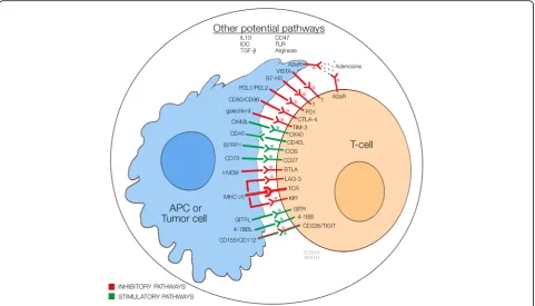
[1]. Figure1 summarizes these molecules and their targets
[1–3]. Immune checkpoint therapy for cancer encompasses
strategies that target these regulatory pathways in order to
enhance immunity activity against tumor cells [4, 5]. The
most broadly studied checkpoints are the inhibitory pathways consisting of cytotoxic T lymphocyte-associated molecule-4 (CTLA-4), programmed cell death receptor-1 (PD-1), and programmed cell death ligand-1 (PD-L1). Ipili-mumab [anti-CTLA-4 monoclonal antibody (mAb)] was the first immune checkpoint inhibitor (ICI) approved by
FDA in 2011 [6]. Many biological agents that target these
molecules are now broadly used in a variety of malignancies. * Correspondence:[email protected]
2Division of Hematology and Oncology, Mayo Clinic, Jacksonville, FL, USA
Full list of author information is available at the end of the article
Currently approved ICIs are only effective in a small fraction of patients and resistance after initial response is a common phenomenon. Nevertheless, new inhibitory and stimulatory pathways have emerged as potential targets for immune checkpoint therapy and immunotherapy is extending even
beyond this approach [7, 8]. Novel immune checkpoint
agents and combination therapies currently under investiga-tion in phase I/II clinical trials are reviewed and discussed in this article.
Methodology
We performed a PubMed search using the keywords and MeSH terms immunotherapy, immune check-point therapy, immune checkcheck-point inhibitors, immune checkpoint agonists, and immune checkpoint adju-vants. We also searched American Society of Clinical Oncology (ASCO) and American Association for Can-cer Research (AACR) meeting abstracts, and Clinical-Trials.gov from June 5, 2016, through January 30, 2018. We focused on phase I and phase II clinical tri-als of new agents in immune checkpoint therapy that were being used alone or in conjunction with other forms of immunotherapy. Data was collected from the trials reviewed with at least preliminary results pub-lished or presented before the date of the search. Ex-clusion criteria included phase III or later stage clinical trials, clinical trials focusing only on anti-CTLA-4 and anti-PD-1/PD-L1, trials focusing on
pediatric population, and non-interventional trials. We have included 62 phase I and 23 phase II clinical
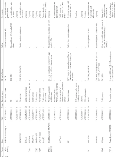
trials in this review. Table 1 summarizes these agents
and clinical trials.
Inhibitory pathways
Binding of CTLA-4 and PD-1/PD-L1 to cancer cell or tumor-microenvironmental ligands leads to T cell at-tenuation, which enables the tumor cells to avoid
immune-mediated destruction [1]. Similarly, other
in-hibitory pathways have been identified and new blocking agents are being developed to induce immune reaction
against malignant cells [4]. These inhibitory pathways
can be classified as T cell associated and non-T cell asso-ciated, as follows.
T cell-associated inhibitory molecules
LAG-3 (CD223)
Lymphocyte activation gene-3 (LAG-3, CD223) is expressed by T cells and natural killer (NK) cells after
major histocompatibility complex (MHC) class II
ligation [9, 10]. Although its mechanism remains
un-clear, its modulation causes a negative regulatory effect over T cell function, preventing tissue damage and auto-immunity. LAG-3 and PD-1 are frequently co-expressed
and upregulated on tumor-infiltrating lymphocytes
(TILs) leading to immune exhaustion and tumor growth
[11]. Thus, LAG-3 blockade not only improves
Table
1
Summary
of
ongoing
phase
1/2
clinical
trials
utilizing
immune
checkpoint
therapy
(Continued)
Category
Target
Drug
Trial
Phase
Type
of
tumor
Clinical
efficacy
Safety
Comments
NCT02543645
I/II
Solid
tumors
––
Terminated
(portfolio
re-prioritization)
CD40
CP-870893
NCT01103635
I
Melanoma
––
Ongoing
NCT02304393
I
Solid
tumors
––
Ongoing
APX005M
NCT02482168
I
NSCLC,
melanoma,
urothelial
cancer,
HNSCC
––
Ongoing
NCT03165994
II
Esophageal
and
gastroesophageal tumors
––
Ongoing
NCT02706353
I/II
Melanoma
––
In
conjunction
with
pembrolizumab. Ongoing
NCT03123783
I/II
Melanoma,
NSCLC
––
In
conjunction
with
nivolumab.
Ongoing
ADC-1013
NCT02379741
I
Solid
Tumors
––
Completed
NCT02829099
I
Solid
Tumors
––
Ongoing
JNJ-64457107
NCT02829099
I
Solid
tumors
––
Ongoing
SEA-CD40
NCT02829099
I
Solid
tumors
and
lymphomas
––
Alone
or
in
conjunction
with
pembrolizumab
RO7009789
NCT02304393
I
Solid
tumors
––
Ongoing
NCT02588443
I
Pancreatic
cancer
––
In
conjunction
with
Nab-paclitaxel
and
gemcitabine.
Ongoing
NCT02760797
I
Solid
Tumors
––
In
conjunction
with
Emactuzumab. Ongoing
NCT02665416
I
Solid
Tumors
––
In
conjunction
with
vanucizumab.
Ongoing
Other pathways
IDO
BMS-986205
NCT02658890
I
Solid
tumors
–
3/42
patients
with
grade
3
autoimmune
hepatitis
In
conjunction
with
nivolumab
Indoximod
NCT02073123
II
Melanoma
ORR
52%
No
significant
toxicities
Used
in
conjunction
with
ipilimumab,
nivolumab,
or
pembrolizumab
NCT02077881
I/II
Pancreatic
cancer
ORR
37%
1/30
patients
with
colitis
Used
in
conjunction
with
gemcitabine
and
nab-paclitaxel
NCT01560923
II
Prostate
cancer
Median
PFS
increased
from
4.1
to
10.3
months
No
significant
AEs
tumor immune responses but also potentiates other forms of immunotherapy given its different mechanism of action mainly mediated by impeding cell cycle
pro-gression [12–14]. Although simultaneous use with
anti-PD-1 therapy is considered synergistic, it remains un-clear whether other immune checkpoint inhibitory mol-ecules in conjunction with anti-LAG-3 therapy will be as
effective [15]. Furthermore, clinical benefits from
com-bination come at the expense of increased incidence of
autoimmune toxicities [1]. Currently two inhibitory
ap-proaches have been developed: a LAG-3-Ig fusion pro-tein (IMP321, Immuntep®) and mAbs targeting LAG-3
[5].
IMP321, a soluble form of LAG-3, upregulates co-stimulatory molecules and increases interleukin (IL)-12 production to enhance tumor immune responses. Two phase I clinical trials using IMP321 in advanced renal cell carcinoma (RCC) and pancreatic adenocarcinoma showed an increase in tumor reactive T cells, but no meaningful
objective response (OR) was observed [16, 17]. Another
phase I clinical trial studied IMP321 in combination with paclitaxel in metastatic breast cancer (BC) and an
object-ive response rate (ORR) of 50% was observed [18]. This
promising result has prompted a phase IIb clinical trial that is currently recruiting patients with metastatic BC (NCT02614833).
Targeting LAG-3 with antagonistic mAbs interferes with the LAG-3 interaction between MCH II molecules expressed by tumor and/or immune cells, promoting
tumor cell apoptosis [19]. A phase I clinical trial is
recruiting melanoma patients to determine the safety of anti-LAG-3 (BMS-986016), with and without nivolumab (NCT01968109). Interim results show promising efficacy with an ORR of 16% and disease control rate (DCR) of 45% among patients who had progressed despite previ-ous therapy with anti-PD-1/PD-L1. The safety profile is
similar to nivolumab alone [20]. LAG525 is another
anti-LAG-3 mAb being studied on a phase I/II clinical trial with metastatic solid malignancies (NCT02460224), and currently no data is available.
TIM-3
T cell immunoglobulin-3 (TIM-3) is a direct negative regulator of T cells and is expressed on NK cells and macrophages. TIM-3 indirectly promotes immunopression by inducing expansion of myeloid-derived sup-pressor cells (MDSCs). Its levels have been found to be particularly elevated on dysfunctional and exhausted T
cells suggesting an important role in malignancy [21].
Presence of TIM-3+ T cells correlates with severity and poor prognosis in non-small cell lung carcinoma
(NSCLC) and follicular lymphoma [11]. On the other
hand, low levels of TIM-3 have been associated with autoimmune processes such as in diabetes or multiple
sclerosis [22]. Similarly, the use of monoclonal
anti-bodies to block TIM-3 causes an increase in T cell pro-liferation and cytokine production which may not only explain its antitumor activity but also its role in
aggra-vating autoimmune diseases [22]. Furthermore, there
has been concern with the use of these antibodies given that TIM-3 could act as an enhancer of CD8 T cells
dur-ing certain acute infections includdur-ing Listeria [23].
Modulation of this pathway occurs through multiple ligands including galectin-9, phosphatidyl serine, and
CEACAM-1 [11]. These molecules play an important
role in carcinogenesis, tumor survival, and even progres-sion of different malignancies including melanoma,
gastrointestinal, and lung cancer [24–26]. As opposed to
other inhibitory pathways that interfere with cellular function, TIM-3 primarily exert its function by
regulat-ing cell apoptosis [27]. This could potentially explain its
enhancing effects when used with other ICIs. However, the best complementary molecule to be used with TIM-3 remains unknown.
Currently, one anti-TIM-3 mAb (MBG453) is being
in-vestigated in phase I–II clinical trial in patients with
ad-vanced malignancies (NCT02608268). No clinical results are yet reported.
TIGIT
T cell immunoglobulin and ITIM domain (TIGIT) is part of the CD28 family-like receptors expressed by NK and T cells. It exerts direct immunosuppressive effects on these cells and indirectly increases the release of immunoregula-tory cytokines (e.g., IL-10), decreases the production of
interferon (IFN)-γand IL-17, and prevents maturation of
DCs [28, 29]. Two agonists, CD155 (poliovirus
receptor-PVR) and CD112 (PVRL2, nectin-2), are expressed by im-mune cells, non-imim-mune cells, and by tumor cells
includ-ing melanoma [30]. Moreover, TILs often express high
levels of TIGIT along with PD-1, TIM-3, and LAG-3,
con-sistent with a dysfunctional phenotype [31].
Initial ex vivo and murine studies targeting dual block-ade of TIGIT and either PD-1 or TIM-3 has shown a syn-ergistic effect in immune cell proliferation, cytokine release, degranulation, and reversal of T cell exhaustion with subsequent tumor rejection and induction of
protect-ive memory responses [11, 32]. Importantly, the
expres-sion of TIGIT appears to be higher in the cells within tumor microenvironment than in those in the periphery, which would theoretically offer the advantage of a more targeted-directed therapy with less systemic autoimmune-like toxicities. Furthermore, TIGIT appears to exert its ef-fects primarily by limiting cytokine competency and CD8 T cell function which would in theory explain its
comple-mentary effects when used with other forms of ICIs [27].
anti-TIGIT mAb OMP-31M32 (NCT03119428). No results are yet available.
VISTA
V-domain Ig suppressor of T cell activation (VISTA), also known as programmed death-1 homolog (PD-1H), is a unique molecule with dual activity. It behaves as a stimulatory ligand for antigen presenting cells (APCs) causing immune activation and as a negative ligand for T cells suppressing activation, proliferation, and cytokine
production [33]. Le Mercier et al. demonstrated that its
blockade improved TIL activation and enhanced tumor-specific T cell responses in the periphery despite the presence of high PD-L1 levels or the lack of expression
of VISTA within tumor cells [34]. Therefore, both
path-ways are considered independent and simultaneous dual blockade of PD-1 and VISTA is often viewed as
synergis-tic [35]. Interestingly, VISTA expression levels appear to
vary among different tumors, often seen as a limitation given theoretical response heterogeneity. However, its blockade has proven to be effective even in the absence of detectable levels which offers the advantage of a broader clinical applicability but poses the challenge of
finding specific biomarkers to predict response [35].
Additionally, this pathway is expressed mainly by TILs which, similar to TIGIT, allow it to be more tumor-specific and less toxic than other pathways.
Two molecules are being tested on phase I clinical trials: JNJ-61610588, a fully human mAb against VISTA, and CA-170, an oral inhibitor of both PD-L1/PD-L2 and VISTA.
Both trials are currently recruiting (NCT02671955,
NCT02812875).
B7-H3 (CD276)
B7 homolog 3 (B7-H3), also known as CD276, is a protein that belongs to the B7-CD28 pathway family and is widely expressed in different solid organs as well as immune cells including APCs, NKs, and B and T cells. It has an inhibi-tory function on T cell activation, proliferation, and
cyto-kine production [36]. Furthermore, this pathway appears
to promote cancer aggressiveness. Thus, blocking this agent would not only offer the advantage of enhancing in-nate immunological responses against malignancy but also would exert a direct effect over tumor behavior. B7-H3 expression is limited on healthy tissues but overexpression is common in multiple malignancies including melanoma, NSCLC, prostate, pancreatic, ovarian, and colorectal
can-cer (CRC) [36, 37]. Therefore, developing strategies to
block this pathway would offer the advantage of exerting more localized effects over malignancies with less promin-ent systemic toxicities. Additionally, given its unique mechanism of action compared to other anticancer strat-egies, B7-H3 appears to have a synergistic effect when
combined with chemotherapy or other ICIs [36].
Enoblituzumab (MGA271) is an engineered Fc human-ized IgG1 monoclonal antibody against B7-H3 with potent anti-tumor activity. Interim results of an ongoing phase I clinical trial using MGA271 in melanoma, prostate cancer, and other solid tumors (NCT01391143) shows that it is overall well-tolerated without dose-limiting toxicities. Dis-ease stabilization and objective responses ranging from 2 to
69% were noted across several tumor types [38]. Another
phase I clinical trial is evaluating the use of enoblituzumab in combination with pembrolizumab (NCT02475213). Both studies are currently recruiting.
The use of dual affinity re-targeting (DART) proteins that bind both CD3 on T cells and B7-H3 on the target cell has been found to recruit T cells to the tumor site and promote
tumor eradication [39]. MGD009 is a humanized DART
protein that is being studied on a phase I clinical study in patients with B7-H3 expressing tumors including
melan-oma, NSCLC, mesothelimelan-oma, and urothelial cancers [40].
The trial is ongoing and recruiting patients (NCT02628535). 8H9 is an antibody against B7-H3 labeled with radio-active iodine (I-131) which, after internalization,
pro-motes cancer cell death [36]. This drug has been tested
on metastatic neuroblastoma in conjunction with
radi-ation therapy and surgery [41]. The ongoing trial is
assigning patients to be treated with either mAbs against B7-H3 or against GD-2 (NCT00089245). Preliminary re-sults revealed that 17/21 patients studied were alive and free of disease after a median follow-up of 33 months
[41]. 8H9 is also being studied on peritoneal cancers,
gli-omas, and advanced central nervous system malignan-cies (NCT01099644, NCT01502917, NCT00089245).
A2aR and CD73
The adenosine pathway encompasses specific adenosine receptors and enzymes that synthetize it. Adenosine A2a receptor (A2aR) is one of the most important factors in
this pathway and is mainly activated by adenosine [1].
A2aR is expressed on immune cells, including T cells, APCs, NK cells, and on endothelial cells. Increased levels of adenosine in tumor microenvironment can promote formation of Treg cells and can dampen the immune re-sponse of multiple effectors including macrophages, NK,
APCs, and neutrophils [42]. CD73, on the other hand, is
widely expressed by most tissues and is thought to serve as an adhesion molecule for lymphocyte binding to the endothelium and to play an important role as a co-signal for T lymphocyte activation. However, it also widely expressed by malignant cells where it acts as an enzyme and promotes the formation of adenosine by the
dephos-phorylation of AMP, favoring tumor progression [43]. Not
surprisingly, often these molecules are overexpressed in various malignancies and usually correlate with poor
over-all prognosis [44]. Given the multiple mechanisms that
microenvironment, different strategies to target both A2aR and CD73 have been developed. The main advan-tage of this approach is the potential use of combination strategies with other forms of therapy including chemo-therapy or other ICIs. Furthermore, the use of combin-ation strategies among the adenosine pathway is an
additional possibility [44]. However, an area of concern
with this approach is the blockade of adenosine synthetic enzymes which may favor the accumulation of ATP, a molecule that can play a pro-tumor role in the tumor
microenvironment [44]. An additional limitation, as with
other forms of immunotherapy, is the lack of clinical or biological markers that help with stratification of patients that are most likely to benefit from this form of therapy.
Blockade of A2aR on mice demonstrated increased prolif-erative capacity and function of T cells, as well as enhanced
immunologic memory [42]. Preliminary results from a
phase I clinical trial evaluating the oral adenosine A2aR an-tagonist CPI-444 alone and in combination with atezolizu-mab for advanced solid cancer showed that 42% of patients (10 of 24) who had been resistant to anti-PD-1/PD-L1 ther-apy, achieved disease control. Furthermore, grade 1 and 2 toxicities were the most common with only one case of
grade 3 autoimmune hemolytic anemia [45]. This trial is
ongoing and recruiting patients (NCT02655822).
MEDI9447 is a monoclonal antibody specific for CD73 that is being studied on a first-in-human clinical trial in patients with advanced solid tumors that have progressed
or are refractory to standard therapy (NCT02503774). No
preliminary results are yet available. Of note, CD73 could play a role in tumor angiogenesis; however, no studies have been designed yet to evaluate a possible synergistic
effect of anti-CD73 and antiangiogenic therapy [46].
BTLA
B and T cell lymphocyte attenuator (BTLA, CD272) is an inhibitory receptor that is structurally and function-ally related to CTLA-4 and PD-1 and is expressed by the majority of lymphocytes. Ligation of BTLA by its ligand, herpes virus entry mediator (HVEM), blocks B and T cell activation, proliferation, and cytokine
pro-duction [47]. Tumor cells exploit this pathway by either
promoting the formation of dysfunctional T cells that persistently express BTLA and render them susceptible to inactivation, or by expressing HVEM, as it has been
found with melanoma [47]. High levels of BTLA/
HVEM on melanoma and gastric cancer patients
cor-relate with poor prognosis [48, 49]. Thus, the
BTLA-HVEM pathway is being considered as a new target for
checkpoint blockade [48]. The main limitation with this
form of therapy has been the complexity of the receptor-ligand system. Additionally, given a different mechanism of action compared to other forms of im-munotherapy, combination with other molecules could
be synergistic but also be associated with an increased
risk of toxicity [47].
Non-T cell-associated inhibitory molecules
TGF-β
Transforming growth factor (TGF)-β is a cytokine that
helps maintain tissue homeostasis by regulating cellular
growth, differentiation, proliferation, and survival [50].
Al-though this pathway is able to control early-stage tumors by promoting cell cycle arrest and apoptosis, in advanced stages, it allows for tumor evasion by suppressing cyto-toxic T cells and promotes cancer cell proliferation, inva-sion, and metastases, a functional switch known as the
“TGF-β paradox” [51, 52]. Malignant cells achieve this
switch through either the inactivation of their TGF-β
re-ceptors, or by selectively disabling the tumor-suppressive arm of this pathway, allowing cancer cells to use the
TGF-β regulatory functions to their advantage by promoting
immune tolerance [53]. In fact, tumors that produce high
levels of TGF-βcan shield themselves from immune
sur-veillance [50]. Consistently, increased TGF-β expression
by NSCLC, CRC, gastric, and prostate cancer has
corre-lated with tumor progression and poor prognosis [50].
Many malignant cells have an abnormal TGF-β
sig-naling pathway and blocking agents exert an indirect action mainly by acting over the cells within the
tumor microenvironment [54]. This allows for
poten-tial combination with other forms of therapy includ-ing immune checkpoint targetinclud-ing and chemotherapy. Some challenges to note with this approach include the lack of biomarkers that allow defining the micro-environment where these agents are most usefuland the potential risk of synchronous occult tumor growth
by inhibiting the TGF-β suppressive action in
early-stage cancers [54]. There are three methods for
blocking the TGF-β pathway: blocking the ligand,
ligand-receptor interaction, or the receptor tyrosine kinase activity. Trabedersen [AP12009], a synthetic antisense oligonucleotide that hybridizes with RNA
sequences and blocks TGF-β translation, has been
tested on patients with glioblastoma multiforme and
anaplastic astrocytoma [55, 56]. It was also tested on
advanced pancreatic cancer where OS improved by
9.9–11.8 months although no improvement on
progression-free survival (PFS) was observed [57].
M7824 is a dual anti-PD-L1 monoclonal antibody
fused with a soluble extracellular domain of TGF-β
re-ceptor II, which acts as a TGF-βtrap. A phase I clinical
trial is being conducted on patients with metastatic or locally advanced solid tumors using this novel chimeric molecule (NCT02517398). Preliminary results from a trial in 16 patients demonstrate an acceptable safety
pro-file with no grade 4–5 adverse events. Preliminary
demonstrating a CR, one with durable PR, one patient with a 25% reduction of target lesions after two doses,
and two cases with prolonged stable disease (SD) [58].
Galusertinib (LY2157299), a blocker of the receptor’s
tyrosine kinase activity was tested in a recent phase II clinical study, but failed to demonstrate improved OS
compared to placebo [59]. This molecule is being
stud-ied on NSCLC, hepatocellular carcinoma (HCC), pan-creatic cancer, and BC (NCT02423343, NCT02734160, and NCT02672475).
KIR
Killer immunoglobulin-like receptors (KIRs, CD158) are a family of transmembrane proteins that promote self-tolerance by dampening lymphocyte activation, cytotoxic activity, and cytokine release. They are expressed by NK cells and some T cells and assist with self-recognition of host cells through the binding of MHC-I. KIR aids in the identification and destruction of cells that have lost their
MHC-I as with many tumor cells, a process termed“
miss-ing self” recognition [60]. Some malignancies, however,
develop mechanisms to evade this pathway by either up-regulating non-classical MHC-I molecules or by changing the tumor microenvironment properties rendering NK
cells dysfunctional [61].
The use of monoclonal antibodies to manipulate the KIR pathway is an active area of investigation as interfering with MHC-I interactions can stimulate NK cells by mimicking
the“missing self”response [62]. The main advantage of
tar-geting KIR is activating mostly NK rather than T cells, which is a potentially synergistic antitumor approach by allowing T cell ligands be available for targeting with other forms of immunotherapy. However, given its importance in self-recognition, NK cell overactivation may lead to a proin-flammatory state and increase the risk for autoimmune
re-actions [63]. Different molecules targeting KIR are under
investigation. Lirilumab, a fully human monoclonal anti-body that blocks KIR2DL1/2L3, is currently being studied in a phase I/II clinical trial with concurrent use of nivolu-mab and ipilimunivolu-mab in patients with squamous cell carcin-oma of the head and neck (NCT01714739). Preliminary results are promising, with an ORR of 24% and a DCR of 52%, and only 8% of patients stopping therapy due to
ad-verse events [64].
KIR3DL2 is frequently expressed by cutaneous T cell lymphomas (CTCL) and has prognostic and diagnostic
features within this population [65]. IPH4102 is a
mono-clonal antibody against kIR3DL2 which is currently be-ing investigated in a phase I clinical trial in patients with relapsed or advanced CTCL (NCT02593045). Prelimin-ary results reveal an ORR of 45%; 10 out of 22 patients with PR, 2 CR in skin, and 5 CR in blood. Six patients
developed grade 3 or more severe adverse events [66].
PI3Kγ
The expression of Phosphoinositide 3-kinase gamma
(PI3Kγ) by macrophages controls a critical switch towards
immune suppression in presence of inflammation and
can-cer. Additionally, PI3Kγseems to play a role in angiogenesis
by affecting the function of tumor-associated macrophages,
major producers of VEGF [67]. Thus, similar to TGF-β,
blocking this pathway exerts an indirect antitumor effect by modifying the microenvironment, improving the immuno-logical function against malignant cells, and affecting the tumor vasculature. Unfortunately, as with other forms of immunotherapy, blocking PI3K enzymes has been associ-ated with multiple autoimmune-like toxicities, and there-fore the use of lower doses in conjunction with other forms
of immunotherapy is often used [67].
IPI-549 is an oral selective inhibitor of PI3Kγ being
studied on a phase I clinical trial as monotherapy or in combination with nivolumab in patients with melanoma, NSCLC, or head and neck cancer (NCT02637531). Pre-liminary results demonstrate no dose-limiting toxicities and only mild adverse events including nausea and fa-tigue. Importantly, 12 out of 15 patients have demon-strated durable clinical benefit, and 50% of patients have
been able to remain on treatment≥16 weeks [68].
CD47
CD47, also known as integrin-associated protein, is a molecule that exerts its action through the signal
regulatory protein alpha (SIRPα). It is ubiquitously
expressed by healthy cells to help with autologous
recognition and avoid inappropriate phagocytosis [69].
Solid tumors (e.g., bladder and BC) and hematologic
cancers (e.g., acute myeloid leukemia and
non-Hodgkin’s lymphoma) overexpress CD47 causing an
in-hibitory effect over macrophages and other myeloid cells and high levels of CD47 correlate with poor
prog-nosis [69]. The blockade of the CD47/ SIRPα axis
re-sults in an increased macrophage recruitment and antitumor activity through phagocytosis and cytokines secretion. However, the use of this pathway has pointed certain limitations mainly derived from the diffuse
ex-pression of CD47. First, a potential“antigen sink”effect
where high doses may be required to achieve an
appro-priate therapeutic blockade [70]. Second, there is an
in-creased risk for “on-target” systemic toxicities over
healthy cells that express CD47. Until now, therapy has been overall well tolerated and anemia has been the
most common adverse event [70]. Hu5F9-G4, a
human-ized monoclonal antibody targeting CD47, is being studied on a phase 1 clinical trial in patients with solid tumors (NCT02216409). In preliminary results, it showed acceptable tolerability and SD in 2 out of 16
patients for 16 and 8 months, respectively [71].
combination with rituximab in patients with relapsed
or refractory B cell non-Hodgkin’s lymphoma is still
recruiting patients (NCT02953509).
TTI-621 (SIRPαFc) is a fully recombinant fusion protein
consisting of a CD47 binding domain linked to the Fc
re-gion of IgG1 to block the CD47 “do not eat me” signal
and engage macrophage Fcγreceptors to enhance
phago-cytosis and antitumor activity [72]. A phase I clinical trial
using TTI-621 in patients with relapsed or refractory per-cutaneously accessible solid tumors and mycosis fungoides is currently recruiting patients (NCT02890368).
Co-stimulatory pathways
As opposed to inhibitory pathways that attenuate the mune system, co-stimulatory molecules augment im-munological responses against malignant cells. Malignant
cells inhibit these pathways to promote tumorigenesis [5].
OX40
OX40 (CD134) is a member of the TNF receptor super family, highly expressed by activated CD4, CD8 T cells, and Tregs, and in a lesser degree by neutrophils and NK cells. This molecule, along with its ligand, OX40L, plays a pivotal role in activation, potentiation, proliferation, and survival of T cells and modulation of NK cell
func-tion [73]. Furthermore, this molecule inhibits the
sup-pressive activity of Tregs by directly interfering with their function and proliferation, and indirectly
antagon-izing their inhibitory byproducts (e.g., TGFβ) [74].
Im-portantly, when tumor antigens are recognized by TILs, its expression of OX40 increases, and not surprisingly, the amount of OX40-expressing TILs correlates with
im-proved prognosis in certain populations [75].
The use of mAbs to activate OX40 has been a strat-egy used to increase the antitumor activity by the im-mune system. Of note, these antibodies have been associated with depletion of TILs through an antibody-dependent cell cytotoxicity. NK cells recognize the anti-bodies bound to antigens over cell surfaces and kill
these cells [76]. However, this only occurs in the
pres-ence of NKs within the tumor, which varies depending on the host and type of malignancy. Another limitation is a potential activation of peripheral lymphocytes ra-ther than TILs when ra-therapy is given systemically. Thus, its intratumoral administration has been
pro-posed as a way to minimize systemic toxicity [76].
Despite its limitations, use of these antibodies has
dem-onstrated tumor regression in several preclinical
models, although often are used in conjunction with
other forms of immunotherapy [75]. 9B12 is a murine
IgG monoclonal agonistic antibody against OX40 that was studied in a phase I clinical trial in 30 patients with
metastatic solid malignancies [77]. Although no
pa-tients achieved PR, SD was achieved in 6 papa-tients.
Adverse events were overall tolerable and limited to grades 1 and 2 except for transient lymphopenia which
was found to be grade 3 or more in 7 patients [77].
MOXR 0916 is a humanized IgG agonistic monoclonal OX40-specific antibody that is currently being tested in combination with atezolizumab in patients with advanced solid malignancies (NCT02410512). Preliminary results show no dose-limiting toxicities but efficacy results are
not yet available [78]. PF-04518600 (PF-8600) is an IgG2
humanized agonistic monoclonal antibody of OX40 that is undergoing a first-in-human trial (NCT02315066). Pre-liminary results in patients with selected advances solid tumors including melanoma and NSCLC revealed no dose-limiting toxicities, and 4 out of 9 patients
demon-strated SD [79].
MEDI6383, MEDI0562, MEDI6469, INCAGN01949, and GSK3174998 are other agonistic monoclonal antibodies that are part of different phase I clinical trials for which no preliminary results are yet available (NCT02221960, NCT02528357, NCT02923349, NCT02705482).
GITR
Glucocorticoid-induced TNF receptor family-related pro-tein (GITR) is a co-stimulatory cell surface receptor that is constitutively expressed by T cells and NK cells, and ex-pression increases markedly following T cell activation. Its ligand, GITRL, is mainly expressed by APCs and endothe-lial cells and appears to have a role in upregulating the
im-mune system, leukocyte adhesion, and migration [80]. The
expression of GITR by TILs in the tumor microenviron-ment has been found to be higher than levels expressed by peripheral lymphocytes, indicating local T cell
activa-tion [80]. Agonizing agents of this pathway have been
con-sidered as a way to increase the immune antitumor activity, although the clinical utility of such agents de-pends on the presence of T cells in the tumor and the sub-set of TILs which may vary among different malignancy
[81]. Thus, selection of patients who will derive the most
benefit from this therapy is still unclear. Immune-related adverse events should also be considered. Preclinical data suggests that GITR therapy appears to be better tolerated
than anti-CTLA4 agents [81].
GITR modulation in the preclinical models has shown promising antitumor activity via significant increase in
effector T cells and decrease in Tregs [80]. TRX-518, an
aglycosylated human mAb that agonizes GITR, is cur-rently undergoing phase I clinical study in various solid malignancies (NCT01239134). Preliminary results dem-onstrate an acceptable safety profile without dose-limiting toxicities and SD in 10% of study patients (4 out
of 40 patients) [82]. BMS-986156 is another anti-GITR
results showed no dose-limiting toxicities, though no
ef-ficacy results were reported [83]. AMG 228, an agonistic
IgG1 monoclonal antibody of GITR, was also recently studied in a first-in-human clinical trial in 30 patients with refractory CRC, head and neck squamous cell
car-cinoma, urothelial carcar-cinoma, and melanoma [84]. None
of the patients demonstrated OR, and no dose-limiting toxicities were identified. Up to 90% of patients (27/30) experienced adverse events consisting of electrolyte
im-balances, anemia, and fever [84].
Other similarly agents including MEDI1873, MK-4166, INCAGN01876, and GWN323 are also being studied in multiple solid and hematologic malignan-cies (NCT02583165, NCT02132754, NCT02697591, NCT03126110, NCT02740270).
ICOS
Inducible stimulator (ICOS), a specific T cell co-stimulatory molecule of the CD28/CTLA-4 family mainly expressed by CD4 T cells, is a co-stimulator of
prolifera-tion and cytokine producprolifera-tion by these cells [85]. Its levels
are upregulated in activated T lymphocytes, especially after the use of anti-CTLA4 therapies, and its expression is considered a biomarker to indicate that anti-CTLA4
agents are binding its target [86]. Increased ICOS
expres-sion on circulating T cells after ipilimumab administration
has been associated with improved clinical outcomes [87].
Interestingly, ICOS appears to be a less potent pathway compared to other forms of immunotherapy mainly be-cause of a predominant CD4 expression. However, its use with other approaches, particularly CTLA4 blockade, can lead to a potent synergistic effect as a result of an increase
in the expression of ICOS after anti-CTLA4 therapy [85].
Some molecules have been developed and are being investigated. JTX-2011 is an agonistic monoclonal anti-body of ICOS that is currently being tested in a phase I/ II clinical trial alone and in combination with nivolumab in patients with advanced solid malignancies including endometrial, breast, lung, pancreatic, and CRC (ICONIC
Trial—NCT02904226). Preliminary results showed no
dose-limiting toxicities, although efficacy is not reported
[88]. Similarly, GSK3359609 is a humanized IgG4
mono-clonal agonistic antibody of ICOS that is undergoing clinical investigation in a phase I clinical trial, alone or in combination with pembrolizumab in patients with ad-vanced solid tumors (INDUCE-1 trial - NCT02723955). Finally, MEDI-570, an agonist monoclonal IgG1 anti-body directed against ICOS is also being studied in a phase I clinical trial in patients with Non-Hodgkin lymphomas (NCT02520791).
4-1BB
4-1BB (CD137) is an inducible co-stimulatory receptor expressed by T cells, NK cells, and APCs. Once expressed,
it binds its ligand (4-1BBL) and triggers subsequent im-mune cell proliferation and activation, particularly of T
and NK cells [89]. The activation of NK cells leads to an
increased antibody-dependent cell-mediated toxicity.
Thus, the use of anti-41BB agonists not only increases immune-mediated antitumor activity but is also consid-ered an ideal agent to use in combination with other monoclonal antibodies such as rituximab and trastuzumab
[89]. Of note, the use of 4-1BB antibodies in conjunction
with other ICIs may lead to an important antitumor re-sponse with potential increased toxicity. In fact, given the diffuse expression of 4-1BB, there is a notorious risk for
“on-target”systemic adverse events [89].
These antibodies have been expanded to clinical studies after demonstrating potent anti-cancer efficacy in murine
models [90]. Utomilumab (PF-05082566), a fully human
mAb that stimulates 4-1BB, has been studied in a phase I clinical trial in combination with pembrolizumab in patients
with advanced solid tumors [91]. No dose-limiting toxicities
were reported and 6 out of 23 patients had either CR or PR. This drug is currently being studied in multiple phase I clin-ical trials: alone or in various combinations with rituximab (NCT01307267), mogamulizumab (NCT02444793), an ex-perimental OX40 agonist (NCT02315066), and avelumab (NCT02554812).
Urelumab is another agonist antibody of 4-1BB that has been studied in various clinical trials in patients with ad-vanced solid tumors. A safety analysis from these trials concluded that this agent can occasionally cause
signifi-cant transaminitis when high doses are used [92].
Cur-rently, this medication is being evaluated in combination with nivolumab in a phase I/II clinical trial in patients
with solid tumors and B cell non-Hodgkin’s lymphoma
(NCT02253992). Preliminary results showed that 6/60 of the patients with lymphoma treated with urelumab
mono-therapy achieved a PR (n= 3) or CR (n= 3), 9/86 patients
who received combination therapy achieved PR although none of the patients with NSCLC or diffuse large B cell lymphoma had reported response. Of note, at least 3% of
patients developed grade 3–4 transaminitis, and 7% of the
123 enrolled patients developed serious adverse events
leading to discontinuation in 5% of study patients [93].
Another phase I clinical trial evaluating urelumab in com-bination with rituximab is being conducted in patients with metastatic solid tumors and refractory NHL (NCT01471210). No results have been yet published.
According to a recent comparison between urelumab and utomilumab, the former seems to exert a more
marked agonistic activity on the receptor [94].
CD27-CD70
cells, and to boost B cells [95]. Despite its wide spectrum of action, this pathway has not demonstrated to be par-ticularly effective in overcoming the immunosuppressive features of the tumor microenvironment. Thus, CD27 is considered most useful as combination rather than monotherapy. Furthermore, its use with other blocking agents like anti-CTLA-4 or anti-PD-1/PD-L1 may not only be synergistic but also associated with less
auto-immune toxicities [96]. When used as monotherapy,
CD27 agonist has been well tolerated and only minor adverse events are reported. An important aspect in this pathway is the identification of CD27 phenotype on tumor, as cancers that express this molecule could
achieve a more favorable outcome [96].
The use of CD27-CD70 agonist agents has been evaluated in various preclinical settings and is being studied in mul-tiple clinical trials. ARGX-110 is an agonistic anti-CD70 monoclonal antibody that has been studied in a phase I
clinical trial in patients with T cell lymphoma [97]. Of note,
2 out of 9 patients had a reduction of malignant clones of > 90%, one patient achieved radiological PR, and 2 patients reached PR in the skin. Currently one phase I clinical trial is
recruiting patients with advanced malignancies
(NCT01813539). BMS-936561 (MDX-1203) is another fully human monoclonal agonistic CD70-specific antibody that
was studied in RCC and B cell lymphoma [98]. Results
demonstrated disease stabilization in 69% of treated individ-uals. Varlilumab, a monoclonal agonistic antibody against CD27, is currently under investigation in a phase I clinical trial with simultaneous use of nivolumab in patients with advanced solid tumors (NCT02335918). Preliminary results showed a notable increase of TILs in post-treatment
biop-sies [99]. Currently this molecule is being studied in other
phase I and II clinical trials in patients with gliomas, mela-nomas, RCC, and other solid tumors (NCT02924038, NCT02302339, NCT02386111, NCT02543645).
CD40
CD40 is a member of the TNF receptor family expressed by APCs and B cells whereas its ligand, CD154, is expressed by activated T cells. Interaction between CD40-CD154 stimulates cytokines secretion of B cells with subsequent T cell activation and tumor cell death
[100]. Despite its potential synergy with other forms of
anticancer therapy, the use of CD40 agonists has also been associated with particular toxicities including cyto-kine release syndrome, thromboembolic events, and tumor angiogenesis. It is probably related to the
expres-sion of CD40 by platelets and endothelial cells [101].
The main challenges that remain with this particular form of therapy include the identification of appropriate combinations and patient population that would benefit from these agents. As of now, eight mAbs have entered
clinical trials: CP-870893, APX005M, ADC-1013,
lucatumumab, Chi Lob 7/4, dacetuzumab, SEA-CD40, and RO7009789. Some of these were recently reviewed
[102, 103]. Others are still under investigation
(NCT02482168, NCT03165994, NCT02706353,
NCT03123783, NCT02829099, NCT02588443,
NCT02760797, NCT02665416, NCT02304393).
Other potential pathways
IDO
Indoleamine 2,3-dioxygenase (IDO) is a tryptophan-degrading enzyme that converts tryptophan to kynure-nines. Kynurenines promote the differentiation and activity of Treg and decrease the amount and activity of CD8 T cells leading to an immunosuppressed environ-ment only worsened by the high levels of PD-1/PD-L1
concurrently present in this milieu [104]. IDO has been
found overexpressed in various tumor cell types includ-ing melanoma, chronic lymphocytic leukemia, ovarian,
CRC, and more recently in sarcomas [104,105].
Further-more, high levels of IDO not only correlate with poor outcomes in some malignancies but may also be in-volved in drug resistance to chemotherapeutic agents
[106]. Though their ability to counterbalance the
im-munosuppressive tumor microenvironment is promising, treatment with IDO inhibitors has also raised specific concerns. First, IDO is induced by inflammatory
mole-cules such as IFNγ. Therefore, the lack of inflammation
in the tumor microenvironment may be associated with
a suboptimal response to anti-IDO agents [106]. Second,
IDO and other similar enzymes are also expressed by healthy tissue, and its inhibition may lead to cross-reaction side effects. Regardless, IDO inhibitors remain a great area of interest among immune checkpoint therapy and different molecules are under investigation.
BMS-986205 is a once-daily, selective, and potent oral IDO1 inhibitor that is currently undergoing in a phase I
clinical trial with concomitant use of nivolumab
(NCT02658890). All reported toxicities have been grades
1–2 except for three cases of grade 3 hepatitis, rash, and
hypophosphatemia. No efficacy was reported [107].
Indoximod is another IDO inhibitor that is being stud-ied in phase II clinical trials in melanoma (NCT02073123), pancreatic cancer (NCT02077881), and castrate-resistant prostate cancer (CRPC) (NCT01560923). Results seem promising. ORR was 52% in patients with melanoma in whom indoximod was given with either ipilimumab,
nivolu-mab, or pembrolizumab [108]. Patients with pancreatic
can-cer had an ORR of 37% with concomitant use of indoximod,
gemcitabine, and nab-paclitaxel [109]. With indoximod,
me-dian PFS has increased from 4.1 to 10.3 months in
meta-static CRPC compared to placebo [110].
NCT02178722). Preliminary results have demonstrated an ORR ranging from 75% in melanoma to 4% in CRC. Its use seems to be safe with pembrolizumab. Although no dose-limiting toxicities have been identified, up to 3% of patients
have discontinued therapy due to adverse events [111,112].
In another completed phase I clinical trial with 52 patients who had advanced solid tumors (INCB024360), treatment with epacadostat demonstrated overall well tolerable ad-verse reactions except for 1/52 grade 3 pneumonitis and 1/ 52 grade 3 fatigue. No OR was reported, but 7/52 patients
achieved SD greater than 16 weeks [113].
TLR
Toll-like receptors (TLRs) are considered critical in the recognition of pathogens and control of the immune re-sponse. However, their role in tumorigenesis is far more complex. Some TLRs, like TLR4, may promote cancer progression by either favoring inflammation in the tumor microenvironment or inducing Tregs or PD-L1. Other TLRs like TLR7/8 and TLR9, induce antitumor responses
by promoting a“danger signal” within the tumor
micro-environment and activating the immune system against
malignant cells [114]. The use of agents to manipulate
these TLRs pathways seem to not only promote an im-mune response against malignancy but also induce
au-tophagy and apoptosis of cancer cells [115]. There are
certain important aspects to note with TLR therapy. First, its non-specific capability of inducing not only cytotoxic T cells but also immunosuppressive cells within the tumor microenvironment leads to an overall attenuated
tumori-cidal effect [116]. Second, an appropriate combination
partner and identification of patients that would benefit the most of these agents remains unclear. It has been established that concomitant use of these molecules with other forms of antitumor therapy including radiation and chemotherapy appears to offer stronger anticancer
re-sponses than either therapy alone [117]. These
combina-tions, unfortunately, may also be associated with an increased frequency of toxicities and autoimmune reac-tions. Despite these challenges, multiple agents are being evaluated in different clinical trials. MEDI9197 is a dual agonist of TLR7/8 that is currently under phase I clinical test in combination with durvalumab and radiation ther-apy in metastatic or locally advanced solid malignancies (NCT02556463). Preliminary results demonstrate that the agent is overall safe with only mild adverse events. No
effi-cacy data has been yet reported [118]. PG545 (pixatimod,
pINN) is an agonist of TLR9/IL-12 that was tested in a phase I clinical trial in patients with advance solid tumors (NCT02042781). Results revealed that 3 out of 23 patients developed dose-limiting toxicities, and the disease control
rate of 38% [119].
Polyinosinic-polycytidylic acid polylysine
carboxy-methylcellulose (poly-ICLC) is a potent TLR3 agonist
that has been recently studied in combination with radi-ation in a phase I clinical trial in patients with HCC not
eligible for surgery [120]. Intratumoral injection of this
agent was found to be overall safe with mostly grade I or II adverse events. A PFS of 66% at 6 months and 28% at 24 months, OS of 69% after 1 year and 38% after 2 years
were demonstrated [120].
IL-2R
IL-2 mediates its immune-enhancing effect through ei-ther a low-affinity dimeric and/or a high-affinity trimeric IL-2 receptor (IL-2R). The dimeric IL-2R consists of
CD122 (also known as IL-2Rβ) and CD132 (also known
as ϒc), whereas the trimeric IL-2R comprises an
add-itional component, the CD25 (also known as IL-2Rα)
which increases the affinity for its ligand [121].
IL-2 has been part of cancer treatment for many de-cades and is considered the first immunotherapy proven
to be effective in human cancer in 1984 [121]. However,
IL-2 has had certain limitations including a dual role en-hancing both T cells and Tregs favoring immunosup-pression, and a short life span with subsequent high doses requirements and potential severe toxicities in-cluding pulmonary edema, hypotension, and vascular
leak syndrome [122]. In need of better strategies, IL-2R
agonists have been developed to potentiate and prolong IL-2 antitumor effects allowing for lower doses and
de-creased toxicities [123]. Furthermore, IL-2R agonists
could also enhance other forms of immunotherapy with-out the associated toxicity provided by IL-2.
NKTR-214, an engineered cytokine that specifically
stimulates through CD122 (IL-2Rβ), is being tested in
solid tumors including melanoma, NSCLC, and BC (NCT02869295, NCT02983045). Studies using both NKTR-214 and nivolumab showed no dose-limiting tox-icities. One patient had a mixed radiographic response with a 40% decrease in LDH, and another patient had an
unconfirmed CR after only 6 weeks of treatment [124].
Another trial showed no dose-limiting toxicities, a tumor shrinkage ranging from 10 to 30% in 6 out of 26 patients (23%) and an increase of T cells and NK cells within the
tumor microenvironment in 100% of patients [125].
Arginase inhibitors
Arginine is an important amino acid for T cell activation and proliferation. High levels of arginase are produced by malignant cells and MDSCs leading to depletion of argin-ine and a subsequent immunosuppressive tumor
micro-environment [126]. The use of arginase inhibitors could
decreasing the availability of substances that favor tumor
growth [127]. Finally, given a higher expression of arginine
among the tumor microenvironment than that in plasma, the use of these molecules could be associated with a more specific and less toxic effect than other forms of immunotherapy.
CB-1158 is a selective arginase inhibitor being studied in a phase I clinical trial alone or in combination with nivolumab in patients with metastatic solid tumors (NCT02903914). Preliminary results show that the drug is well tolerated with no dose-limiting toxicities, > 90% of arginase inhibition, and up to a fourfold increase in
plasma arginine levels [128].
Oncolytic peptides
Lactoferrin-derived lytic peptide LTX-315 is a cytotoxic chemotherapeutic peptide that permeabilizes mithocondrial membrane and triggers caspase-independent necrosis
[129]. This agent modifies the tumor microenvironment by
decreasing immunosuppressive cells and increasing T cells
[130]. Intratumoral injection of this agent leads to tumor
antigen release, with subsequent increase of TIL activity. This form of administration makes it an attractive way to limit systemic toxicities, but it also limits its applicability to more localized malignancies. Another important aspect of LTX-315 is the substantial increase of CTLA-4 expres-sion following its administration. This suggests that this form of therapy may be particularly useful when used in
conjunction with anti-CLTA-4 agents [131].
A phase I clinical trial using this molecule as mono-therapy or in combination with ipilimumab or pembroli-zumab is being conducted in patients with metastatic
solid tumors, particularly melanoma and BC
(NCT01986426). Preliminary results showed that 2/28 patients achieved a CR, 5 patients had a decreased of >
50% of tumor size, and 8 patients achieved SD [132].
IL-10
IL-10 inhibits secretion of proinflammatory cytokines
(e.g., IFNγ, TNFα, IL-1β, IL-6) and also inhibits the
ex-pression of MHC molecules and costimulatory mole-cules at several levels, leading to inhibition of T cell
function [133]. Recently, IL-10 was also found to play
some antitumor role by inducing the activation and pro-liferation of CD8. CD8 cells expressing IL-10 has been associated with a favorable prognosis in patients with
lung cancer [134]. However, similar to other interleukins
like IL-2, its effects are pleotropic and this raises con-cern for potential systemic toxicity. Other unresolved is-sues similar to IL-2 therapy include determining the patient population that could benefit the most from this form of therapy and the most appropriate therapeutic
combinations [135]. In this regard, both PD-1 and IL-10
receptors are upregulated in TILs and therefore the
combined use of these molecules is reasonable [136].
AM0010 is a PEGylated recombinant human IL-10 that is currently being studied in combination with pembroli-zumab in melanoma patients in a phase I clinical trial (NCT02009449). Preliminary results revealed that 11 out of 25 recruited patients developed grade 3 or 4 adverse events including fatigue, thrombocytopenia, and anemia. Although no objective tumor response was seen, DCR
was 45% [137].
Limitations and challenges of immune checkpoint therapy
Although immune checkpoint therapy has been a great advancement in cancer treatment, several challenges such as immune-associated toxicity, treatment resist-ance, and clinical benefit limited to only a fraction of pa-tients remain unresolved.
Immune checkpoint therapies are often associated with a set of toxicities known as immune-related adverse events, a form of autoimmune-like reactions resulting from an in-creased activity of the immune system. These toxicities can manifest as generalized symptoms including fatigue or fever, or can produce organ-specific damage leading to rash, colitis, pneumonitis, and adrenal or thyroid insufficiency,
among many others [138–140]. Thus, using immune
checkpoint therapy mandates a comprehensive understand-ing of these adverse events from clinicians as a way to pre-vent, recognize, and appropriately treat each specific reaction. Most adverse events are resolved with interrup-tion of treatment and short course of steroids. Serious pneumonitis and colitis refractory of steroids may require
use of biological agents, like infliximab [141–144].
Despite durable response rates observed with im-mune checkpoint therapy, the majority of patients do not benefit from the treatment (primary resistance), and some responders develop cancer progression after
initial response (acquired resistance) [145]. Even
within the same patient, heterogeneous responses have been observed in different metastatic lesions. Both tumor intrinsic and micro-environmental extrin-sic factors contribute to this resistance. Tumor intrin-sic mechanisms for resistance include the absence of tumor antigen, loss or downregulation of MHC, alter-ation of antigen presenting machinery such as beta-2 microglobulin mutation, alteration of pathways that prevent immune cell infiltration or function
(mitogen-activated protein kinase, PI3K, WNT/b-catenin,
Interferon-gamma pathways), and escape mutations in
IFN signaling [145, 146]. Resistance can also be
de-rived from extrinsic factors from tumor
microenviron-ment. Regulatory T cells (Treg), MDSCs, M2
macrophages, and other inhibitory immune check-points may all contribute to inhibition of anti-tumor
mechanisms will assist with the process of designing new strategies to overcome resistance and provide the rationale for combination of different forms of
im-munotherapy [145, 147].
Clinical responses to checkpoint immunotherapy are variable. The identification of biomarkers to predict re-sponse and treatment-mediated toxicity remains an im-portant unresolved issue. A number of biomarkers have been found promising. For example, immunohistochem-ical determination of PD-L1 expression, high mutational load, selective CD8+ T cell infiltration, and distribution at tumor invasive margins correlate with clinical
re-sponse to anti-PD-1/PD-L1 treatment [148, 149]. A
study demonstrated that the presence of epithelial-mesenchymal transition correlates with a distinct tumor microenvironment in lung cancer consisting of elevated inflammatory signals and multiple immune checkpoints
[150]. Specific genes involved in chromatin remodeling
may also serve as markers of response. As an example,
the loss of function of thePBRM1gene encoding for the
chromatin remodeling complex SWI/SNF was recently found to correlate with response to anti-PD-1 therapy in
patients with clear cell RCC [151]. A recently developed
model using malignancy-specific neoantigens appears to predict tumor response to ICI therapy in patients with melanoma and lung cancer receiving anti-CTLA-4 and
anti-PD-1 therapy, respectively [152]. Furthermore, this
model may also be useful to identify acquired resistance to therapy.
Lastly, immunotherapy is expensive and the cost per-quality life-year gained can be prohibitive in many developing countries, limiting its access to the eligible patients.
Conclusions
Significant advances have been made in cancer immuno-therapy in the last decade. Immune checkpoint immuno-therapy, particularly CTLA4, PD-1, or PD-L1 anti-bodies, has revolutionized oncology care and quickly has become the standard of care in multiple malignancies. Immunotherapy targeting immune checkpoints is often better tolerated than traditional chemotherapy and dur-able responses are frequently seen. However, the clinical benefit has been limited to a subset of cancer patients. Furthermore, some who initially respond to treatment often relapse due to cancer resistance. Expanding clinical benefit to the majority of patients and preventing cancer resistance requires a better understanding of the mecha-nisms that lead to an effective anti-tumor response. The discovery of new immune inhibitory, stimulatory path-ways, and rational combination strategies as discussed in this article will likely shed the light to the next step to-wards improvement of cancer immunotherapy.
Abbreviations
A2aR:Adenosine A2a receptor; AACR: American Association for Cancer Research; APCs: Antigen presenting cells; ASCO: American Society of Clinical Oncology; B7-H3: B7 homolog 3; BC: Breast cancer; BTLA: B and T cell lymphocyte attenuator; CRC: Colorectal cancer; CRPC: Castrate-resistant prostate cancer; CTCL: Cutaneous T cell lymphomas; CTLA-4: Cytotoxic T lymphocyte-associated molecule-4; DART: Dual affinity re-targeting; DCR: Disease control rate; HCC: Hepatocellular carcinoma; HVEM: Herpes virus entry mediator; I-131: Radioactive iodine; ICIs: Immune checkpoint inhibitors; ICOS: Inducible co-stimulator; IDO: Indoleamine 2,3-dioxygenase; IFN: Interferon; IL: Interleukin; IL-2R: IL-2 receptor; KIRs: Killer immunoglobulin-like receptors; LAG-3: Lymphocyte activation gene-3; MDSCs: Myeloid-derived suppressor cells; MHC: Major Histocompatibility Complex; NK: Natural killer; NSCLC: Non-small cell lung carcinoma; OR: Objective responses;
ORR: Objective response rate; 1: Programmed cell death receptor-1; PD-1H: Programmed death-1 homolog; PD-L1: Programmed cell death ligand-1;
PFS: Progression-free survival; PI3Kγ: Phosphoinositide 3-kinase gamma;
Poly-ICLC: Polyinosinic-polycytidylic acid polylysine carboxymethylcellulose;
RCC: Renal cell carcinoma; SD: Stable disease; TGFβ: Transforming growth
factor-β; TIGIT: T cell immunoglobulin and ITIM domain; TILs:
Tumor-infiltrating lymphocytes; TIM-3: T cell immunoglobulin-3; TLRs: Toll-like receptors; VISTA: V-domain Ig suppressor of T cell activation
Acknowledgements Not applicable.
Funding Not applicable.
Availability of data and materials
All data generated or analyzed in this study are included in this published article.
Authors’contributions
JMA drafted the manuscript; YL designed and supervised the study, as well as edited the manuscript; BD, AS, SC, and KK reviewed and edited the text. All authors reviewed and approved the final version of the manuscript.
Ethics approval and consent to participate Not applicable.
Consent for publication Not applicable.
Competing interests
The authors declare that they have no competing interests.
Publisher’s Note
Springer Nature remains neutral with regard to jurisdictional claims in published maps and institutional affiliations.
Author details
1Department of Internal Medicine, Mayo Clinic, Jacksonville, FL, USA. 2Division of Hematology and Oncology, Mayo Clinic, Jacksonville, FL, USA. 3Present Address: Department of Blood and Marrow Transplantation and
Cellular Immunotherapy, Moffitt Cancer Center, Tampa, FL, USA.4Division of Immunology, Mayo Clinic, Jacksonville, FL, USA.
Received: 6 December 2017 Accepted: 1 March 2018
References
1. Pardoll DM. The blockade of immune checkpoints in cancer
immunotherapy. Nat Rev Cancer. 2012;12:252–64.
2. Drake CG, Lipson EJ, Brahmer JR. Breathing new life into
immunotherapy: review of melanoma, lung and kidney cancer. Nat Rev
Clin Oncol. 2014;11:24–37.
3. Chen L, Flies DB. Molecular mechanisms of T cell stimulation and
co-inhibition. Nat Rev Immunol. 2013;13:227–42.
4. Sharma P, Allison JP. The future of immune checkpoint therapy. Science.