INTRODUCTION
Plants of Hibiscus cannabinus Linn. were raised from seeds collected from naturally growing plants of different places in Nagpur and its environs. They were allowed to grow till they attained the flowering and at this stage plants were plants were sprayed with different concentrations of 2,4-D.
The aqueous solution of herbicide ranging from 100 to 5000 ppm was prepared. Ten pots for each concentrations (100 to 2000 ppm) containing 2 to 3 plants were sprayed. If 2000 ppm was found higher; the lower concentrations were tried to determined lethal dose. Asppe- poly sprayer of one litter capacity did spraying. A small quantity of sodium lauryl sulphasate as a surfactant added in the herbicide solution. The Spraying was started in month of October 1996 and same experiments were repeated next year also. Spraying was done twice in an hour to make it more effective in the evening hours, when the wind was slow and temperature comparatively lower than that of the day. This help in less evaporation and more absorption of herbicide
Biosciences, Biotechnology Research Asia Vol. 5(1),401-406 (2008)
Effect of spray application of 2, 4-D on anatomical
characters of Hibiscus cannabinus Linn.
SANJAY I. KAMBLE
Department of Botany and Coordinator, Department of Seed Technology, Phulsing Naik Mahavidyalaya, Pusad, Dist. Yavatmal (India)
(Received: March 19, 2008; Accepted: May 30, 2008)
ABSTRACT
In present study, the herbicidal activities of 2,4-D on Hibiscus cannabinus Linn. has been studied. The anatomical response might produce some light on the manner by which this compound affected on plant. The plants were sprayed with aqueous solution of different concentrations of herbicide from 100 to 10,000 ppm.
The herbicide induced some anatomical changes like proliferation of cambium and phloem in the stem to form large masses of meristematic cells and due to the proliferation of cortical and pith cells after treatment became crushed. In the leaves, desiccation of cells, proliferation of cambium in the midrib region and distortion of vascular elements was common feature observed of 2,4-D treatment.
Key words: Herbicide, 2,4-D, Spray application, Anatomical characters, Hibiscus cannabinus Linn.
solution by the leaves. To avoid contamination of different concentrations of herbicide, cardboard was used at the time of spraying application. Six pots were sprayed with water used as control. Field trials were conducted on naturally growing plants in randomly designed plots of size approximately 353 feet’s.
RESULTS Control:
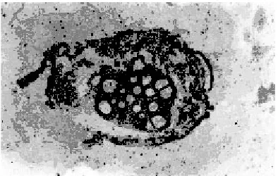
The control stem showed defined single layered epidermis followed by ground tissue. It was divided in to three zones; the outer zone was chlorenchymatous which was made up of 2 to 3 layers of parenchymatous cells containing chloroplast. This zone was followed by 2 to 3 layers collenchymas were immediately followed by 3 to 4 layers of parenchymatous cells. Endodermis and pericycle were distinct. Vascular tissues were arranged in a r ing and enclose large parenchymatous pith (Fig. 1).
The root showed secondary growth and consisted of an outer epidermis, parenchymatous
cortex followed by ring of secondary phloem and xylem. The distinct metaxylam was observed with a large vessels (Fig. 2).
The outer layer of petiole was made up of epidermis. Hypodermis was below the epidermis. Vascular bundles consisted of xylem and phloem (Fig. 3).
The leaf was composed of three types of tissue system, the epidermal, mesophyll and vascular. The lamina has upper and lower epidermis. The mesophyll comprises upper palisade parenchyma and lower spongy parenchyma. The midrib comprises of parenchymatous tissue embedding vascular elements (Fig. 4).
Fig. 1: Stem, T. S. of control X= 15.75 Fig. 2: Root, T. S. of control X= 31.5
2,4-D
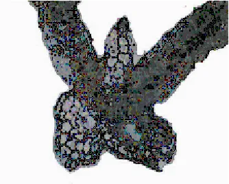
2,4-D induced anatomical changes in stem, root, petiole and leaf of Hibiscus cannabinus Linn. The stem at 100 and 200 ppm showed an abnormal meristematic activity of cambium. Owing to periclinal and anticlinal divisions of cambium, the phloem was pushed outwards and crushed; whereas at 300 and 400 ppm the phloem and rays cells proliferated which results in the formation of meristematic masses. These masses of meristematic cells encroach centrifugally upon the fundamental parenchymatous cortex, disrupting it and finally ruptured the epidermis at many places (Fig. 5).
The root at 100 and 200 ppm showed proliferated masses in the cortical region developed from the cells of cambium and phloem. However at 300 and 400 ppm these proliferated masses grow
in cortex, crushing its cell and forming lacunae. Subsequently, the cortex and epidermis were disorganized and lost their identity (Fig. 6).
The petiole at 100 and 200 ppm showed proliferation of cambium and phloem parenchyma calls. Owing to formation of meristematic masses, the pressure exerted on the cortical cells, lead to disorganization of cor tex and destr uction of epidermis (Fig. 7).
The leaves were severely injured at all concentrations of herbicide. At 100 and 200 ppm leaf showed desiccation of tissues and it lost cellular identity of epidermis. In midrib the parenchymatous cells of phloem were destroyed at 300 and 400 ppm and the mesophylls of the lamina also lost their identity (Fig. 8).
Fig. 5: Stem, T. S. at 300 ppm of 2,4-D. X= 20.16
Fig. 6: Root, T. S. at 400 ppm of 2,4-D. X= 31.5
Fig. 8: Leaf, T. S. at 200 ppm of 2,4-D. X= 31.5 Fig. 7: Petiole, T. S. at 400 ppm
of 2,4-D. X= 25.2
DISCUSSION
This herbicide induced some anatomical changes in stem, root, leaves and petiole at all concentrations. Stem of the plants showed proliferation of phloem for ming meristematic masses and disorganization of cor tical cells were observed. In addition to it ruptured epidermis was observed at all concentrations and pith cells were destroyed and lost their identity. Rubin and Gritsaenta (1968) on Amaranths retroflexux and Chenopodium album reported the proliferation of cambium tissue due to application of 2,4-D. Similar results have been reported by Dnyansager and Khosla (1968) on Cassia tora, Kiepal (1970) on Sinapis arvensis, Bequest (1971) on Myr iophyllum spicatum, Hadke, (1980) on Psoralea corylifolia and Euphorbia geniculata, Deshmukh (1981) on Cassia occidentalis, Corchor us olitor ious, and Lagasca molis, Srinivasu (1986) on Parthenium hysterophorus, Bobde (1993) on Crotalaria juncea , Jain (1993) on Chenopodium album and Gopal (1993) on Medicago sativa.
In the leaves disorganization of mesophylls and destruction of phloem cells in midrib was observed due to application of 2,4-D. Tukey et.al. (1945) on bind weed reported the plasmolysis of mesophyll cells and lost their contents. Loustalot and Muzik (1953) on Stizolobium decringianum reported destruction in mesophyll cells. Similarly, Scifres and Mc Carty (1968) on Vernonia spp., Bakale (1972) on Alternanthera polygonoides and Cressa cretica, Walsh et.al. (1972) on Rhizophora maingle, White and Hemphill (1972) on Tobacco. Bakale and Dnyansagar (1977) on Xanthium strumarium, Kolhe (1979) on Tephrosia hamiltonii, Solanum surrattense, and Celosia argentea, Hadke (1980) on Psoralea corylifolia and Euphorbia geniculata. Deshmukh (1981) on Cassia occidentalis, Corchorus olitorious and Lagasca mollis, Srinivasu (1986) on Par thenium hysterophorus, Bobde (1993) on Crotalaria juncea, Jain (1993) on Chenopodium album and Gopal (1993) on Medicago sativa reported disorganization
of mesophyll and destruction of phloem in mid vein.
In root 2,4-D induced certain anatomical changes such as formation of huge masses of meristematic cells developed from cambium and phloem, destruction of cortical and pith cells were observed at all concentrations of herbicide. Similar results were reported by Tukey et.al. (1945) on bind weed, Leroux (1957) on Nigella damascena, Kilpatrick et.al. (1963) on Trifolium spp., Audus (1964) on some weeds, Calhan and Engel (1965) on Agrostis fenuis, Kolhe (1979) on Tephrosia hamiltonii, Solanum surrattense, and Celosia argentea, Hadke (1980) on Psoralea corylifolia and Euphorbia geniculata. Deshmukh (1981) on Cassia occidentalis, Corchorus olitorious and Lagasca mollis, Srinivasu (1986) on Par thenium hysterophorus, Bobde (1993) on Crotalaria juncea, Jain (1993) on Chenopodium album and Gopal (1993) on Medicago sativa due to application of 2,4-D.
The petiole showed Proliferation of cambium, destr uction of phloem cells, disorganization of cortex and endodermis in the present study. Many workers like Bradley et.al. (1968) on Apricot spp., Dnyansagar and Khosla (1968) on Cassia tora, Kolhe (1979) on Tephrosia hamiltonii, Solanum surrattense, and Celosia argentea, Hadke (1980) on Psoralea corylifolia and Euphorbia geniculata. Deshmukh (1981) on Cassia occidentalis, Corchorus olitorious and Lagasca mollis, Srinivasu (1986) on Parthenium hysterophorus, Bobde (1993) on Crotalaria juncea, Jain (1993) on Chenopodium album and Gopal (1993) on Medicago sativa reported similar results.
REFERENCES
1. Audus, L.S. Studies on pH relationship of
root growth and its inhibition by 2,4-D
and coumarin. New Phytol. 48: 97-144 (1964).
2. Bakale, V.L., Effect of folliar application of
weedicide on Alternanthera polygonoides var.eracta Linn. The Botanique. 10(1-4): 89-101 (1972).
3. Bakale, V.L. and Dnyansagar, V.R., Effect of
weedicide on Xanthium strumarium Linn. J. Indian. Bot. Soc. 56(2): 116-120 (1977).
4. Brequest, E.T., Ecological and Morphological
effects on Myriophyllum spicatum Linn. In a Tennessee valley reservoir. Diss. Abstr. Internet B. 32(116): 137 (1971).
5. Bobde, S.N., Comparative effects of
herbicides on Crotalaria juncea Linn. Ph.D. Thesis, Nagpur Univ., Nagpur (1993). 6. Bradley, M.V., Crane, J.C. and Marie, N.,
Some histological effects 2,4,5-T applied to
mature Apricot leaves. Bot. Gaz. 129: 231- 238 (1968).
7. Calhan, L.H. and Engel, Tissue abnormalities
induced in root of Agrostis fenuis by phenoxy alkyl carboxylic acid herbicide. Weeds. 13: 336-338 (1965).
8. Deshmukh, V.R., Effect of weedicides on
cytomorphology of weeds. Ph.D. Thesis, Nagpur Univ., Nagpur (1981).
9. Dnyansager, V.R. and Khosla, S.N., Effects
of 2,4-D on anatomical characters of some
weeds. Proc. Ind. Acad. Sci. 703: 287-294 (1968).
10. Gopal, K.R, Herbicidal effects on
cytomorphology of weeds Medicago sativa.. Ph.D. Thesis, Nagpur Univ., Nagpur (1993).
11. Hadke, S.M., Effect of herbicide on
cytomorphology of weeds Psoralea corylifolia and Euphorbia geniculata. Ph.D. Thesis, Nagpur Univ., Nagpur (1980). 12. Jain, S.B., Cytomorphological effects of
weedicides on weed Chenopodium album. Ph.D. Thesis, Nagpur Univ., Nagpur (1993).
13. Kiepal, Z., Pathological anatomy of Sinapis
arvensis Linn. Stem histological, anatomical changes caused by herbicides in the
auxin type. Acta Agrobotanica. 23: 73-84 (1970).
14. Kilpatrick, R.A., Chen, A.E. and Rodriguez,
Z., Root symptoms and anatomical changes
in Trifolium spp. by 2,4-D. Pl. Diss. Reptr.
47: 497-501 (1963).
15. Kolhe, R.R., Effect of herbicides on the
cytomorphology of farm weeds. Ph.D. Thesis, Nagpur Univ., Nagpur (1979).
16. Leroux, R., Research on morphological and
histological modification on Nigella damascena Linn. Caused by 2,4-D. Rev. Gen. Bot. 64: 299-306 (1957).
17. Loustalot, A.J. and Muzik, T.J., Effect of
2,4-D on apparent photosynthesis and
developmental morphology of Stizolobium decringianum. Bot. Gaz. 115: 56 (1953). 18. Rubin, S.S. and Gritsaenta, A.M., The effect
of 2,4-D on the structure of plants. Bot. Gaz. 53: 377-378 (1968).
19. Scifres, C.J. and Mc Carty, M.K., Reaction
of western ironweed (Vernonia spp.,) leaf tissue to picloram. Weed Science. 34: 347-349 (1968).
20. Sr inivasu, T., Effect of weedicide on
Par thenium hysterophorus Linn. Ph.D. Thesis, Nagpur Univ., Nagpur (1986).
21. Tukey, H.B., Hamner, C.L. and Imhofe, B.,
Histological changes in bind weed
sowthistle concentrations. Bot. Gaz. 107: 62-73 (1945).
22. Walsh, G.E., Cook, G.H. and Barvett, R. . Rhizophora maingle Linn. Effect and
translocation of 2,4-D and piloram. Abstr. Meet. Weed Sci. Soc. America. 11
(1972).
23. White, J.A. and Hemphill, D., An ultra