GLOBAL JOURNAL OF ADVANCED ENGINEERING TECHNOLOGIES AND
SCIENCES
AUTOMATIC LASER INTERSTITIAL THERMAL THERAPY FOR
ROBOT-ASSISTED SURGERY
Mhamed Nour*, Michel Gendreau**, Ahmed Lakhssassi*
*
Université du Québec en Outaouais, Québec, Canada
**Polytechnique Montréal, Québec, Canada
DOI: 10.5281/zenodo.1195103
ABSTRACT
The performance of the minimally invasive surgery (MIS) is enhanced by new Robotic surgical assistants (RSAs) because of the many advantages including small incisions, decreased blood loss, less pain, quicker heating time and the ability to pinpoint locations very precisely [1].
In order to maximize therapeutic effects of the LITT (Laser interstitial thermal therapy) while minimizing side effects, thermal sensors need to be installed at the border between healthy and tumorous tissues. These thermal switches will send a signal to stop the heat process as soon as the temperature is exceeding a trigger. A mathematical formulation of the laser ablation is proposed. Such procedure which defines the laser power distribution and the ablation position can be used for robot-assisted surgery. Such smart procedure will use a tissue damage prediction tool and bang-bang switch at the edges between healthy and unhealthy tissue to prevent side effect (laser ablation of the healthy tissue).
We used a system on chip to control the robotic arm and laser led in order to proceed with the automatic LITT procedure.
KEYWORDS: Laser Interstitial Thermal Therapy, thermal damage, brain cancer, bio heat transfer simulation, Thermal sensor, minimally invasive surgery, Robotic surgical assistants, Robotic arm, Raspberry Pi B+, Matlab, Comsol.
INTRODUCTION
The brain tumor tissue is modelled as a 3D domain with a structure of spheres after the laser ablation. In this paper, the form of the deformation caused by the laser ablation is assumed to be as a sphere. The volume of the sphere will be defined during the simulation with respect to the temperature limit at the border between healthy and tumor tissues.
Each sphere represents the volume deformation of the tissue caused by a laser ablation of its sphere volume. Any sphere ablation will be represented with a sphere volume, sphere radium, laser power distribution through a time limit.
Since the tumor tissue is surrounded at the edge with thermal sensors, so they will not exceed a temperature limit, each sphere ablation assigned to the structure should verify this side effect constraint.
The laser ablation process is defined in the following steps:
A. Assignment of the laser ablation point with spheres structure. The sphere ablation shouldn’t have any side effect.
B. An automatic ablation process will visit the structure within certain order and place the laser led at the center of the structure and proceed with the laser ablation of the spheres attached.
Figure 1. System Overview: The Planner relies on the FEM simulator. At each laser firing point, the planner will call the FEM.
The system has three modules as described fellow (figure 1): Planner.
First convert the MRI Stack to stl format, then the algorithm with construct the firing graph, and generate the laser ablation firing order. For each firing step call the FEM.
FEM.
Simulation software is used to simulate the mathematical model which use thermal conduction based on Fourier’s law and constant blood perfusion. Predecting the results in term of volume damage to the tissu will improve the health care system. This module will send back the laser distrubution power so that no side effect will occurs.
Robotic Arm.
The robotic arm will receive the laser power distribution and the coodinate of the laser ablation point. Then put the laser led at the specified point and fire with the respect of the distribution during the specified time.
As stated in [1], the planning process is divided into three steps :
- The selection of the number of beams and the direction from which to focus the laser ablation on the patient tumor tissue (geometry problem or beam angle optimization problem),
- The selection of intensity patterns for the directions selected in Phase 1 (intensity problem or fluence map optim,ization problem), and
- The selection de a delivery sequence that efficiently administers the treatment (realization problem or segmentation problem).
GENERAL MATHEMATICAL FORMULATION
The following paragraphs will specify the mathematical formulation of the automatic LITT, the description of the hexagonal close-packed structure, the graph contruction and the algoritm for the automatic ablation process.
Mathematical formulation of the Automatic LITT.
Let us assume that the laser beams of power p = 1, . . , n are available for a treatment and that each laser beam is fired on the tumor tissue to form a sphere with radium r = 1, ..,f at a specific point P with coordinates (x,y,z) with an angle α = 1, . . , a to the z axis and within an exposition time t = 1, . . , m to kill the tissue in the volume of that sphere.
We denote D(x,y,z),α,p,r,t the dose deposited at Point P with coordinate (x,y,z), with an angle α, and with an exposition time of t.
We assume that there is a process which will divide the stl tissue format to as many as box.Then for ecah one we will execute the following automatic LITT procedure.
.Figure 2. The tissue brain geometry is represented as a box.
We will represent the laser ablation point by a seven component vector :
(𝑥𝑖, 𝑦𝑖, 𝑧𝑖, 𝑝𝑖𝑡𝑖, 𝛼𝑖, 𝑉𝑖) (1)
Where xi, yi, zi represented the three dimensional coordinates, Pi is the power, ti is the time, a is the angle and Vi is the binary variable between points that correspond to a new laser ablation entry from another.
𝑉𝑖= 1, for any new laser ablation entry. 𝑉𝑖= 0 , else.
Rule 1. Belonging to the same axle tire:
Whenever the point x2, y2, z2, P2, t2, a2, v2 belong to the same entry laser ablation point as x1, y1, z1, P1, t1, a1, v1 then one must have (x2, y2) = (x1, y1), furthermore the coordinate of the entry point in the two dimensions, then z2 must be less than z1 and Z2=0.
𝑆𝑖 𝑥1= 𝑥2 𝑒𝑡 𝑦1= 𝑦2 𝑎𝑙𝑜𝑟𝑠 𝑧2 < 𝑧1 𝑒𝑡 𝑉2= 0 (2)
Rule 2. X 1 is the smallest of the x-axis coordinate.
We number the ablation point in such a way that xi is the smallest x coordinate. Forthermore, we made the assumption that the ablation points are numbered in the increasing order of the x coordinate.
𝑥𝑖+1≥ 𝑥𝑖 is always true. (3)
Condition 1. Limit size of the tissue in axis x.
𝑥𝑖+1− 𝑥𝑖≤ 𝑉𝑖+1𝐷𝑥 (4)
Observation. If the (i+1)th point correspond to a new entry (Xi+1 Greater than Xi) then the Y coordinate of this point is free.
There is not necessarily 𝑦𝑖+1≥ 𝑦𝑖
𝐼𝑓 𝑥𝑖+1> 𝑥𝑖 𝑡ℎ𝑒𝑛 𝑦𝑖+1 𝑖𝑠 𝑓𝑟𝑒𝑒 (5)
Rule 3.
1. 𝐼𝑓 𝑥𝑖+1= 𝑥𝑖𝑡ℎ𝑒𝑛 𝑦𝑖+1> 𝑦𝑖 (6)
2. 𝐼𝑓 𝐷𝑖+1= 0 𝑡ℎ𝑒𝑛 𝑦𝑖+1> 𝑦𝑖 (7)
3. 𝑦𝑖− 𝑦𝑖+1≤ 𝐷𝑖+1𝐷𝑦 (8)
𝐼𝑓 𝐷𝑖+1= 1 𝑡ℎ𝑒𝑛 𝑦𝑖 −𝑦𝑖+1≤ 𝐷𝑦 (9)
Condition 2 Belonging to the same axis shooting.
𝑊𝑒 𝑘𝑛𝑜𝑤 𝑡ℎ𝑎𝑡 𝑖𝑓 𝑉𝑖+1= 0 𝑡ℎ𝑒𝑛
𝐼𝑓 𝑧𝑖+1< 𝑧𝑖 𝑡ℎ𝑒𝑛 𝑧𝑖+1− 𝑧𝑖 ≤ 𝑣𝑖+1 𝐷𝑧 (11)
Condition 3.
𝑦𝑖− 𝑦𝑖+1 ≤ 𝑣𝑖+1 𝐷𝑦 And
yi+1-yi ≤ vi+1 Dy (12)
𝐼𝑓 𝑉𝑖+1= 0 𝑇ℎ𝑒𝑛 𝑦𝑖> 𝑦𝑖+1 (13)
Condition 5. Total covering the Tumor tissue:
For any volume v of the Tumor Tissue, v should be part of at least one or many VD within the total Tumor Tissue.
Constraint of coverage: case of the spherical approximation.
Let the center of the sphere
(𝑥𝑖, 𝑦𝑖, 𝑧𝑖+𝑟) and the laser ablation (𝑥𝑖, 𝑦𝑖, 𝑧𝑖, 𝑝𝑖, 𝑡𝑖, 𝛼𝑖) The question is: did we did the laser ablation at this point (x,y,z)?(𝑥𝑙− 𝑥𝑖) 2+ (𝑦𝑙− 𝑦𝑖) 2+ (𝑧𝑙− (𝑧𝑖+ 𝑟)) 2≤ 𝑟2+ (1 − 𝑤𝑖𝑙) 𝐾 𝑘 = 𝐷𝑥2+ 𝐷𝑦2+ 𝐷𝑧2
The constraint of coverage is :
We have ∑𝑖𝜀𝐼𝑤𝑖𝑙 ≥ 1 𝑟𝑒𝑔𝑎𝑟𝑑𝑙𝑒𝑠𝑠 𝑜𝑓 𝑙 ∈ 𝑇
With 𝑤𝑖𝑙= 1 𝑖𝑓 𝑣𝑜𝑥𝑒𝑙 𝑙 𝑖𝑠 𝑏𝑢𝑟𝑛𝑒𝑑 𝑏𝑦 𝑡ℎ𝑒 𝑙𝑎𝑠𝑒𝑟 𝑎𝑏𝑙𝑎𝑡𝑖𝑜𝑛 𝑖 With 𝑤𝑖𝑙= 0 𝑒𝑙𝑠𝑒.
Covering cubes constraint of coverage: case of the cubic approximation. Cube in the sphere and point in (𝑥𝑖, 𝑦𝑖, 𝑧𝑖)
(𝑥, 𝑦, 𝑧) with 𝑥 ∈ (𝑥𝑖− 𝑟, 𝑥𝑖+ 𝑟) and 𝑦 ∈ (𝑦𝑖− 𝑟, 𝑦𝑖+ 𝑟) and 𝑧 ∈ (𝑧𝑖, 𝑧𝑖+ 2𝑟)
We have ∆𝑥= |𝑥 − 𝑥𝑖| then ∆𝑥≥ 𝑥 − 𝑥𝑖 and ∆𝑥≥ 𝑥𝑖−𝑥 We have ∆𝑦= |𝑦 − 𝑦𝑖| then ∆𝑦≥ 𝑦− 𝑦𝑖 and ∆𝑦≥ 𝑦𝑖−𝑦
We have ∆𝑧= |𝑧 − (𝑧𝑖+ 𝑟)| then ∆𝑧≥ 𝑧− 𝑧𝑖 -r and ∆𝑧≥ 𝑧𝑖+𝑟 − 2
If ∆𝑥+ ∆𝑦+ ∆𝑧≤ 𝑟𝑖 then (x,y,z) belongs to a cube. The cube coverage constraint is
∆𝑥𝑙+ ∆𝑦𝑙+ ∆𝑧𝑙≤ 𝑟+ (1 − 𝑤𝑖𝑙)𝐾
Condition 6. Side effect constraint:
At each laser ablation point, and when firing the laser, there should be no violation at the thermal switches located at the edge points.
For each thermal switch TS:
∀ 𝑡, 𝑇𝑒𝑚𝑝(𝑇𝑠) ≤ 𝑇𝑙𝑖𝑚𝑖𝑡 (14)
The objective function is to minimise the number of laser ablations:
U is the number of reprints, and V the number of axes of tires, and B is a parameter.
Definition of the Thermal damage:
We used the first order Arrhenius equation to compute the damage integral [3]:
dt (16)
where is the original concentration of undamaged cells, is the concentration of the remaining living cells after time t, the treatment time, A is the frequency factor, is the activation energy and R is the universal gas constant. (R=8.314 J mol-1 K-1).
A damage integral of , corresponds to 63% percent probability of cell death, and damage integral of , corresponds to 99% percent probability of cell death at a specific point.
DESCRIPTIOMN OF THE AUTOMATIC ABLATION PROCESS
Either the number of diviison on nx x axis. NX = x/a.
The same way, we can calculate Ny and Nz.
Then a plan of treatment with (Nx.Ny.Nz) cubes. With Nx.Ny.Nz is equal the number of reprints. If we vary the edge has, we'll have another tire plan.
Among the plans drawn, you evaluate them based on the side effect the objective function and offer the best.
To the edges of the fabric, we will propose a refinement.
To optimize the number of ablation, a combination of Side Fire and Full fire will be proposed as follow: When the firing point is in the center of the Tissue, The NeuroBlate diffusing laser (Full Fire) is designed to provide fast, volumetric ablation in a concentric zone of hyperthermia.
While the firing point is near the edges of the Tissue, The NeuroBlate directional laser (SideFire) is the preferred tool for contoured ablation of targets while sparing adjacent healthy tissue.
All full Fire and Side Fire have to verify the side effect constraints.
MATERIALS AND METHODS
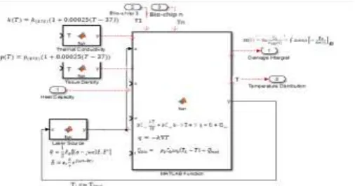
A. Modeling in COMSOL Multiphysics A.1. Heat distribution and damage estimation.
Figure 3. Validation process and damage estimation tool.
A.2 Bang-bang Controller
In [2], visual thermal sensors were implemented at the border between healthy and tumorous brain tissues. Events interface to control the heating process, by either allowing or stopping the source function depending on a temperature limit. As soon as the thermal controller reaches the temperature limit at the border, it will send a signal to stop the heating process immediately.
B Matlab, Comsol simulation and robotic arm. B1. Matlab codes to access the Comsol simulation. model = mphload('busbar.mph');
Then working with the geometry, working with meshes, modeling physics, executing the comsol program within matlab.
A matlab loop will go through the geometry and call Comsol simulation using thermal switches at the edges of the tumor tissue. If the power proposed does not permit the temperature to exceed the temperature limit, the robotic arm can proceed with the laser ablation.
B.2 Matlab codes to control the laser LED through a Raspberry PI B+
The following program will check the temperature at the edges, if temperature limit is exceeded, the program will display ‘do not proceed’ else the program will display ‘Please proceed with the laser ablation.’ Figure 4 shows the Led simulating the laser ablation process.
clear;
mypi=raspi;%
filename=fullfile('C:\users\mhamed\desktop\max-op2.txt'); fid = fopen(filename, 'r') ;
%Opensourcefile. fori=1:8
fgetl(fid) ; %
Read/discardline. end;
buffer=fread(fid, Inf) ; %Readrestofthefile. fclose(fid)
fid = fopen('C:\users\mhamed\desktop\max-
output.txt', 'w') ; % Open destination file. fwrite(fid, buffer) ; % Save to file.
fclose(fid) ; %
filename = fullfile('C:\users\mhamed\desktop\max-output.txt'); T1 = readtable(filename);
C = table2cell(T1); C
max([C{:, :}]) if any([C{:, :}] > 37)
disp('There is at least one value above the limit. Please do not proceed !!!') else
disp('All values are below the limit. Please Proceed.') end
% %
filename = fullfile('C:\users\mhamed\desktop\donne-binaire.txt'); fid = fopen(filename, 'r') ;
% Open source file. for i=1:8
fgetl(fid) ; % Read/discard line.
end;
% Read rest of the file. fclose(fid)
fid = fopen('C:\users\mhamed\desktop\donne-output.txt', 'w') ; % Open destination file. fwrite(fid, buffer) ;
% Save to file. fclose(fid) ; %
filename = fullfile('C:\users\mhamed\desktop\donne-output.txt'); T = readtable(filename);
for i=1:30
writeDigitalPin(mypi, 25,T(i,2));
pause(0.03); end;
Figure 4. Raspberry Pi and Laser ablation.
B3. Matlab codes to control the Robotic Arm.
To control the 6-axis robotic arm, there are manual and an automatic option:
Option 1 is to use the March 3 CNC controller software to send the coordinates of the firing points to the robotic arm. The MATLAB script will execute the COMSOL program, then via the CNC controller, we will control the robotic arm using the Mach 3 program.
Option 2 is to automatically control the robotic arm by using ABB robot Studio. Selecting the robot and tool to simulate the robotic arm and led. The Rapid program includes the path (set of positions) received from the Matlab. The command MoveL with data from the file as positions with move to different positions in order to proceed with the laser ablation. The syntax of the Move l is MoveL RelTool (p1, 0, 0, 100), v100, fine, tool1; the robot is moved to a position that is 100 mm from p1 in the z direction of the tool.
CONCLUSION
In this paper, a mathematical formulation of the automatic laser ablation process was proposed, which include all steps from the calculation of the temperature distribution and tissu dammage, the control of the temperature at the edges, to the safe automatic ablataion process with no side effects. Next step will be the implentation of the whole framework.
REFERENCES
[1] Sachin, Ron Alterovitz, Automated Tissue Retraction for Robot-Assisted Surgical Surgical Procedures, Department of Computer Science, University of North Carolina at Chapel Hill, USA.
[2] Mhamed Nour, Mohammed Bougataya Ahmed Lakhssassi, , Using Virtual Bang-Bang Controllers to Optimize Treatment of Brain Tumors, 1st International Conference on Advanced Research (IACR
2017) Manama, Bahrein, Jan 25-26, 2016.
[3] Mhamed Nour, Aziz Oukaira, Mohammed Bougataya, and Ahmed Lakhssassi, "Thermal Damage Modeling Analysis and Validation during Treatment of Tissue Tumors," International Journal of Pharma Medicine and Biological Sciences, Vol. 6, No. 4, pp. 98-104, October 2017. doi: 10.18178/ijpmbs.6.4.98-104