1535-9778/06/$08.00⫹0 doi:10.1128/EC.00151-06
Copyright © 2006, American Society for Microbiology. All Rights Reserved.
Tcc1p, a Novel Protein Containing the Tetratricopeptide Repeat
Motif, Interacts with Tup1p To Regulate Morphological
Transition and Virulence in
Candida albicans
䌤
†
Aki Kaneko, Takashi Umeyama, Yuki Utena-Abe, Satoshi Yamagoe,
Masakazu Niimi, and Yoshimasa Uehara*
Department of Bioactive Molecules, National Institute of Infectious Diseases, Tokyo 162-8640, Japan
Received 25 May 2006/Accepted 28 August 2006
The transcriptional factor CaTup1p represses many genes involved in intracellular processes, including the
yeast-hypha transition, in the human fungal pathogenCandida albicans. Using tandem affinity purification
technology, we identified a novel protein that interacts with CaTup1p, named Tcc1p (Tup1p complex
compo-nent). Tcc1p is aC. albicans-specific protein with a 736-amino-acid polypeptide with four tetratricopeptide
repeat (TPR) motifs in the N-terminal portion. Tcc1p formed a protein complex with CaTup1p via the TPR
domain of Tcc1p, independently of CaSsn6p-CaTup1p. Thetcc1⌬disruptant showed filamentous growth under
conditions inducing the yeast form, as is true of the Catup1⌬mutant. Consistent with this result, the common
set of hypha-specific genes was negatively regulated by both TCC1and CaTUP1. These observations will
provide new insights into CaTup1p-dependent transcriptional gene regulation inC. albicans.
Gene-specific transcriptional repression plays an important role in gene regulation of a broad range of organisms, from prokaryotes to higher eukaryotes. For example, gene repres-sion is involved in timely regulation of growth, spatial restric-tion in differentiarestric-tion, or responses to environmental changes. In the gene repression system conserved from lower to higher eukaryotes, the assembly of a multiprotein complex termed a “repressosome” has been under a great deal of study (reviewed in reference 9). In theSaccharomyces cerevisiaemodel, a cen-tral core complex contained in a typical repressosome com-prises ScTup1p and ScSsn6p (Cyc8), orthologs of which have been found in humans, flies, worms, slime molds, and fungi (reviewed in reference 26).
ScTup1p and ScSsn6p form a protein complex to act as a global repressor inS. cerevisiae. This complex is targeted to promoters by DNA-binding proteins specific for the different classes of repressed genes (26). ScTUP1was first identified as a mutant that was able to incorporate deoxythymidine (32). Subsequently, a number of distinct phenotypes of the Sctup1
mutant have been observed, including slow growth, floccula-tion, loss of mating in alpha strains, poor sporulafloccula-tion, and loss of some aspects of glucose repression. Scssn6was first identi-fied as a suppressor mutation of the snf1 mutant: Snf1p is required to derepress the expression of many glucose-repress-ible genes, including theSUC2invertase gene, and the Scssn6
mutation causes constitutive invertase synthesis (8). The
Sc-ssn6 mutations are allelic to the cyc8 mutation (8), which causes increased production of iso-2-cytochromec(23). Dele-tion of the ScSSN6gene results in many phenotypes, most of
which are identical to those of the Sctup1mutant. From the viewpoint of protein structure, ScTup1p contains seven copies of a WD40 repeat, named after two amino acids, tryptophan and aspartic acid, commonly found in the repeat and its length. The seven repeats fold into a propeller-like structure, which is hypothesized to bind the homeodomain protein ␣2 (17). ScSsn6p includes 10 copies of the tetratricopeptide repeat (TPR), comprising the 34 amino acids that make up the basic repeat (10), which is related to the interaction of ScSsn6p-ScTup1p (29) or ScSsn6p-␣2 (27). Generally, TPR motifs have been found in a wide variety of proteins from all organisms, from humans to prokaryotes. They mediate mo-lecular recognition and protein-protein interactions. While 22 proteins containing the TPR motif have been found en-coded in the yeast genome, only three proteins are involved in transcriptional regulation: Ctr9p, Tfc4p, and ScSsn6p (10). Of the 10 copies of TPRs in ScSsn6p, the first to the third TPR motifs are known to be responsible for ScTup1 binding, whereas combinations of the other TPRs mediate interactions with different repressor proteins specific for each gene family regulated by the ScTup1p-ScSsn6p com-plex (29).
Recently, studies of Tup1-dependent gene repression in
Candida albicanshave been undertaken by many scientists.C. albicansis an opportunistic fungal pathogen in humans and can cause either systemic or mucosal infection. In immunocompro-mised patients, infection with this organism can progress to severe systemic invasion, leading to life-threatening circum-stances (20, 21).C. albicansis a polymorphic fungus capable of converting its cell shape from budding yeast to a filamentous form, including pseudohyphae and true hyphae. This morpho-logical transition has been strongly associated with pathoge-nicity (6).
TheC. albicans TUP1gene was first isolated and disrupted by Braun and Johnson (4). Since then, several research groups have reported that Candida Tup1p represses hypha-specific
* Corresponding author. Mailing address: Department of Bioactive Molecules, National Institute of Infectious Diseases, 1-23-1 Toyama, Shinjuku-ku, Tokyo 162-8640, Japan. Phone: 81 3 5285-1111. Fax: 81 3 5285-1175. E-mail: [email protected].
† Supplemental material for this article may be found at http://ec .asm.org/.
䌤Published ahead of print on 22 September 2006.
1894
on September 8, 2020 by guest
http://ec.asm.org/
genes (HSGs) under conditions inducing the yeast form, as suggested by the exclusive filamentation of the gene disruptant. CaTup1p may require the DNA-binding protein CaNrg1p for the repression of hypha-specific genes in a pathway that pro-motes yeast form growth, because the Canrg1⌬mutant displays constitutive filamentation similar to that of the Catup1⌬ mu-tant (5, 19). However, whetherC. albicansTup1p and CaNrg1p interact directly with each other remains unknown. The bind-ing partner of CaTup1 has also been thought to be an Ssn6p homolog in C. albicans, analogous to the S. cerevisiae para-digm. However, the phenotypes of the CaSSN6and CaTUP1
gene disruptants are definitely different (12, 14). A recent, excellent study based on DNA microarray analysis by Garcia-Sanchez et al. (12) has shown that almost no hypha-specific genes that were induced by Catup1deletion overlapped with any genes that were upregulated by Cassn6deletion, implying the existence of a CaTup1p-binding partner other than CaSsn6p with respect to morphogenesis regulation.
In this report, we identified a novel protein interacting with Tup1p in C. albicans by using tandem affinity purification (TAP) technology. The protein, termed Tcc1p, a Tup1p com-plex component, formed a protein comcom-plex with CaTup1p in-dependently of the CaSsn6p-CaTup1p complex. Deletion of the TCC1 gene resulted in pseudohyphal morphology under conditions inducing yeast form and attenuated virulence, sim-ilar to the phenotype of the Catup1 deletion mutant. These observations will give new insights into Tup1p-dependent tran-scriptional gene regulation inC. albicans.
MATERIALS AND METHODS
Strains, growth conditions, and basic techniques.Table 1 lists theC. albicans
strains used in this study. Cells were grown in yeast-peptone-dextrose (YPD; adjusted to pH 5.6 [Qbiogene Inc.]), SD-Ura (6.7 g liter⫺1
yeast nitrogen base without amino acids [Difco], 2% glucose, CSM-Ura [Qbiogene Inc.]), or SD-AU (the same as SD-Ura except using CSM-Arg-Ura [Qbiogene Inc.] instead of CSM-Ura) with shaking to induce the yeast form or in YPD (adjusted to pH 7.2) plus 10% serum or Spider medium (1% mannitol, 1% Difco nutrient broth, 0.2% K2HPO4) at 37°C with shaking to induce hyphae. The rate of growth was measured by determining the optical density at 660 nm using a model TN-1506 Biophotorecorder (Advantec, Japan). For filamentous growth on the solid me-dium, strains were grown for 7 days at 37°C on 10% calf serum solidified with the addition of 2% agar or at 30°C on Spider medium solidified with the addition of 1.4% agar.
Escherichia coliXL1-Blue and cloning vectors pUC18 and pUC19 were used for DNA manipulation. General recombinant DNA procedures were performed as described by Sambrook and Russell (24).C. albicanswas transformed by the method described by Umeyama et al. (30). An Applied Biosystems model 3100 automated capillary sequencer was used for nucleotide sequencing.
Northern hybridization and quantitative real-time reverse transcription (RT)-PCR were performed as previously described (13). All primers used for the amplification of Northern probes and real-time PCR are listed in Table S1 in the supplemental material.
Immunostaining was performed as described previously (30). Microscopic observation was performed using a conventional fluorescence microscope (model IX81; Olympus, Japan) equipped with a model DP70 digital camera (Olympus, Japan).
Animal experiments were performed as described previously (30). For each group, five male CD-1 (ICR) mice aged 4 weeks (Charles River, Japan) were inoculated with 106
CFU by intravenous injection. Kaplan-Meier survival curves were compared using the log rank test. APvalue of⬍0.05 was considered significant.
Plasmid and strain construction.All primers used in this study are listed in Table S1 in the supplemental material.
(i) p3HA-ARG4 vector.The p3HA-ARG4 plasmid was designed to fuse a protein with three tandem repeats of the hemagglutinin (3⫻HA) tag at the C terminus and to contain theARG4marker. A KpnI-SacI fragment digested from
plasmid pRS-Arg4⌬SpeI (33) was cloned into the KpnI-SacI sites of pUC18 to yield pUC18-ARG4. Then, a 3.8-kb HindIII-PvuI fragment containing theARG4
marker of pUC18-ARG4 and a 3.2-kb HindIII-PvuI fragment containing the 3⫻HA tag and theACT1 terminator of p3HA-ACT1 were ligated to yield p3HA-ARG4.
(ii) pMyc-SAT1 and p3HA-SAT1 vectors.The p3Myc-SAT1 plasmid was de-signed to facilitate the constitutive expression of a protein fused with a three-tandem repeat of the Myc (3⫻Myc) tag at the C terminus and to contain a nourseothricin resistance marker, SAT1. To generate the DNA fragment 3⫻Myc-A, which encodes the N-terminal portion of the tag sequence, two oligonucleotides, 3⫻Myc-1 and 3⫻Myc-4, were annealed by boiling them for 3 min and then allowing them to cool to room temperature. To generate the DNA fragment 3⫻Myc-B, which encodes the C-terminal portion of the tag sequence, two oligonucleotides, 3⫻Myc-2 and 3⫻Myc-3, were annealed by the above-described method. The two DNA fragments 3⫻Myc-A and 3⫻Myc-B were then ligated, gel purified, and inserted into the XhoI-SphI sites of pFLAG-ACT1 (31) to yield p3Myc-ACT1 (30). A HindIII-PstI fragment digested from plasmid pSFS1A (22) was cloned into the HindIII-PstI sites of pUC18 to yield pUC18-SAT1. Then, a 2.9-kb HindIII-PvuI fragment containing anSAT1marker of pUC18-SAT1 and a 3.2-kb HindIII-PvuI fragment containing a 3⫻Myc tag and anACT1terminator of p3Myc-ACT1 were ligated to yield p3Myc-SAT1. p3HA-ACT1 was used instead of p3Myc-p3HA-ACT1 to yield p3HA-SAT1.
(iii) p6HF-Met3 vector.The p6HF-Met3 plasmid was designed to facilitate the conditional expression of a protein fused with a six-histidine–FLAG (HF) tag at the C terminus. A 5.6-kb XhoI-EcoRI fragment containing theMET3promoter of p3HA-MET3 (30) and a 1-kb XhoI-EcoRI fragment containing the HF tag and theACT1terminator of p6HF-ACT1 (16) were ligated to yield p6HF-Met3.
(iv) TCC1 disruption. Gene disruption of TCC1 was performed using a method similar to that described previously (13). Briefly, two fragments, dis-TCC1-A and disTCC1-B, were amplified using primers disTCC1-1 and -2 and disTCC1-3 and -4, respectively, and used as a flanking homology region for a gene disruption cassette. The PCR-amplified disruption cassette containing an
hph200-URA3-hph200orARG4marker was transformed into the TUA4arg4⫺ ura3⫺strain (hph200represents a 200-bp portion ofhph). Finally, both alleles of theTCC1locus were replaced withhph200andARG4, yielding strain TCC103. Direct-colony PCR and genomic PCR were performed to verify the strain con-struction in each step.
(v) TCC1 revertant. For a complementation test, the DNA fragment TCC1comp was amplified from TUA4 chromosomal DNA using primers disTCC1-1 and TCC1comp-C. A mixture of the amplified DNA fragments TCC1comp and fragTCC1-HF (used for HF tagging, as described below) was introduced into TCC103 to generate TCC107, in which one allele has the wild-type open reading frame tagged with His6-FLAG at the C terminus and the other allele is replaced by anARG4marker. At first, as a negative control, the DNA fragment TCC1comp-nega-C was amplified using primers TCC1comp-nega5⬘
and TCC1comp-C and TUA4 chromosomal DNA as a template. Next, the DNA fragment TCC1comp-nega was amplified using primers disTCC1-1 and TCC1comp-C and DNA fragments disTCC1-A and TCC1comp-nega-C. A mix-ture of the resultant DNA fragments TCC1comp-nega and fragTCC1-HF was introduced into TCC103 to generate TCC106, in which one allele has an incom-pleteTCC1open reading frame with the region between methionine-1 and proline-704 deleted and the other allele is theARG4marker. Direct-colony PCR and genomic PCR were performed to verify the strain construction in each step.
(vi)TUP1,SSN6, andNRG1disruptions.Gene disruption ofTUP1,SSN6, and
NRG1was performed in a manner similar to that described above. A primer set consisting of disTUP1-1, -2, -3, and -4 or disNRG1-1, -2, -3, and -4 for theTUP1
or theNRG1gene, respectively, was used for the creation of a disruption cassette. A disruption cassette forSSN6was amplified using 120-mer-long prim-ers, disSSN6-5⬘and iSSN6-3⬘. Both alleles of each of theTUP1,SSN6, orNRG1
genes were replaced with theARG4marker and a Ura-blaster cassette,hph200
-URA3-hph200(forSSN6) orhisG200-URA3-hisG200(forTUP1andNRG1), in strain TUA4, to generate the homozygous mutant TUP102, SSN602, or NRG102, respectively. SSN602 cells were plated on 5-fluoroorotic acid-contain-ing medium to isolate theura⫺segregants (SSN603). Direct-colony PCR and genomic PCR were performed to verify the strain construction in each step.
(vii)HGC1disruption.Single or double disruptions ofHGC1and/orTCC1
were performed using a method similar to that described above. Briefly, for the first allele, the two fragments disHGC1-A and disHGC1-B were amplified using primers disHGC1-1 and -2 and disHGC1-3 and -4, respectively, and used as flanking homology regions for a gene disruption cassette with pC220-URA3 (30). The PCR-amplified disruption cassette containing theC220-URA3marker was transformed into the TUA4arg4⫺ ura3⫺ strain or TCC103 (tcc1⌬), yielding HGC101 or HGC111, respectively. Then, strain HGC101 and HGC111 were
on September 8, 2020 by guest
http://ec.asm.org/
TABLE 1. Strains used and constructed in this study
Strain Parent Genotype Reference
CAI4 SC5314 ura3⌬::imm434/ura3⌬::imm434 11
TUA4 CAI4 ura3⌬::imm434/ura3⌬::imm434 arg4⌬::hisG200/arg4⌬::hisG200 16 TUA6 TUA5 ura3⌬::imm434/ura3⌬::imm434 arg4⌬::hig200/ARG4 RP10::p3HA-ACT1 13 TCC103 TUA4 ura3⌬::imm434/ura3⌬::imm434 arg4⌬::hisG200/arg4⌬::hisG200 This study
tcc1⌬::hph200/tcc1⌬::ARG4
TCC106 TCC103 ura3⌬::imm434/ura3⌬::imm434 arg4⌬::hisG200/arg4⌬::hisG200 This study TCC1⌬Met1-Pro704/tcc1⌬::ARG4
TCC107 TCC103 ura3⌬::imm434/ura3⌬::imm434 arg4⌬::hisG200/arg4⌬::hisG200 This study TCC1-HF/tcc1⌬::ARG4
TUP102 TUA4 ura3⌬::imm434/ura3⌬::imm434 arg4⌬::hisG200/arg4⌬::hisG200 This study Catup1⌬::hisG200-URA3-hisG200/Catup1⌬::ARG4
NRG102 TUA4 ura3⌬::imm434/ura3⌬::imm434 arg4⌬::hisG200/arg4⌬::hisG200 This study Canrg1⌬::hisG200-URA3-hisG200/Canrg1⌬::ARG4
SSN602 TUA4 ura3⌬::imm434/ura3⌬::imm434 arg4⌬::hisG200/arg4⌬::hisG200 This study Cassn6⌬::hph200-URA3-hph200/Cassn6⌬::ARG4
SSN603 SSN602 ura3⌬::imm434/ura3⌬::imm434 arg4⌬::hisG200/arg4⌬::hisG200 This study Cassn6⌬::hph200/Cassn6⌬::ARG4
HGC101 TUA4 ura3⌬::imm434/ura3⌬::imm434 arg4⌬::hisG200/arg4⌬::hisG200 This study hgc1⌬::C220-URA3/HGC1
HGC102 HGC101 ura3⌬::imm434/ura3⌬::imm434 arg4⌬::hisG200/arg4⌬::hisG200 This study hgc1⌬::C220/HGC1
HGC104 HGC102 ura3⌬::imm434/ura3⌬::imm434 arg4⌬::hisG200/arg4⌬::hisG200 This study hgc1⌬::C220/hgc1⌬::URA3
HGC111 TCC103 ura3⌬::imm434/ura3⌬::imm434 arg4⌬::hisG200/arg4⌬::hisG200 This study tcc1⌬::hph200/tcc1⌬::ARG4 hgc1⌬::C220-URA3/HGC1
HGC112 HGC111 ura3⌬::imm434/ura3⌬::imm434 arg4⌬::hisG200/arg4⌬::hisG200 This study tcc1⌬::hph200/tcc1⌬::ARG4 hgc1⌬::C220/HGC1
HGC114 HGC112 ura3⌬::imm434/ura3⌬::imm434 arg4⌬::hisG200/arg4⌬::hisG200 This study tcc1⌬::hph200/tcc1⌬::ARG4 hgc1⌬::C220/hgc1⌬::URA3
iTUP1-HF TUA4 ura3⌬::imm434/ura3⌬::imm434 arg4⌬::hisG200/arg4⌬::hisG200 This study TUP1/TUP1-His6-FLAG
iTUP1-HA-ARG4 TUA4 ura3⌬::imm434/ura3⌬::imm434 arg4⌬::hisG200/arg4⌬::hisG200 This study TUP1/TUP1-3⫻HA-ARG4
iTCC1-HF TUA4 ura3⌬::imm434/ura3⌬::imm434 arg4⌬::hisG200/arg4⌬::hisG200 This study TCC1/TCC1-His6-FLAG
iTCC1-HA TUA4 ura3⌬::imm434/ura3⌬::imm434 arg4⌬::hisG200/arg4⌬::hisG200 This study TCC1/TCC1-3⫻HA
iSSN6-HF TUA4 ura3⌬::imm434/ura3⌬::imm434 arg4⌬::hisG200/arg4⌬::hisG200 This study SSN6/SSN6-His6-FLAG
iSSN6-HA TUA4 ura3⌬::imm434/ura3⌬::imm434 arg4⌬::hisG200/arg4⌬::hisG200 This study SSN6/SSN6-3⫻HA
iSSN6-Myc TUA4 ura3⌬::imm434/ura3⌬::imm434 arg4⌬::hisG200/arg4⌬::hisG200 This study SSN6/SSN6-3⫻Myc
2TAG-TT iTUP1-HF ura3⌬::imm434/ura3⌬::imm434 arg4⌬::hisG200/arg4⌬::hisG200 This study TUP1/TUP1-His6-FLAG TCC1/TCC1-3⫻HA
3TAG-TTS 2TAG-TT ura3⌬::imm434/ura3⌬::imm434 arg4⌬::hisG200/arg4⌬::hisG200 This study TUP1/TUP1-His6-FLAG TCC1/TCC1-3⫻HA SSN6/SSN6-3⫻Myc
D2T05 SSN603 ura3⌬::imm434/ura3⌬::imm434 arg4⌬::hisG200/arg4⌬::hisG200 This study ssn6⌬::hph200/ssn6⌬::ARG4 TUP1/TUP1-3⫻HA-ARG4
D2T06ACT1 D2T05 ura3⌬::imm434/ura3⌬::imm434 arg4⌬::hisG200/arg4⌬::hisG200 This study ssn6⌬::hph200/ssn6⌬::ARG4 TUP1/TUP1-3⫻HA-ARG4 RP10::p3HA-SAT1-TCC1
DelTCC1VEC iTUP1-3HA-ARG4 ura3⌬::imm434/ura3⌬::imm434 arg4⌬::hisG200/arg4⌬::hisG200 This study TUP1/TUP1-3⫻HA-ARG4 RP10::p6HF-ACT1
DelTCC1-W iTUP1-3HA-ARG4 ura3⌬::imm434/ura3⌬::imm434 arg4⌬::hisG200/arg4⌬::hisG200 This study TUP1/TUP1-3⫻HA-ARG4 RP10::p6HF-MET3-TCC1
DelTCC1-N1 iTUP1-3HA-ARG4 ura3⌬::imm434/ura3⌬::imm434 arg4⌬::hisG200/arg4⌬::hisG200 This study TUP1/TUP1-3⫻HA-ARG4 RP10::p6HF-ACT1-TCC1-N
DelTCC1-N2 iTUP1-3HA-ARG4 ura3⌬::imm434/ura3⌬::imm434 arg4⌬::hisG200/arg4⌬::hisG200 This study TUP1/TUP1-3⫻HA-ARG4 RP10::p6HF-ACT1-TCC1-N2
DelTCC1-C1 iTUP1-3HA-ARG4 ura3⌬::imm434/ura3⌬::imm434 arg4⌬::hisG200/arg4⌬::hisG200 This study TUP1/TUP1-3⫻HA-ARG4 RP10::p6HF-MET3-TCC1-C1
DelTCC1-C2 iTUP1-3HA-ARG4 ura3⌬::imm434/ura3⌬::imm434 arg4⌬::hisG200/arg4⌬::hisG200 This study TUP1/TUP1-3⫻HA-ARG4 RP10::p6HF-MET3-TCC1-C2
iTCC1-3HA-ARG3 TUA4 ura3⌬::imm434/ura3⌬::imm434 arg4⌬::hisG200/arg4⌬::hisG200 This study TCC1/TCC1-3⫻HA-ARG4
TUP111 iTCC1-3HA-ARG4 ura3⌬::imm434/ura3⌬::imm434 arg4⌬::hisG200/arg4⌬::hisG200 This study TCC1/TCC1-3⫻HA-ARG4Catup1⌬::SAT1/CaTUP1
TUP112 TUP111 ura3⌬::imm434/ura3⌬::imm434 arg4⌬::hisG200/arg4⌬::hisG200 This study TCC1/TCC1-3⫻HA-ARG4Catup1⌬::SAT1/Catup1⌬::MET3-CaTUP1
on September 8, 2020 by guest
http://ec.asm.org/
plated on a medium containing 5-fluoroorotic acid to isolate HGC102 and HGC112, respectively. For another allele of HGC1, the DNA fragment disHGC1-C was amplified using primers disHGC1-5 and disHGC1-6; we used disHGC1-A and disHGC1-C as a gene disruption cassette with pUC19-URA3KX (30). A second transformation using the amplified DNA led to the isolation of anhgc1⌬single mutant (HGC104) or anhgc1⌬tcc1⌬double mutant (HGC114). Direct-colony PCR and genomic PCR were performed to verify the strain construction in each step.
(viii)TCC1deletion series.To generate plasmids expressing the full length of Tcc1p, a DNA fragment was amplified by primers TCC1-FL-5⬘and TCC1-FL-3⬘, using the TUA4 chromosome as a template, digested with PstI and XhoI, and cloned into the PstI-XhoI sites of p6HF-MET3, yielding p6HF-MET3-TCC1. To generate plasmids expressing a deletion series of Tcc1p, a DNA fragment cor-responding to amino acid positions 1 to 475 or 1 to 250 of Tcc1p was amplified by primers TCC1-FL-5⬘and TCC-N1-3⬘or by TCC1-FL-5⬘and TCC1-N2-3⬘, using the p6HF-MET3-TCC1 plasmid as a template, digested with PstI and XhoI, and cloned into the PstI-XhoI sites of p6HF-ACT1, yielding p6HF-ACT1-TCC1-N1 or p6HF-ACT1-TCC1-N2, respectively. A DNA fragment correspond-ing to amino acid positions 251 to 736 or 476 to 736 of Tcc1p was amplified by primers TCC1-C1-5⬘and TCC-FL-3⬘or by TCC1-C2-5⬘and TCC1-FL-3⬘, using the p6HF-MET3-TCC1 plasmid as a template, digested with PstI and XhoI, and cloned into the PstI-XhoI sites of p6HF-MET3, yielding p6HF-MET3-TCC1-C1 or p6HF-MET3-TCC1-C2, respectively.
To generate Ura-auxotrophic strain iTUP1-HA-ARG4 as a host for theTCC1
deletion series, a DNA fragment encoding a 3⫻HA tag and anARG4marker was amplified by primers iTUP1-5⬘and iTUP1-3⬘, using p3HA-ARG4 as a template, and introduced into TUA4. To verify the strain construction, direct-colony PCR was performed, after which the nucleotide sequence of the PCR fragment was confirmed. Plasmid p6HF-ACT1, p6HF-MET3-TCC1, p6HF-ACT1-TCC1-N1, p6HF-ACT1-TCC1-N2, p6HF-MET3-TCC1-C1, or p6HF-MET3-TCC1-C2 was introduced into iTUP1-3HA-ARG4 to yield DelTCC1VEC, DelTCC1-W, DelTCC1-N1, DelTCC1-N2, DelTCC1-C1, or DelTCC1-C2, respectively.
(ix) Epitope tagging.To tag CaTup1p with the HF epitope in the genomic locus, a DNA fragment containing the 3⬘region ofTUP1, the HF tag sequence, theACT1terminator, theURA3marker, and the downstream region ofTUP1
was amplified by PCR with primers iTUP1-5⬘and iTUP1-3⬘, using p6HF-ACT1 (16) as a template, and introduced into TUA4 to generate iTUP1-HF. Similarly, in order to tag Tcc1p or CaSsn6p with the HF epitope, primers iTCC1-5⬘and iTCC1-3⬘or primers iSSN6-5⬘and iSSN6-3⬘were used for the amplification of fragTCC1-HF or fragSSN6-HF to yield the iTCC1-HF or iSSN6-HF DNA cas-sette, respectively.
To generate strain iTCC1-HA, a DNA fragment containing a 3⫻HA tag, an
ACT1terminator, and aURA3marker was amplified by primers iTCC1-5⬘and iTCC1-3⬘, using p3HA-ACT1 as a template, and introduced into TUA4.
To generate strain iSSN6-HA, a DNA fragment containing a 3⫻HA tag, an
ACT1terminator, and aURA3marker was amplified by primers iSSN6-5⬘and iSSN6-3⬘, using p3HA-ACT1 as a template, and introduced into TUA4.
To generate strain iSSN6-Myc, a DNA fragment containing a 3⫻Myc tag, an
ACT1terminator, and anSAT1marker was amplified by primers iSSN6-5⬘and iSSN6-3⬘, using p3Myc-SAT1 as a template, and introduced into TUA6. Nourseothricin-resistant clones were selected on YPD medium containing 200
g/ml clonNAT (WERNER BioAgents, Germany).
Strain 3TAG-TTS was designed to simultaneously express three tagged pro-teins, CaTup1p-HF, Tcc1p-HA, and CaSsn6p-Myc. A DNA fragment containing a 3⫻HA tag and anARG4marker was amplified by primers iTCC1-5⬘and iTCC1-3⬘, using p3HA-ARG4 as a template, and introduced into iTUP1-HF to yield 2TAG-TT. Next, a DNA fragment containing a 3⫻Myc tag and anSAT1
marker was amplified by primers iSSN6-5⬘and iSSN6-3⬘, using p3Myc-SAT1 as a template, and introduced into 2TAG-TT to yield 3TAG-TTS.
Strain D2T06ACT1 was designed to simultaneously express two tagged pro-teins, CaTup1p-HF and Tcc1p-HA, in anssn6⌬genetic background. The same PCR-amplified DNA cassette as that used for the iTUP1-HF construction was introduced into SSN603 to generate D2T05. The p6HF-TCC1 plasmid was di-gested with PstI and XhoI and cloned into the PstI-XhoI sites of p3HA-SAT1 to yield p3HA-SAT1-TCC1. The StuI-digested p3HA-SAT1-TCC1 plasmid was transformed into D2T05 to yield D2T06ACT1, in which Tcc1p-HA is expressed from theACT1promoter.
Strain TUP112 was designed to express Tcc1p-HA protein in a strain in which the CaTUP1gene can be repressed in the presence of methionine and cysteine. To generate Ura-auxotrophic strain iTCC1-HA-ARG4 as a host for CaTUP1
depletion, a DNA fragment encoding a 3⫻HA tag and anARG4marker was amplified by primers iTCC1-5⬘and iTCC1-3⬘, using p3HA-ARG4 as a template, and introduced into TUA4. Gene replacement of the first allele of CaTUP1was
performed in a manner similar to that described above. A primer set consisting of disTUP1-1, -2, -3, and -4 was used to create a disruption cassette, which was amplified using pUC18-SAT1 as a template. The first allele was replaced with the
SAT1marker in strain iTCC1-HA-ARG4 to yield TUP111. To construct pCaDis-TUP1, DNA fragment A was amplified with primers N and TUP1-SacI-3⬘, DNA fragment TUP1-B was amplified with primers TUP1-SacI-5⬘and TUP1-400-3⬘, and each DNA fragment was gel purified. A 400-bp portion of CaTUP1containing an artificial SacI site was amplified by mixing the two DNA fragments (A and B) and the two primers (N and TUP1-400-3⬘), digested by BamHI and SphI, and then cloned into the BamHI-SphI sites of pCaDis (7). The SacI-digested pCaDis-TUP1 was transformed into a heterozygous strain, TUP111, to generate TUP112.
To verify the strain construction, direct-colony PCR was performed, after which the nucleotide sequence of the PCR fragment was confirmed.
Yeast two-hybrid assay.The MATCHMAKER two-hybrid system 3 (Clon-tech) was used for the yeast two-hybrid assay.
A DNA fragment corresponding to amino acid positions 1 to 130 of CaTup1p was amplified by primers THTUP1-5⬘and THTUP1-3⬘, using TUA4 chromo-somal DNA as a template, digested with BamHI and PstI, and cloned into the BamHI-PstI sites of pGADT7 (Clontech), yielding pGADT7-TUP1-N.
A DNA fragment corresponding to amino acid positions 1 to 475 of the Tcc1p codon, optimized forS. cerevisiae, where two CUG codons were changed to TCG, was obtained by a three-step PCR. First, three DNA fragments were amplified using p6HF-TCC1-N as a template with primers THTCC1-5⬘ and THTCC1-mut1-3⬘, primers THTCC1-mut1-5⬘and THTCC1-mut2-3⬘, and prim-ers THTCC1-mut2-5⬘and THTCC1-3⬘to generate NT-A, THTCC1-NT-B, and THTCC1-NT-C, respectively. Each fragment was purified by agarose gel electrophoresis to prevent contamination of the template DNA. A second PCR was performed with two DNA fragments, NT-A and THTCC1-NT-B, and two primers, THTCC1-5⬘and THTCC1-mut2-3⬘, in the same tube to generate THTCC1-NT-AB. A third PCR was performed with two DNA frag-ments, THTCC1-NT-AB and THTCC1-NT-C, and two primers, THTCC1-5⬘ and THTCC1-3⬘. A resultant DNA fragment containing the substitutions at Ser182 and Ser258 (changed from CTG to TCG) was digested with EcoRI and PstI and cloned into the EcoRI-PstI sites of pGBKT7 (Clontech) to generate pGBKT7-TCC1-N.
As shown in Fig. 4, both pGBKT7 and pGADT7 derivative plasmids were simultaneously introduced intoS. cerevisiaeAH109 (Clontech), and the trans-formants were checked for the expression ofMEL1, which encodes␣ -galactosi-dase, according to the manufacturer’s protocol. Plasmids pGADT7-T and pGBKT7-53 were supplied as positive controls in the MATCHMAKER two-hybrid system 3 (Clontech).
Preparation of total cell lysates, purification, and Western blotting.Cells were collected and disrupted with glass beads in NP-40 buffer (10 mM Tris HCl [pH 8], 1 mM EDTA, 150 mM NaCl, 10% glycerol, 1% NP-40) using Bead Shocker (Yasui Kikai, Japan). After centrifugation at 10,000⫻gfor 10 min, the super-natant was extracted for Western blotting and purification. Tandem affinity purification was performed as described by Kaneko et al. (16). Western analysis was performed as described by Umeyama et al. (30). Anti-FLAG M2 monoclonal antibody and agarose were purchased from Sigma. Anti-HA F-7 (monoclonal; horseradish peroxidase conjugate), anti-Myc 9E10 (monoclonal; horseradish peroxidase conjugate), and anti-PSTAIRE (polyclonal) antibodies were pur-chased from Santa Cruz. Anti-HA and anti-Myc agarose were purpur-chased from Sigma, and anti-histone H4 polyclonal antibody was purchased from Upstate.
Subcellular fractionation.Cells were harvested and treated to obtain sphero-plasts with Zymolyase 100T in Zymolyase buffer (50 mM Tris HCl [pH 7.5], 10 mM MgCl2, 1 M sorbitol, 1 mM dithiothreitol) at 30°C for 40 min with mild shaking. The spheroplast suspension was introduced drop by drop into a beaker containing Ficoll buffer (18% [wt/vol] Ficoll-400, 10 mM Tris HCl [pH 7.5], 20 mM KCl, 5 mM MgCl2, 3 mM dithiothreitol, 1 mM EDTA). The diluted solution was centrifuged at 20,000⫻gfor 20 min at 4°C, and the supernatants were used for Western analysis as a cytoplasmic fraction. The resultant pellets were resus-pended in Ficoll buffer in a volume equal to the supernatant and used for Western analysis as a nuclear fraction.
Nucleotide sequence accession number.The newly determined sequence for
TCC1was deposited in GenBank under accession number AB252688.
RESULTS
Identification of proteins interacting with CaTup1p.In the
budding yeast, S. cerevisiae, Tup1p forms a protein complex with Ssn6p to act as a global repressor. Until a few years ago,
on September 8, 2020 by guest
http://ec.asm.org/
it had been believed that C. albicans Ssn6p would form a complex with CaTup1p to regulate its morphogenesis, on the basis of the S. cerevisiae paradigm. However, recent reports (12, 14) have shown that morphological phenotypes and mor-phology-related gene regulation of the Cassn6mutant barely overlap those of the Catup1mutant, indicating that CaTup1p and CaSsn6p act independently on morphological transition. Therefore, to identify a CaTup1p-binding partner involved in transcriptional repression for a hyphal program other than CaSsn6p, we performed TAP. Crude extracts prepared from a strain in which CaTup1p was tagged with the His6-FLAG
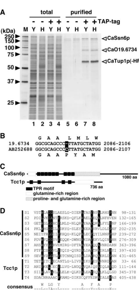
epitope sequence were subjected to TAP procedures, leading to the detection by sodium dodecyl sulfate-polyacrylamide gel electrophoresis of proteins composing a CaTup1p complex (Fig. 1A). We identified three major proteins of the purified protein complex by peptide mass fingerprinting using matrix-assisted laser desorption ionization–time of flight mass spec-trometry. Two proteins approximately 60 kDa and 160 kDa in size were expected to be CaTup1p and CaSsn6p, respectively. There is the possibility that a gel band corresponding to CaTup1p includes tagged or native protein. Peptide finger-printing of the 80-kDa protein demonstrated that this band was presumed to be a novel protein corresponding to CaO19.6734 and Ca19.14026 (http://www-sequence.stanford.edu/group /candida). Since no protein homologous to this novel protein was found in theS. cerevisiae genome database (http://www .yeastgenome.org/) or other fungal genome databases, we called Tcc1p the Tup1p-complex component. Reciprocally, TAP procedures with strain iTCC1-HF or iSSN6-HF, in which a single genomic locus forTCC1or CaSSN6was tagged with His6-FLAG, identified CaTup1p as a binding protein (data not
shown). At the start, we used the database sequence of CaO19.6734 to construct a C terminus-tagged protein. How-ever, we could not detect a tagged protein by Western blotting. In fact, experimental nucleotide sequencing of a PCR-ampli-fied DNA fragment corresponding to CaO19.6734 revealed that the sequence that we had determined had one base pair insertion compared to the coding sequences of CaO19.6734 in theCandidagenome database; this frameshift leads to differ-ent lengths of the deduced polypeptides (Fig. 1B). Since we were able to detect an appropriate size for the Tcc1p protein epitope tagged at the C terminus based on the newly deter-mined sequences (accession no. AB252688), we used this sequence for further analysis.
On the basis of the nucleotide sequences that we deter-mined, the deduced open reading frame encoded a polypep-tide of 736 amino acids with a calculated molecular mass of 80,177 Da. Tcc1p contains four copies of the TPR motif in the N-terminal portion and glutamine-rich regions in the C-termi-nal portion (Fig. 1C). In general, the TPR motif is involved in
FIG. 1. Identification of a protein encoded by Orf19.6734 as a component of the CaTup1p complex. (A) The CaTup1p complex was purified by tandem affinity purification (⫺or⫹TAP-tag) using anti-FLAG and Ni-nitrilotriacetic acid (Ni-NTA) agarose. Protein fractions of crude extracts (lanes 1 to 4) and purified samples (lanes 5 to 8) were separated by 10% sodium dodecyl sulfate-polyacrylamide gel electro-phoresis and visualized by silver staining. Open arrowheads indicate the component of the CaTup1p complex identified by matrix-assisted laser desorption ionization–time of flight mass spectrometry. Yeast cells (Y) of strains CAF2-1 (lanes 1 and 5) and iTUP1-HF (lanes 3 and 7) were grown at 30°C for 4 h in YPD medium (pH 5.6). Hyphal cells (H) of strains CAF2-1 (lanes 2 and 6) and iTUP1-HF (lanes 4 and 8) were grown at 37°C for 4 h in YPD medium (pH 7.2) containing 10% calf serum. M, molecular size marker. (B) Differences in nucleotide sequences between the database sequence (19.6734) and the analyzed sequence (AB252688). The altered nucleotide is indicated as boxed characters. The nucleotide positions of each open reading frame are indicated on the right. (C) Schematic diagram showing the sequence
conservation of CaSsn6p and Tcc1p. (D) Sequence alignment of the TPR motifs of CaSsn6p and Tcc1p. The numbering on the right side refers to the position of each sequence in the protein. The TPR consensus sequence (3) is shown below the alignment, with the motif numbering indicated below the sequence. Eight amino acid residues (W/L/F, L/I/M, G/A/S, Y/L/F, A/S/E, F/Y/L, A/S/L, and P/K/E) show a high frequency of conservation. Boxed residues in the alignment indicate amino acids matching the consensus sequence shown below.
on September 8, 2020 by guest
http://ec.asm.org/
protein-protein interaction (10). C. albicans Ssn6p also has nine copies of the TPR motif; the amino acid alignment of the TPR motif in the sequences of Tcc1p and CaSsn6p is shown in Fig. 1D with TPR consensus sequences. In addition, although Tcc1p was predicted to be a nuclear protein because it inter-acted with CaTup1p, it was found to contain neither nuclear localization signals nor nuclear exporting signals. Thus, Tcc1p, which was identified as a CaTup1p-binding partner, is a novel
C. albicans-specific protein with a TPR motif.
Tcc1p and CaSsn6p interact independently with CaTup1p.
To confirm that CaTup1p and Tcc1p bind to each other and to analyze the relationships among CaTup1p, Tcc1p, and CaSsn6p, we constructed a strain in which CaTup1p, Tcc1p, and CaSsn6p were tagged with disparate epitope tags (His6
-FLAG, 3⫻HA, and 3⫻Myc, respectively) and performed im-munoprecipitation, followed by Western blotting. We also con-structed three strains, in each of which His6-FLAG-tagged
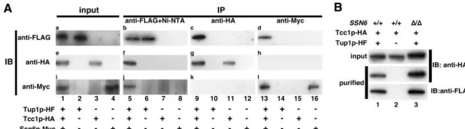
CaTup1p (iTup1p-HF), 3⫻HA-tagged Tcc1p (iTcc1p-HA), or 3⫻Myc-tagged CaSsn6p (iSsn6p-Myc) was expressed under the control of each native promoter. Western analysis con-firmed that each protein was expressed with each epitope tag as expected (Fig. 2A, lanes 1 to 4). Cell extracts from each strain were then subjected to tandem affinity purification. Western blotting using anti-FLAG, anti-HA, and anti-Myc an-tibodies with the immunoprecipitant from each strain demon-strated that Tcc1p-HA and CaSsn6p-Myc were detected in the fraction of strain 3TAG-TTS (Fig. 2A, lane 5), indicating that the CaTup1p binds to Tcc1p and CaSsn6p. When a similar immunoprecipitation using anti-HA antibody agarose was per-formed, only CaTup1p-HF and Tcc1p-HA were detected and CaSsn6p-Myc was below the detectable level (Fig. 2A, lane 9). Similarly, an immunoprecipitation experiment using anti-Myc antibody agarose showed interaction between CaTup1p-HF and CaSsn6p-Myc and no interaction of Tcc1p-HA (Fig. 2A, lane 13). These reciprocal experiments indicated that CaTup1p-Tcc1p and CaTup1p-CaSsn6p were independent complexes.
To verify this independence more clearly, we investigated
Tcc1p-CaTup1p interaction in a Cassn6deletion mutant back-ground. For this purpose, tandem affinity purification was per-formed using the wild type and Cassn6⌬ cells expressing Tcc1p-HA and CaTup1p-HF (Fig. 2B). Western blotting using anti-HA antibody revealed that Tcc1p-HA was copurified with CaTup1p-HF, even in the absence of CaSSN6(Fig. 2B, lane 3), suggesting that CaSsn6p might have no effect on CaTup1p-Tcc1p complex formation.
The TPR domain of Tcc1p contributes to interaction with
CaTup1p.To determine which region in Tcc1p is necessary for
CaTup1p binding, we used an immunoprecipitation experi-ment and a yeast two-hybrid system. For immunoprecipitation, a series of His6-FLAG-tagged deletion mutants of the Tcc1p
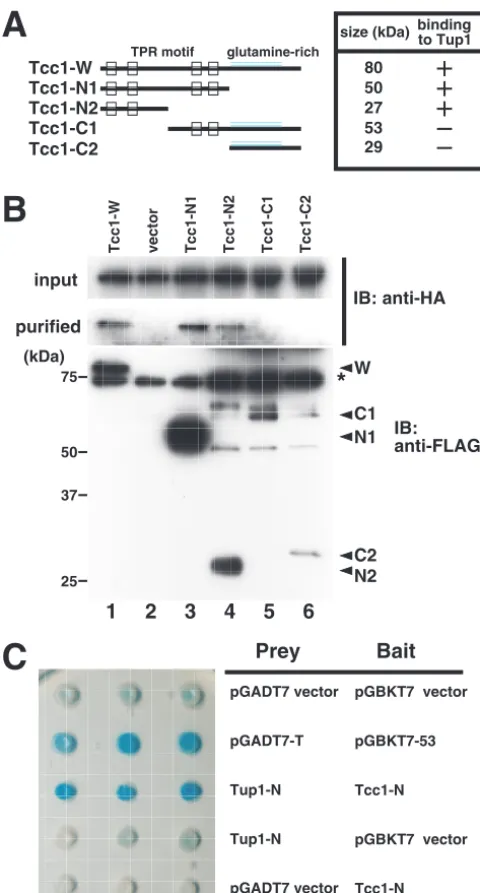
protein were expressed from theACT1orMET3promoter in the C. albicans cells that contain HA-tagged CaTup1p. By performing TAP, the Tcc1p mutant that contains TPR motifs corresponding to the amino acid positions 1 to 475 (Tcc1-N1) or 1 to 250 (Tcc1-N2) was detected as a protein interacting with HA-tagged CaTup1p (Fig. 3B, lanes 3 and 4, respectively). CaTup1p-HA was not detected in the immunoprecipitated complex with the C-terminal domains of Tcc1p, corresponding to amino acid positions 251 to 736 (Tcc1-C1) and 476 to 736 (Tcc1-C2), although only a small amount of the Tcc1p mutant was present (Fig. 3B, lanes 5 and 6). Even when three times as much crude extract as indicated in Fig. 3B was used for puri-fication, no signals of CaTup1p-HA were detected (data not shown). The faint signals of the Tcc1-N2, Tcc1-C1, and Tcc1-C2 deletion mutants could possibly be due to protein instability. These results suggest that the first two TPR do-mains of Tcc1p might serve an important role in binding to CaTup1p, although the possibility that the C-terminal glu-tamine-rich region can bind to CaTup1p could not be denied. To confirm the importance of the TPR domain in the CaTup1p-Tcc1p interaction in vivo, we designed a yeast two-hybrid system. The effect of the repressive activity of CaTup1p was avoided by using the portion of CaTup1p comprising amino acids 1 to 130, which contains no WD repeats, because
FIG. 2. Tcc1p interacts with CaTup1p independently of CaSsn6p. (A) Tcc1p-CaTup1p and CaSsn6p-CaTup1p are contained in different complexes. Immunoprecipitation (IP) and Western blotting (IB) were performed. Cells from strain 3TAG-TTS (lanes 1, 5, 9, and 13), iTUP1-HF (lanes 2, 6, 10, and 14), iTCC1-HA (lanes 3, 7, 11, and 15), and iSSN6-Myc (lanes 4, 8, 12, and 16) were cultured at 30°C for 3 h in YPD medium (pH 5.6). CaTup1p-HF, Tcc1p-HA, and CaSsn6p-Myc were expressed under the control of each native promoter in one cell of the 3TAG-TTS strain. Blots for total proteins (a, e, and i) and immunoprecipitated fractions (b, c, d, f, g, h, j, k, and l) were probed with anti-FLAG (a, b, c, and d), anti-HA (e, f, g, and h), and anti-Myc (i, j, k, and l) antibodies. Immunoprecipitation was performed with anti-FLAG agarose, followed by Ni-NTA agarose (b, f, and j), anti-HA agarose (c, g, and k), and anti-Myc agarose (d, h, and l). Panels e and h and panels i and k were originally derived from the same blot. (B) Tcc1p forms a complex with CaTup1p in Cassn6⌬cells. Cells from 2TAG-TT (lane 1), iTCC1-HA (lane 2), and SSN622 (lane 3) were cultured at 30°C for 3 h in YPD medium (pH 5.6). Then, tandem affinity purification and Western blotting were performed. Blots for total proteins (upper panels) and purified fractions (middle panels) were probed with anti-HA antibody. Blots for purified fractions were probed with anti-FLAG antibody (lower panels).
on September 8, 2020 by guest
http://ec.asm.org/
the full-length CaTup1p fused to the Gal4 DNA-binding do-main itself could repress the reporter activity. The CaTup1p mutant without this N-terminal portion could not bind to Tcc1p in the above-described immunoprecipitation experiment (our unpublished results). It is important to note thatTCC1
contained two CUG codons, which encode serine inC. albicans
but leucine in S. cerevisiae (25), in the sequence coding for amino acid positions 1 to 475. To expressTCC1functionally in
S. cerevisiae, we changed both these codons into TCG by PCR-mediated mutagenesis, and the N-terminal portion of codon-optimized Tcc1p was fused to the Gal4-activating domain. When the Gal4-binding-domain-fused N-terminal portion of CaTup1p (amino acids 1 to 130) and the Gal4-activation do-main-fused N-terminal portion of Tcc1p (amino acids 1 to 475) were simultaneously expressed in a host strain, AH109, that possesses an ␣-galactosidase MEL1 gene as a reporter, the constructed strain took on a blue color on agar medium con-taining X-␣-Gal (5-bromo-4-chloro-3-indolyl-␣-D
-galacto-pyranoside) (Fig. 3C), indicating an in vivo protein-protein interaction. Integrating the results of immunoprecipitation and the yeast two-hybrid assay suggests that the TPR domain of Tcc1p and the N-terminal glutamine-rich domain of CaTup1p contribute to Tcc1p-CaTup1p binding.
Expression profile and nuclear localization of Tcc1p. To
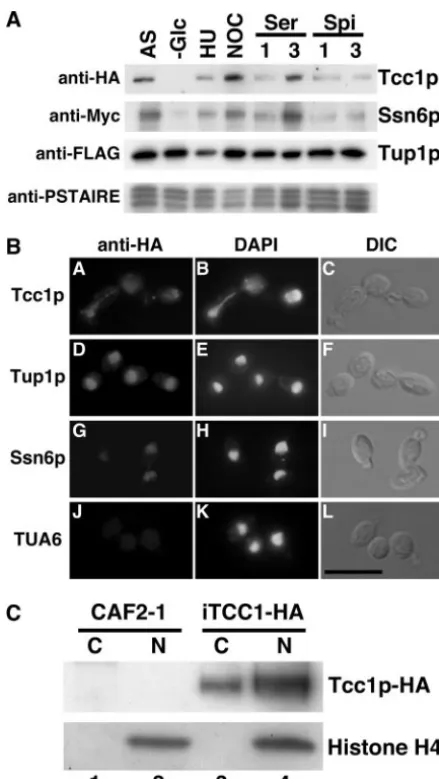
determine whether Tcc1p expression alters in response to nu-trient depletion, cell cycle toxins, or hyphal induction and whether the expression profile overlaps that of CaTup1p, crude extracts were prepared from yeast or hyphal cells of strain 3TAG-TTS in which CaTup1p, Tcc1p, or CaSsn6p was separately tagged in one cell for Western blotting analysis. Western blotting using anti-FLAG antibody demonstrated that CaTup1p was expressed under all conditions examined (Fig. 4A). Tcc1p-HA was not detected in the G1 phase but was
expressed under both yeast and hyphal growth conditions. The expression levels reached a peak when cells were arrested with nocodazole at the G2/M phase. Analysis by anti-Myc antibody
Western blotting showed that the profile of the CaSsn6p-Myc expression was similar to that of Tcc1p-HA. Furthermore, we analyzed the CaTup1p protein complex immunoprecipitated from the samples under each condition. Western blotting with Tcc1p-HA and CaSsn6p-Myc showed that the alterations of protein interacting with CaTup1p were similar to the profiles of expression (data not shown). That is, the independent com-plexes CaTup1p-Tcc1p and CaTup1p-CaSsn6p could exist in similar stages of the cell cycle.
To examine the cellular localization of Tcc1p, CaTup1p, or CaSsn6p, each protein was tagged with three tandem repeats of HA at the C terminus, and cells expressing HA-tagged proteins were immunostained with anti-HA antibody. CaTup1p-HA and CaSsn6p-HA localized in the nucleus as expected (Fig. 4B). Immunostaining with anti-HA antibody also demonstrated nu-clear localization of Tcc1p (Fig. 4B). Under conditions that induce hyphae, while obvious nuclear localization of
CaTup1p-FIG. 3. TPR domain within Tcc1p is necessary for binding to CaTup1p. (A) Diagram of Tcc1p TPR motif in the wild type and the deletion derivatives used for interaction assays. (B) Immunopre-cipitation of CaTup1p-HA by the Tcc1p deletion mutant. Cells from strains DelTCC1-W (Tcc1-W; lane 1), DelTCC1VEC (vector; lane 2), DelTCC1-N1 (Tcc1-N1; lane 3), DelTCC1-N2 (Tcc1-N2; lane 4), DelTCC1-C1 (Tcc1-C1; lane 5), and DelTCC1-C2 (Tcc1-C2; lane 6) were cultured at 30°C for 4 h in YPD medium (pH 5.6). Crude extracts (lanes 1 to 3, 300g; lane 4, 600g; lanes 5 and 6, 1,000g each) were subjected to tandem affinity purification using anti-FLAG agarose and Ni-NTA agarose. Total protein (upper panel) and purified fractions (middle panel) were probed with anti-HA antibody. The same purified fractions were immunoblotted (IB) with anti-FLAG antibody (lower panel). The signals for the deletion mutants (W, N1, N2, C1, and C2) are indicated by arrowheads, while the 75-kDa nonspecific signal is indicated by an asterisk. (C) The interaction between the N-terminal portion of the codon-optimized Tcc1p and the N-terminal portion of CaTup1p was detected in the yeast two-hybrid system. Cell suspensions of strains harboring pGADT7 and pGBKT7 derivative plasmids were spotted onto an X-␣-Gal plate assay. Tup1-N is a pGADT7 derivative plasmid that contains the DNA fragment corresponding to amino acid positions 1 to 130 of CaTup1p. Tcc1-N is a pGBKT7 derivative plasmid
that contains the codon-optimized DNA fragment corresponding to amino acid positions 1 to 475 of Tcc1p. The pGADT7-T and pGBKT7-53 plasmids, which encode the simian virus 40 large T antigen and the murine p53 protein, respectively, served as positive controls.
on September 8, 2020 by guest
http://ec.asm.org/
HA and CaSsn6p-HA was observed, no signal of Tcc1p-HA was detected (data not shown), suggesting that the accumula-tion of Tcc1p in the nucleus might be dependent on morphol-ogy. To confirm the nuclear localization biochemically, subcel-lular fractionation was performed. A successful fractionation was verified by Western blotting probed with anti-histone H4 antibody. Tcc1p-HA was detected in both the nuclear fraction and the cytoplasmic fraction (Fig. 4C). To investigate whether the accumulation of Tcc1p in the nucleus depends on CaTup1p, HA-tagged Tcc1p was expressed in a CaTUP1 con-ditional mutant, in which CaTUP1lies under the regulation of the MET3 promoter, and subcellular fractionation was per-formed. The repression of CaTUP1in the presence of methi-onine and cysteine was verified by microscopic observation and quantitative RT-PCR of CaTUP1andECE1mRNA (data not shown). In the presence of methionine and cysteine (with the
MET3promoter off), CaTUP1expression was 3% of that of the wild type and, as a consequence, a hypha-specific gene,HYR1, was elevated significantly. Even if the expression of CaTup1p was depressed, Tcc1p-HA was detected in both the nucleus and the cytoplasm (data not shown), indicating that the nuclear localization of Tcc1p may not depend on CaTup1p.
tcc1⌬disruptant shows cell elongation phenotype.To
inves-tigate the cellular functions of Tcc1p inC. albicans, we deleted both copies. If Tcc1p functions as a CaTup1p-mediated tran-scriptional repressor, phenotypes of thetcc1⌬mutant would at least partly overlap those of the Catup1⌬ mutant. The two copies ofTCC1were sequentially replaced withARG4and a 200-bp portion ofhph(hph200) in strain TUA4. The ability to generate a viable tcc1/tcc1 null mutant strain indicates that
TCC1 is not an essential gene in C. albicans. There are no significant differences between the wild type and the tcc1⌬
mutant in their susceptibilities to fluconazole, calcofluor white, sodium chloride, or hydrogen peroxide (data not shown), in-dicating thatTCC1might not play an important role in drug or stress resistance. To confirm that the loss of theTCC1function was responsible for any of the observed phenotypes, a PCR-amplified fragment containing aTCC1complete open reading frame was used to replace thehph200locus of thetcc1⌬ mu-tant TCC103 to generate the reconstituted strain TCC107, in which Tcc1p is tagged with His6-FLAG at the C terminus. As
a negative control strain, a PCR-amplified fragment containing only a 100-bp C-terminal portion of theTCC1gene was used to replace thehph200locus of TCC103 to generate null mutant TCC106. Both the null mutant TCC106 and the reconstituted strain TCC107, which have a single copy of URA3 at their respectiveTCC1loci, were used for the following experiments to compare phenotypes of morphology and virulence.
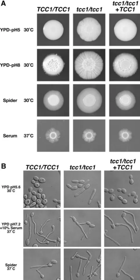
The effect of the TCC1 deletion on the phenotype of the
tcc1⌬mutant was studied under conditions that promote both yeast and hyphal growth inC. albicans(Fig. 5A). On a serum agar medium that induces hyphal growth, there were no sig-nificant differences among the wild type, thetcc1⌬null mutant, and the revertant. On a YPD agar medium adjusted to pH 5, under conditions in which the wild type and the revertant exhibit smooth colonies and the Catup1⌬and Canrg1⌬mutants should show filamentous growth, as reported previously (4, 5, 19), theTCC1disruptant exhibited rough colonies, indicating more filamentous growth in the disruptant. The filamentous phenotype of thetcc1⌬mutant was enhanced by an alkaline
FIG. 4. Expression profile and nuclear localization of Tcc1p. (A) Western blotting for the detection of Tcc1p-HA. 3TAG-TTS cells expressing CaTup1p-HF, Tcc1p-HA, or CaSsn6p-Myc from each na-tive promoter were used. Each lane was processed using a total cell extract with the following culture conditions: AS, asynchronous cells cultured for 3 h at 30°C;⫺Glc, unbudded cells collected in yeast nitrogen base medium (without glucose); HU, synchronized cells with 0.1 M hydroxyurea at the G1/S phase; NOC, synchronized cells with 20
g/ml nocodazole at the G2/M phase; Ser, hyphal cells cultured in YPD containing 10% serum for 1 or 3 h (represented by 1 and 3); Spi, hyphal cells cultured in Spider medium for 1 or 3 h (represented by 1 and 3). After cells were grown under each condition, Western blotting using anti-FLAG, anti-HA, or anti-Myc antibody was performed. Western blotting using anti-PSTAIRE antibody was performed as a loading control. (B) Fluorescence microscopy for the detection of Tcc1p-HA. TUA4 cells expressing HA-tagged Tcc1p, CaTup1p, and CaSsn6p were grown overnight at 30°C, inoculated into fresh YPD (pH 5.6) medium, fixed in 3% formaldehyde, treated with anti-HA for a primary antibody and anti-rabbit immunoglobulin G antibody con-jugated with Alexa Fluor 594 for a secondary antibody, and viewed with a fluorescence microscope and differential interference contrast optics. Immunostained, 4⬘,6⬘-diamidino-2-phenylindole (DAPI)-stained, and differential interference contrast images in the same field of view are shown. Bar, 10m. (C) Subcellular fractionation was analyzed by West-ern blotting with anti-HA and anti-histone H4 antibodies. Cell extracts from strains CAF2-1 (not tagged; lanes 1 and 2) and iTCC1-HA (Tcc1p-HA; lanes 3 and 4) were separated into cytoplasmic fractions (lanes 1 and 3) and nuclear fractions (lanes 2 and 4).
on September 8, 2020 by guest
http://ec.asm.org/
condition. In addition, on Spider medium that induces filamen-tation, the growth zone of the tcc1⌬ mutant that indicates filamentation was larger than that of the wild type. These observations suggest that the deletion ofTCC1may enhance the filamentation ofC. albicansunder the conditions examined in this study.
We then observed cell morphology in liquid yeast- and hypha-inducing media (Fig. 5B). In the YPD medium that sup-ports yeast growth, cell elongation was observed with tcc1⌬
cells, whereas the length oftcc1⌬cells was shorter than the previously reported lengths of Catup1⌬and Canrg1⌬cells (4, 5, 19). Calculation of the axial growth rates (length/width) sup-ported the fact thattcc1⌬ cells exhibited the cell elongation phenotype during yeast growth (TUA6, 1.18⫾0.31 [n⫽204]; TCC106, 2.47⫾0.85 [n⫽221]; and TCC107, 1.44⫾0.26 [n⫽
206]). Additionally,tcc1⌬cells did not exhibit a Catup1⌬-like constitutive filamentation without constriction. There were no significant differences between the wild type and the tcc1⌬
mutant grown in Spider medium or in serum medium, which induces hyphal growth. The phenotypes described above were restored in the reconstituted strain TCC107, indicating that filamentous phenotypes were caused solely by the deletion of theTCC1gene.
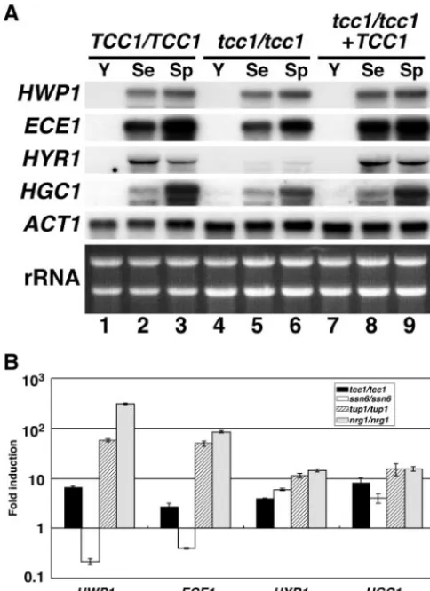
Effects of TCC1disruption on the transcription of
hypha-specific genes. To determine whether the transcription of
HSGs is induced under yeast growth conditions byTCC1 dis-ruption as well as by CaTUP1 or CaNRG1 disruption, we compared the expression levels of HSGs such asHWP1(28),
ECE1(2),HYR1(1), andHGC1(34) by Northern hybridiza-tion and quantitative real-time RT-PCR. If Tcc1p served as a transcriptional repressor for HSGs, as does CaTup1p, HSGs would be upregulated by Tcc1p depletion. The wild type, the disruptant, and the reconstituted strain were cultured under yeast or hyphal growth conditions and subjected to Northern blotting analysis (Fig. 6A). A comparison of yeast growth (Fig. 6A, lane 1) and hyphal growth (Fig. 6A, lane 2 or 3) confirmed the successful detection of HSGs. There were no significant differences between the wild type and thetcc1⌬disruptant in the HWP1, the ECE1, or the HGC1 gene expression levels, based on Northern analysis. The expression ofHYR1was de-creased by TCC1 deletion under hyphal growth conditions, consistent with previous data indicating thatHYR1mRNA in the Catup1⌬mutant under such conditions was lower than that in the wild type (15). This implies that the CaTup1p-Tcc1p complex might regulate the activation or repression of a tran-scriptional repressor in the HYR1 gene during filamentous growth. However, Northern analysis indicated that the yeast growth conditions did not seem to induce any significant HSGs.
As described above, the filamentous phenotype of thetcc1⌬
mutant was less than that of the Catup1⌬or Canrg1⌬mutant (Fig. 5). Therefore, since it remains a possibility that derepres-sion byTCC1deletion might be weaker than that by CaTUP1
or CaNRG1deletion, we attempted to detect the expression of HSGs by quantitative real-time RT-PCR, which has a higher sensitivity than Northern analysis. Compared to the results reported so far, theHWP1andECE1mRNA were derepressed in Catup1⌬and Canrg1⌬cells but not in the Cassn6mutant (Fig. 6B), consistent with the report by Garcia-Sanchez et al. (12). Interestingly,HYR1mRNA andHGC1mRNA were de-repressed in Catup1⌬ and Canrg1⌬ cells and even in the Cassn6⌬cells. However, Garcia-Sanchez et al. (12) reported thatHYR1mRNA was not elevated in Canrg1⌬and Cassn6⌬
cells and thatHGC1mRNA was not elevated in Cassn6⌬cells. These discrepancies are probably caused by an experimental difference between microarray analysis and quantitative
RT-FIG. 5. Morphology ofC. albicansstrains grown on solid agar me-dium (A) and in liquid meme-dium (B). (A) Cells from strains TUA6 (TCC1/TCC1), TCC106 (tcc1/tcc1), and TCC107 (tcc1/tcc1 TCC1) were grown overnight at 30°C. Then, 106cells were spotted onto the indicated agar plate and grown for 7 days at 30°C on YPD medium and Spider medium and at 37°C on agar medium containing 10% serum. (B) Cells from strains TUA6 (TCC1/TCC1), TCC106 (tcc1/tcc1), and TCC107 (tcc1/tcc1 TCC1) were grown for 2 h under the conditions indicated at the left. Bar, 10m.
on September 8, 2020 by guest
http://ec.asm.org/
PCR. These results indicate that HWP1/ECE1 and HYR1/
HGC1might be transcriptionally regulated in a different man-ner and that CaSsn6p may regulate HYR1 and HGC1
negatively. Moreover, all HSGs tested were derepressed by the deletion ofTCC1, but the expression level of theTCC1mutant was lower than that of the Catup1⌬or Canrg1⌬mutant (Fig. 6B). Therefore, these results further support that Tcc1p, ac-companied by CaTup1p, might function as a transcriptional repressor involved in the morphological transition ofC. albi-cans.
The filamentous phenotype of thetcc1⌬mutant is restored
by HGC1disruption. In the previous paragraph, we
demon-strated that Tcc1p might repress the expression of the hypha-specific G1 cyclin Hgc1p under conditions that induce yeast
growth. The concept thatHGC1acts downstream of CaTup1p
regulation has been supported by the previous report that constitutive filamentation is blocked by the deletion ofHGC1
(34). To examine whether Tcc1p is also linked to the function ofHGC1, we deletedHGC1together withTCC1. The obser-vation that the hgc1⌬ single mutant was deficient in hyphal formation (Fig. 7) was consistent with a previous report (34). The double mutant did not show filamentation on a serum medium (Fig. 7), in that the deletion ofTCC1did not induce filamentation ofhgc1⌬cells and the deletion ofHGC1reduced filamentation of tcc1⌬ cells. On a YPD agar medium, the deletion of HGC1 eliminated the filamentous phenotype of
tcc1⌬cells as well as that of Catup1⌬cells. This reinforced the idea that CaTup1p and Tcc1p might coordinate in regulating the function ofHGC1, followed by the repression of hyphal formation.
Attenuated virulence in the tcc1⌬disruptant. CaTUP1 or
CaNRG1disruption causes reduced virulence in a mouse model
FIG. 8. Thetcc1⌬mutant exhibits markedly reduced virulence.C. albicans cells from TUA6 (TCC1/TCC1), TCC106 (tcc1/tcc1), and TCC107 (tcc1/tcc1⫹TCC1) were grown overnight in YPD. Each mouse was injected via the tail vein with 106CFU and monitored for survival.
FIG. 6. Expression of hypha-specific genes in thetcc1⌬ mutant. (A) Northern blotting. Cells were grown for 3 h in the indicated media (Y, YPD, pH 5.6, at 30°C; Se, YPD, pH 7.2, plus 10% calf serum at 37°C; Sp, Spider medium at 37°C). RNA was prepared from each strain, and Northern analysis was performed using probes for the indicated genes. The ethidium bromide-stained rRNAs are presented below the hybridization patterns to demonstrate comparable loading. (B) Quantitative real-time RT-PCR analysis of hypha-specific mRNAs. Strains TUA6 (the wild type), TCC106 (tcc1/tcc1), SSN602 (ssn6/ssn6), TUP102 (tup1/tup1), and NRG102 (nrg1/nrg1) were used. Values normalized toACT1mRNA and fold induction relative to the values of the TUA6 wild-type strain are shown for four HSGs (HWP1, ECE1, HYR1, and HGC1). Data are shown as means ⫾ standard deviations of results from at least three experiments.
FIG. 7. Filamentation of thetcc1⌬mutant was restored byHGC1 disruption. Cells from strains TUA6 (TCC1/TCC1 HGC1/HGC1), TCC106 (tcc1/tcc1 HGC1/HGC1), HGC104 (TCC1/TCC1 hgc1/hgc1), and HGC114 (tcc1/tcc1 hgc1/hgc1) were grown overnight at 30°C. Then, 106cells were spotted onto the indicated agar plate and grown for 7 days at 30°C on YPD medium and at 37°C on agar medium containing 10% serum. Photographs of colony edges were taken with a phase-contrast microscope at⫻20 magnification.
on September 8, 2020 by guest
http://ec.asm.org/
of systemic candidiasis (19). To determine whether Tcc1p plays an important role in pathogenicity, mice were intravenously in-jected with the wild type (TUA6), the tcc1⌬ null mutant (TCC106), and the revertant (TCC107) and monitored for sur-vival. We found that mice infected with 106CFU of thetcc1⌬
mutant did not die until 31 days postinfection, with a sustained-survival curve, whereas the same-size inocula of the wild-type or the reconstructed strain killed all infected mice within 11 days (Fig. 8). The mean survival times for the mice infected with the wild type, the null mutant, and the revertant were 7.5⫾ 3.78 days, 21.2⫾8.07 days, and 9⫾2.35 days, respectively (TCC106 versus TUA6,P⬍0.01; TCC106 versus TCC107,
P ⬍ 0.02). This demonstrates that TCC1is involved in C. albicans virulence and supports, in part, the concept that Tcc1p might function with CaTup1p inC. albicans.
DISCUSSION
In this study, we identified proteins that form complexes with CaTup1p, a global transcriptional repressor. Analogous to theS. cerevisiae homolog, CaSsn6p has long been regarded as a CaTup1p-binding partner for mediation of the negative regula-tion of yeast-hypha morphological transiregula-tion inC. albicans. How-ever, whether CaSsn6p actually interacts with CaTup1p remains unproven. Furthermore, a recent report (12) in which DNA mi-croarray analysis was performed to compare the wild type and the Cassn6mutant indicates that genes repressed by CaTup1p do not necessarily correspond to those repressed by CaSsn6p. In other words, it is highly possible that CaSsn6p may not be a CaTup1p-binding partner, at least in the morphological transition ofC. albicans. In order to isolate a real binding partner that chiefly regulates morphogenesis, we used the so-called TAP technique, which we previously used for the purification of a septin complex (16). We found that complexes purified by the TAP technique contained CaSsn6p (Fig. 1). Moreover, immunoprecipitation/ Western analysis of strains expressing CaTup1p and CaSsn6p tagged with a different epitope demonstrated that CaSsn6p inter-acted with CaTup1p (Fig. 2). Therefore, we were able to provide evidence that CaTup1p and CaSsn6p of C. albicans formed a protein complex, as did those ofS. cerevisiae.
To identify a binding partner that actually regulates the mor-phology of C. albicans together with CaTup1p, we purified a protein complex including CaTup1p. One of the components contained in the CaTup1p complex was identified asTCC1, a
Tup1p-complexcomponent. In addition, we demonstrated that CaTup1p independently interacted with Tcc1p or CaSsn6p. How-ever, Tcc1p and CaSsn6p share common properties and behav-iors as described below. Their first similarity is the existence of a TPR domain: Tcc1p contains four copies of the TPR motif, and nine TPRs are located within theC. albicansSsn6p polypeptide. Since both Tcc1p and CaSsn6p possess the TPR motif, Tcc1p probably interacts with CaTup1p in a mode similar to that of CaSsn6p. Actually, we demonstrated that the TPR domains of Tcc1p are responsible for CaTup1p binding by using immunopre-cipitation and yeast two-hybrid analysis (Fig. 3). Their second behavior in common is their expression profiles. The expression levels of Tcc1p and CaSsn6p, which were tagged with different epitopes in one cell, were altered in response to cell cycle toxins and glucose depletion: the expression levels reached a peak when cells were arrested at the G2/M phase and decreased to an
un-detectable level in the unbudded G1cells (Fig. 4A). In addition,
the complex formation with CaTup1p in response to cell cycle toxins was no different between Tcc1p and CaSsn6p (data not shown). Their third similarity is localization to the nucleus. Im-munostaining with anti-HA antibody demonstrated that Tcc1p and CaSsn6p localize to the nucleus (Fig. 4B), although both were predicted to contain no nuclear localization signals. Whereas sub-cellular fractionation further supports the nuclear localization of Tcc1p, Tcc1p was also detected in the cytoplasmic fraction. Tcc1p localization to the nucleus was not altered in a CaTUP1-depleted cell, suggesting that Tcc1p localization may not depend on CaTup1p. Since the nuclear localization of Tcc1p was not ob-served in hyphal cells, shuttling between the cytoplasm and the nucleus would be regulated by some signaling associated with morphology. Despite these common characteristics, Tcc1p and CaSsn6p seem to function differently: the phenotypes of the mu-tants and the transcriptional control ofHWP1andECE1were contradictory between Tcc1p and CaSsn6p. One of the reasons that the functions of Tcc1p and CaSsn6p have different effects is assumed to be because they might require disparate sequence-specific DNA-binding proteins. Nevertheless, derepression of
HYR1andHGC1mRNA intcc1⌬and Cassn6⌬cells might indi-cate the existence of a common DNA-binding protein. Future global transcriptional analysis using a DNA microarray might give clues for solving the question of why the depletion of each protein resulted in opposing phenotypes, despite the common binding partner, the similar expression profiles, and the nuclear localiza-tion. Also, study of a double deletion ofTCC1and CaSSN6would explain how the two products share roles.
The Catup1⌬null mutant demonstrates constitutive filamenta-tion even under condifilamenta-tions that induce the yeast form (4). The Cassn6⌬homozygous mutant shows a higher rate for phenotype switching (12) and no filamentation on serum or Spider agar medium (data not shown). When both alleles ofTCC1identified in this study were deleted, the disruptant grew in a pseudohyphal form under conditions that induce the yeast form. Obviously, the morphological phenotype of tcc1⌬ is more similar to that of Catup1⌬than to that of Cassn6⌬from the viewpoint of the cell elongation phenotype. Taken together with the results of the immunoprecipitation experiment and morphological observation, it is highly possible that the protein complex consisting of CaTup1p and Tcc1p might behave independently of CaTup1p-CaSsn6p. However, the filamentous phenotype of the tcc1⌬
mutant was not as severe as that of the Catup1⌬or Canrg1⌬
mutant, and thetcc1⌬ mutant showed a relatively mild cell elongation. This reduced severity is probably associated with the degrees of HSG repression. For example, the elevation of derepressed HSG transcription by the deletion ofTCC1was not stronger than that in the Catup1⌬or Canrg1⌬mutant (Fig. 6). In addition, although we did not compare them directly, the virulence demonstrated by the tcc1⌬, Catup1⌬, or Canrg1⌬
mutant indicates a decrease or attenuation: alltcc1⌬ mutant-infected mice were killed within 31 days (Fig. 8), while almost no mice inoculated with the strain from which CaTUP1 or CaNRG1was deleted were dead within the time period exam-ined (4, 5, 19). These results suggest that the degree of cell elongation, the increased quantity of HSG transcription, and the attenuated virulence, which are caused by gene disruption, are closely connected to each other. While the Catup1⌬mutant was demonstrated to be more sensitive to hydrogen peroxide