*Corresponding author: E-mail: ([email protected]), ([email protected]), Department of Chemistry, Faculty of Science, Golestan University, Gorgan, Iran, Tel:+981732245882
Chemical Methodologies 3(2019) 643-650
Chemical Methodologies
Journal homepage: http://chemmethod.com
Original Research article
Vanadium(IV) Schiff base Complex: Synthesis,
Characterization, Crystal Structure and Thermal
Decomposition into V
2O
5Particles
Aliakbar Dehno Khalajia, Maryam Ghorbanib, Seyyed Javad Peyghounb, Norollah Feizib, Alireza Akbarib, Wolfgang Hornfeckc, Michal Dusekc, Vaclav Eignerc
aDepartment of Chemistry, Faculty of Science, Golestan University, Gorgan, Iran bDepartment of Chemistry, Payame Noor University, PO Box 19395-3697 Tehran, Iran
cInstitute of Physic of the Czech Academy of Sciences, Na Slovance 2, 182 21 Prague, Czech Republic
A R T I C L E I NF O R MA T I O N A B S T R A C T
Received: 23 April 2019
Received in revised: 23 May 2019 Accepted: 01 June 2019
Available online: 29 July 2019
DOI: 10.33945/SAMI/CHEMM.2019.6.3
Vanadium(IV) complex [VO((MeO-bph)2en)] (1), (MeO-bph)2en=N,N
-bis(2-hydroxy-4-methoxybenzophenone)-1,2-ethanediamine, was synthesized and characterized by elemental analyses (CHN), FT-IR spectroscopy, thermogravimetry, SEM and single crystal X-ray diffraction. The title complex 1 was prepared by the reaction of VO(acac)2, 1,2-ethanediamine
and 2-hydroxy-4-methoxybenzophenone (molar ratio 1:1:2). The single-crystal X-ray analysis of 1 shows that the vanadium(IV) ion is located in a distorted square pyramid (N2O3) environment with the tetradentate Schiff
base ligand coordinated in equatorial positions and one oxygen atom in the axial position. Thermogravimetric analysis shows that the complex 1
decomposes in two stages. Finally, the complex was calcinated at 500 C for 3 h and the V2O5 products characterized by FT-IR and SEM.
KEYWORDS
Vanadium(IV) complex Distorted square pyramid Spectroscopy
Graphical Abstract
Introduction
Schiff basses have played an important role in the development of the V(IV) complexes, because they are capable of giving the complexes interesting structural features, suitable properties and applications [1-3]. The interest in the vanadium Schiff base complexes has grown due to their catalytic applications and biological activity [4-6]. The vanadium(IV) Schiff base complexes are colored either green with a monomeric structure [7] or orange with a polymeric linear chain structure [8]. Recently, Grivani et al., have focused on the coordination chemistry of vanadium Schiff base complexes because of their structural features and catalytic application [7-10]. In this paper, mononuclear vanadium(IV) Schiff base complex [VO((MeO-bph)2en)] (1) (Scheme 1) was prepared and characterized.
Scheme 1. The preparation procedure of tetradentate Schiff base ligand (MeO-bph)2en and its vanadium(IV)
complex
Experimental
General
results agreed with calculated values. FT-IR spectra were recorded as a KBr disk on a FT-IR PerkinElmer spectrophotometer. The TG/DTA were performed on a PerkinElmer TG/DTA lab system 1 (technology by SII) in argon atmosphere with a heating rate of 20 °C/min in the temperature span of 50–800 °C. The scanning electron microscopy (SEM) images were obtained from a Philips XL-30ESEM.
Preparation of 1
A solution of 1,2-ethanediamine (0.1 mmol) in 15 mL methanol is added drop-wise to a methanolic solution of 2-hydroxy-4-methoxybenzophenone (0.2 mmol) under stirring condition. The reaction mixture is then refluxed for 1 h during which the solution color turns yellow. Then, a solution of VO(acac)2 (0.1 mmol in 15 mL methanol) was added drop-wise. After the addition was completed, the stirring was continued at reflux for 3 h. After several days by slow evaporation of solvent, dark green crystals of the complex were filtered and washed with cold methanol and dried at room temperature. Anal. Calcd. for C30H26N2O5V: C, 65.99.; H, 4.77.; N, 5.13%. Found; C, 66.04.; H, 4.82.; N, 5.08%. FT-IR (KBr, cm-1): 2925-3033 (CH aromatic and aliphatic), 2844 (CH=N), 1587 (C=N), 1465-1542 (C=C aromatic).
Preparation of V2O5 particles
About 0.5 g of the title compound was loaded into a platinum crucible and placed in an oven and heated at a rate of 10 C min-1 in air. The black product was obtained at 500 after 3 h, washed with ethanol to remove impurities, dried at room temperature and characterized by FT-IR and SEM.
X-ray structure determination
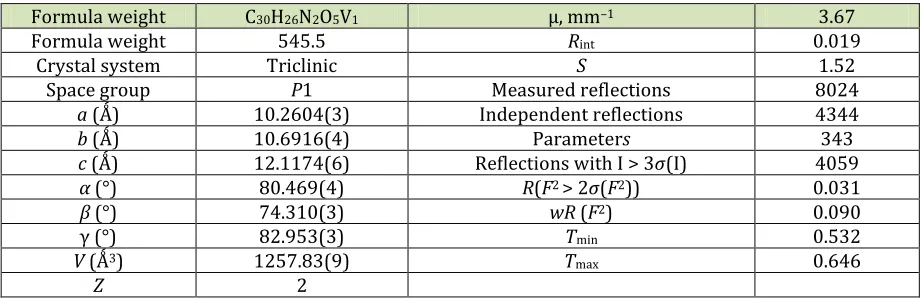
Crystallographic data and details of the data collection and structure solution and refinements are listed in Table 1.
Results and discussion
Syntheses and characterization
From the one-pot reaction of VO(acac)2, 1,2-ethanediamine and 2-hydroxy-4-methoxybenzophenone in methanol, vanadium(IV) Schiff base complex [VO((MeO-bph)2en)] (1) was prepared and characterized. In the FT-IR spectra of 1, the band at 1580 cm-1 was assigned to -C=N-group. Also, another band at 945 cm-1 was assigned to V=O group (Grivani et al., 2015). The vibrational frequency of the OH group is absent in the complexes (Grivani et al., 2015).
The complex 1 was thermally decomposed in an electric furnace. The FT-IR spectra of the final residue (Figure 1) confirmed the formation of V2O5 [14, 15], by showing peaks at about at 1076, 990, 758 and 434 cm-1 assigned to V=O stretching mode [14]. The peaks at 1609 and 3461 cm-1 in the FT-IR spectra assigned to H2O molecules were adsorbed on the surface of particles [16].
Table 1. Crystallographic data and structural refinement details of 1
Formula weight C30H26N2O5V1 µ, mm–1 3.67
Formula weight 545.5 Rint 0.019
Crystal system Triclinic S 1.52
Space group P1 Measured reflections 8024
a (Ǻ) 10.2604(3) Independent reflections 4344
b (Ǻ) 10.6916(4) Parameters 343
c (Ǻ) 12.1174(6) Reflections with I > 3σ(I) 4059
α (°) 80.469(4) R(F2 > 2σ(F2)) 0.031
β (°) 74.310(3) wR (F2) 0.090
γ (°) 82.953(3) Tmin 0.532
V (Ǻ3) 1257.83(9) Tmax 0.646
Z 2
The TG curve of 1 (Figure 2) shows that there is no detectable change up to 130 C, and then during further heating the complex undergoes decomposition in three stages (130–285 (11.5%), 285-400
Figure 1. FT-IR spectrum of the V2O5 particles obtained from 1
Figure 2. TG curve of 1
The morphology of the vanadium(IV) Schiff base complex [VO((MeO-bph)2en)] (1) and the obtained V2O5 particles were further investigated by SEM. Figure 3 shows the SEM images of 1 and the obtained particles.
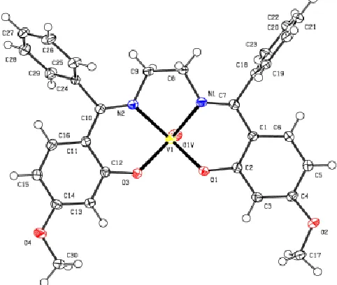
Description of the structure of 1
The coordination sphere of vanadium is completed by two iminic nitrogen atoms N1, N2, two phenoxy oxygen atoms O1, O3 of the Schiff base ligand, and a non-bridging oxo atom O1 v. The V1-N1, V1-N2, V-O1, V1-O3 single bond lengths and the V1=O1 v double bond length are respectively 2.0604 (15), 2.0535 (14), 1.9118 (12), 1.9155 (11) and 1.6015 (14) Å, and they are similar like in other mononuclear square-pyramid vanadium(IV) complexes [7, 8]. The chelating angles N1-V1-N2, N1-V1-O1 and N1-V1-O3 of 80.85 (6), 87.23 (5) and 87.45 (5), respectively, and also other angles around the vanadium center (see Table 2) indicate the distortion of the square-pyramidal geometry around the vanadium atom [7, 8].
(a)
(b)Figure 3. SEM images of 1 (a) and the V2O5 particles obtained from 1 (b)
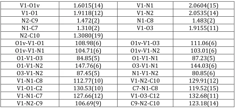
Table 2. Selected bond distances (Å) and angles (°) of 1
V1-O1v 1.6015(14) V1-N1 2.0604(15)
V1-O1 1.9118(12) V1-N2 2.0535(14)
N2-C9 1.472(2) N1-C8 1.483(2)
N1-C7 1.310(2) V1-O3 1.9155(11)
N2-C10 1.3080(19)
O1v-V1-O1 108.98(6) O1v-V1-O3 111.06(6) O1v-V1-N1 104.71(6) O1v-V1-N2 103.01(6) O1-V1-O3 84.85(5) O1-V1-N1 87.23(5) O1-V1-N2 147.76(6) O3-V1-N1 144.03(6)
O3-V1-N2 87.45(5) N1-V1-N2 80.85(6)
V1-N1-C8 112.77(10) V1-N2-C10 129.91(12) V1-O1-C2 130.53(10) C7-N1-C8 119.52(15) V1-N1-C7 127.66(12) V1-O3-C12 132.68(11) V1-N2-C9 106.69(9) C9-N2-C10 123.18(14)
Conclusions
In conclusion, we synthesized the mononuclear complex [VO((MeO-bph)2en)] (1) in simple procedure. X-ray result showed that the vanadium center had a distorted square pyramid geometry. In addition, thermal decomposition of the complex was investigated at the temperature about 600 °C. Finally, the analysis (FT-IR and SEM) of the obtained solid showed the formation of the V2O5 particles.
Acknowledgments
We are grateful to the Payame Noor and Golestan University for financial support of this work. The crystallography was supported by the project 15-12653S of the Czech Science Foundation using instruments of the ASTRA lab established within the Operation program Prague Competitiveness - project CZ.2.16/3.1.00/24510.
Supplementary data
Crystallographic data (excluding structure factors) for the structures reported in this paper has been deposited with the Cambridge Crystallographic Center, CCDC No. 1543319 (1). Copies of the data can be obtained free of charge on [email protected] or http:www.ccdc.cam.ac.uk.
References
[1] Leelavathy L., Anbu S., Kandaswamy M., KarthikeyanN., Mohan N. Polyhedron, 2009, 28:903 [2] Rayati S., Ghaemi A., Sadeghzadeh N. Cat. Commun., 2010, 11:792
[4] Rayati S., Torabi N., Ghaemi A., Mohebbi S., Wojtczak A., Kozakiewicz A. Inorg. Chim. Acta, 2008,
361:1239
[5] Ando R., Ono H., Yagyn T., Maeda M. Inorg. Chim. Acta, 2004, 357:2237
[6] Y. Dong Y., R.K. Narla R.K., E. Sudbeck E., F.M. Uckun F.M. J. Inorg. Biochem., 2000, 78:321 [7] Grivani G., Ghavmi A., Eigner V., Dusek M., Khalaji A.D. Chin. Chem. Lett., 2015, 26:779
[8] Grivani G., Delkhosh S., Fejfarova K., Dusek M., Khalaji A.D. Inorg. Chem. Commun., 2013, 27:82 [9] Grivani G., Khalaji A.D., Tahmasebi V., Gotoh K., Ishida H. Polyhedron, 2012, 31:265
[10] Grivani G., Tahmasebi V., Khalaji A.D. Polyhedron, 2014, 68:144 [11] Palatinus L., Chapuis G. J. Appl. Crystallogr., 2007, 40:786 [12] Petricek V., Dusek M., Palatinus L. Z. Kristallogr., 2014, 229:345
[13] Diamond - Crystal and Molecular Structure Visualization. Crystal Impact - K. Brandenburg & H. Putz GbR, Rathausgasse 30, D-53111 Bonn
[14] Farahmandjou M., Abaeiyan N. Int. J. Bio-Inorg. Hybr. Nanomater., 2015, 4:243 [15] Alan I., Kriza A., Badea M., Stanica N. J. Therm. Anal. Calorim. 2013, 111:483 [16] Khalaji A.D., Nikookar M., Das D. J. Therm. Anal. Calorim. 2014, 115:409