Review
Osteoclasts and Microgravity
J. Kelly Smith
Departments of Academic Affairs and Biomedical Sciences, James H Quillen College of Medicine, East Tennessee State University, PO Box 70300, Johnson City, TN 37614, USA.
Correspondence: [email protected]; Tel: +1-423-794-7425
Abstract: Astronauts are at risk of losing 1.0 to 1.5% of their bone mass for every month they spend in space despite their adherence to high impact exercise training programs and dietary regimens designed to preserve their musculoskeletal system. This loss is the result of microgravity-related impairment of osteocyte and osteoblast function and the consequent upregulation of osteoclast-mediated bone resorption. This review describes the ontogeny of osteoclast hematopoietic stem cells, the contributions of macrophage colony stimulating factor, activator of NFkB and the calcineurin pathways make in osteoclast differentiation, and provides details of bone formation, the osteoclast cytoskeleton, the immune regulation of osteoclasts, and osteoclast mechanotransduction on Earth, in the microgravity of space, and in conditions of simulated microgravity. The article discusses the need to better understand how osteoclasts are able to function in zero gravity and reviews current and prospective therapies that may be used to treat osteoclast-mediated bone disease.
Keywords: Osteoclasts; microgravity; spaceflight; osteoblasts; osteocytes; M-CSF; RANKL; bone; microgravity; cytokines
1. Introduction
The skeletal system of vertebrates has had millions of years to adapt to the force of gravity on Earth (9.8 m/sec2) and to allow osteocytes and components of the innate and adaptive immune system to balance the activities of bone forming osteoblasts and bone resorbing osteoclasts. This osteoimmunological system is complex and involves commonly shared osteoclastogenic factors such as receptor activator of NF-kB ligand (RANKL) and macrophage colony stimulating factor (M-CSF), as well as cytokines and immune cells that inhibit or enhance osteoclast ontogeny [1]. This adaptation has involved the construction of cytoskeletons supported by actin and intermediate filaments and microtubules [2,3], intracellular adhesion molecules such as integrins, matrix clusters of cell extension kinases [4,5], mechanosensors shuttling between plasma membranes and nuclei [6], and the use of thermal convection in which heated fluids rise to the top of the gravity vector and then are exchanged by cooler fluids, establishing a convection system that dissipates heat, renews nutrient supplies, and removes waste materials [7].
Man’s venture into the vacuum of space where the force of gravity is one millionth of that on Earth has resulted in adverse effects on the osteoimmunological system, particularly bone homeostasis [8]. Astronauts are at risk of losing 1.0 to 1.5% of their bone mass for every month they spend in space despite their adherence to high impact exercise programs and diets high in nutrients, potassium, calcium, and vitamin D, all designed to preserve the skeletal system [9-13]. Unfortunately, the adverse effects of space travel on the skeletal system may last for years after returning to Earth [8].
In this review, I describes the ontogeny of osteoclast hematopoietic stem cells, the contributions of macrophage colony stimulating factor, activator of NFkB and the calcineurin pathways make in osteoclast differentiation, and provide details of bone formation, the osteoclast cytoskeleton, the
immune regulation of osteoclasts, and osteoclast mechanotransduction on Earth, in the microgravity of space, and in conditions of simulated microgravity. The article discusses the need to better understand how osteoclasts are able to function in zero gravity and reviews current and prospective therapies that may be used to treat osteoclast-mediated bone disease.
2.0. Osteoclast Stem Cells
2.1. Osteoclast Stem Cell Niche
Goto and associates have provided evidence that a small population of CXCR4+ CD45- bone marrow cells provide a niche for osteoclastogenesis. These cells express low levels of receptor activator of NFkB (RANK) and its ligand RANKL, but high levels of essential chemokines including stromal cell derived factor 1 (SDF-1), chemokine X-C motif) ligand 7 (CXCL7), and chemokine (C-X3-C motif) ligand 1 (CX3CL1). Their findings suggest that CXCR4+ CD45- cells support an appropriate microenvironment for osteoclastogenesis with a direct effect on cells expressing SDF-1, CXCL7, and CX3CL1 receptors [14].
2.2. Osteoclast Stem Cell Circulation
Bone marrow osteoclast hematopoietic stem cells (HSC) expressing type-2 receptors for sphingosine-1-phosphate (S1PR2) enter the circulation by binding sphingosine-1-phosphate (S1P), a chemotactic lysophospholipid normally present in high concentrations in blood [15,16]. Circulating CXCR4 expressing HSC are attracted to bone surfaces by gradients of SDF-1 (CXCL12) secreted by CXCR4+ CD45-, stromal, and endothelial marrow cells [14,17]. HSC may then be recycled to the bone marrow by binding S1P to type-1 receptors (S1PR1) or stay at bone surfaces where they evolve into mature osteoclasts by binding macrophage colony stimulating factor (M-CSF) and RANKL produced primarily by osteoblasts and osteocytes [15,16] (see Figure 1).
RANK+CD45- stromal marrow cells. HSC may then be recycled to the bone marrow by binding S1P
to type-1 receptors (S1PR1) or stay at bone surfaces where they evolve into mature bone resorbing osteoclasts by binding M-CSF and RANKL produced primarily by osteoblasts and osteocytes. .
3.0. Osteoclast Differentiation
3.1. Macrophage Colony Stimulating Factor (M-CSF) Pathway
Differentiation of HSC into osteoclast precursors requires the expression of spleen focus-forming virus proviral integrin 1 (PU.1), heterodimeric complex of micropthalmia-associated transcription factor (MITF), and transcription factor E3 (TfE3) which initiate the expression of the M-CSF receptor C-Fms [18]. Ligation of M-CSF to C-Fms initiates the expression of RANK, the cognate receptor for RANKL. M-CSF ligation also initiates the expression of several cytokines and their receptors including interleukin (IL)-1α, IL-18, interferon (IFN)-β, IL-11Rα2, IL-6/11R gp130, and IFN-γ-R; also expressed are factors involved in the cell’s response to RANKL ligation, including tumor necrosis factor receptor-associated factor 2A (TRAF2A), phosphatidylinositol 3-kinase (PI3K), mitogen-activated protein kinase/extracellular signal-regulated kinase kinase kinase 3 (
MEKK3
), and receptor-interacting serine-threonine kinase 1 (RIPK1) [19].3.2. RANKL Pathway
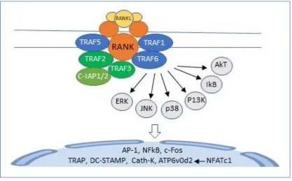
Figure 2. Binding of RANKL to RANK activates TRAFs 1,2,3,5 and 6. TRAF 6 recruits and activates a kinase cascade which includes ERK, JNK, p38, PI3K, IkB and AkT. This cascade initiates the transcription of AP-1, c-Fos, NFkB, and NFATc1 with consequent induction of the osteoclast specific genes, TRAP, cathepsin K, DC-STAMP and ATP6v0d2.
3.3. Calcineurin Pathway
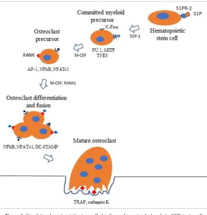
Figure 4. Circulating hematopoietic stem cells binding sphingosine-1-phosphate (S1P) to type 2 receptors (S1PR2) are attracted to bone surfaces by chemokines such as stromal-derived factor-1 (SDF-1). Here they differentiate into committed myeloid precursors expressing PU.1, MITF, and TfE3, transcription factors that induce the expression of C-Fms, the receptor for M-CSF. Binding of M-CSF to C-Fms results the expression of transcription factors AP-1, NFkB, NFATc1, and RANK, the receptor for RANKL. Binding of M-CSF and RANKL to their cognate receptors promotes further differentiation into osteoclast precursors expressing transcription factors NFkB, NFATc1, and DC-STAMP. These cells fuse, forming mature TRAP, cathepsin K positive osteoclasts.
4.0. Osteoclast Cytoskeleton
4.1. Cytoskeleton Elements
The osteocyte cytoskeleton is made of filamentous structures that belong to one of four categories: polarized actin filaments; polarized microtubules; non-polarized intermediate filaments; or non-polarized septin filaments. The cytoskeleton fulfills essential functions including cell adhesion, migration, contractility, division, vesicular transport, and bone resorption [2-6].
4.1.1. The Sealing Zone
In the center of the sealing zone the osteoclast membrane differentiates into a ruffled border where it secretes hydrochloric acid and proteases (cathepsin-K, MMP9 and MMP14) to dissolve hydroxyapatite crystals and bone matrix, respectively [2].
4.1.2. The Actin Cytoskeleton
The core domain of podosomes consists of branched actin that polymerizes below the plasma membrane. The core is surrounded by unbranched actinomysin filaments which connect to integrins and link neighboring podosomes. The core also contains adhesion protein CD44. Although the typical half-life of a podosomes is measured in minutes, the podosomes belt is made of thousands of podosomes and can last for hours [2].
The tyrosine kinase Src is a key controller of podosomes dynamics and organization. Src binds to the tyrosine kinase Pyk2 resulting in their activation and regulation of podosome dynamics largely through small GTPases of the Rho family [2].
4.1.3. Crosstalk between Actin and Microtubular Networks
The actin motor protein unconventional myosin X (Myo10) can bind actin, microtubules, and integrins, and has been proposed to crosslink actin cytoskeleton and microtubules in osteoclasts [2].
4.1.4. Intermediate and Septin Filaments
Intermediate filaments include vimentin, plectin, and fimbrin; they connect the nuclear and plasma membranes with microtubules and actin filaments. Vimentin filaments are found along microtubules and the podosomal belt, and plectin and fimbrin are both podosomal proteins. Plectin is required for microtubule acetylation and Src and Pyk2 activities, and, along with fimbrin, connects vimentin to actin filaments [2].
Relatively little is known about the functions of the 13 septin filaments. Septin 9 links septin filaments to other cytoskeletal elements and membranes, bundles microtubules, and inhibits the activity of myosin and cofilin. It is associated with actin filaments and microtubules in the sealing zone and its inhibition is detrimental for bone resorption [2] (see Figure 5).
circular network above the belt. Hydrochloric acid and proteases are secreted inside the sealing zone to resorb bone. The typical lifespan of an osteocyte is two weeks.
5.0. Bone formation
Osteoclast precursors are recruited from the bone marrow to the bloodstream by chemokines where they circulate until attracted back to bone marrow by ligation of sphingosine-1-phosphate receptor (SIPR)-2 or to bone surfaces (bone remodeling units, BRUs) by osteoblast, osteoclast and stromal cell secretion of M-CSF and RANKL. At BRUs, osteocyte precursors undergo differentiation, forming mature TRAP, DC-Stamp, ATP6v0d2 positive multinucleated osteoclasts. The newly formed osteoclasts secrete hydrochloric acid, cathepsin K, and metalloproteinases degrading surface bone and bone matrix and releasing imbedded growth factors, including bone morphogenic proteins (BMPs), transforming growth factor (TGF)-β, and Insulin-like growth factor (IGF)-1. Osteoclasts form Howship’s lacunae in trabecular bone and a cutting zone in cortical bone; once these cavities reach a certain size, they undergo apoptosis, terminating bone resorption. The newly liberated growth factors stimulate osteoblasts to evolve from their mesenchymal stem cell precursors to control bone mineralization and secrete collagen to ossify the bone matrix. In addition to type 1 collagen and hydroxyapatite (Ca10 (PO4)6 (OH)2), the matrix contains osteopontin and osteocalcin. In the final phase of bone remodeling, osteoblasts trapped in the bone matrix evolve into osteocytes, cells that connect with one another, osteoblasts, and osteoclasts through a myriad of dendritic processes that constitute the lacunar-canalicular network. Osteocytes are the most abundant cell type in bone (90-95% of all bone cells); they respond to hormonal and mechanical signals and are the primarily cell responsible for the control of bone homeostasis [8] (see Figure 6).
Figure 6. The cycle of bone formation. Hematopoietic stem cell (HSC) ligation of M-CSF and RANKL initiates their differentiation into mature bone resorbing osteoclasts. Osteoclast-mediated bone resorption releases osteoblast growth factors transforming growth factor (TGF)-β, bone morphogenic proteins (BMP) and insulin-like growth factor (IGF)-1 resulting in osteoblast differentiation from mesenchymal stem cells with subsequent bone formation and mineralization. Osteoblasts trapped in bone matrix evolve into osteocytes, the most abundant cells in bone and the primary regulators of bone homeostasis.
Hematopoietic
stem cells
+ M-CSF, RANKL
Mature Osteoclasts
Bone resorption
TGF-β, BMP, IGF-1 release
Osteoblast
proliferation
Bone formation
Osteoblast to
osteocyte
transformation
Osteocyte
6.0. Immunoregulation
6.1. Cytokines
Osteoclasts are immunologically reactive cells that have taken over the duties of balancing bone resorption with bone formation. As such, they are responsive to a variety of pro-inflammatory cytokines, which generally increase their resorptive capacity, and anti-inflammatory cytokines which generally diminish their ability to resorb bone. These interactions play a critical role in inflammatory bone and joint diseases such as rheumatoid arthritis, ankylosing spondylitis and psoriatic arthritis. The following lists cytokines based on whether their predominant effect on osteoclastogenesis is favorable or inhibitory.
6.1.1. Osteoclastogenic Cytokines
Foremost among the osteoclastogenic cytokines is TNF-α, a potent stimulator of osteoclastogenesis and a dominant cytokine in most bone and joint inflammatory diseases. TNF-α induces osteoclastogenesis in RANKL and M-CSF positive hematopoietic cell precursors [29, 30] where its effects are mediated by TNF-α receptors type 1 (p55r) and type 2 (p75r), the former being the most effective in inducing the differentiation of TRAP-positive multinucleated cells [31]. In addition, TNF-α has the capacity to inhibit osteoblastogenesis by downregulating the expression of insulin-like growth factor-1 (IGF-1) in mesenchymal stem cell precursors [32]
Also prominent among the osteoclastogenic cytokines are IL-1α and IL-1β. Tanabe and associates examined the effect of IL-1α on cultures of rat osteoblasts and hematopoietic stem cells and found that it stimulated osteoclastogenesis by upregulating M-CSF and PGE2 production and by decreasing OPG production in osteoblasts [33]. Azuma and associated found that IL-1β enhanced the ability of TNF-α to upregulate bone resorption by TRAP-positive multinucleated cells [31]. However, IL-1β has also been reported to suppress osteoclast formation by upregulating OPG production by chondrocytes [34].
Transforming growth factor (TGF)-β, which is released from bone matrix during bone resorption, has been shown to enhance osteoclast differentiation in RANKL and M-CSF stimulated hematopoietic stem cell cultures [35]. Bone morphogenic protein-1 in conjunction with RANKL has been shown to increase the differentiation and survival of osteoclasts [36]. And IL-7 + RANKL produced by activated T cells is reported to stimulate osteoclastogenesis in cultures of peripheral blood mononuclear cells [37]. IL-34 produced by osteoblasts recognizes the receptor for M-CSF (c-fms) on osteoclast progenitors thereby promoting osteoclastogenesis [38]. In association with inflammatory joint diseases such as rheumatoid arthritis, IL-17 produced by T17 lymphocytes serves as a potent osteoclastogenic cytokine [1].
6.1.2. Anti-osteoclastogenic Cytokines
IL-6, IL-12, IL-18, and interferon (IFN)-γ also possess anti-osteoclastogenic properties. Honda studied cultures of chondrocytes containing IL-6 and its soluble receptor sIL-6r for up to 28 days; they then tested the culture supernatant for its ability to induce differentiation of RAW264.7 cells into osteoclast precursors. He found that IL-6 and IL-6r suppressed the differentiation of osteoclasts by inducing chondrocytic PGE2 [44]. Using mouse bone marrow cultures, Kitaura and associates found that IL-12 when added to TNF-α-stimulated cultures induced osteoclast apoptosis by upregulating their expression of Fas/Fas ligands [45]. Using injections of TNF-α alone and with 18 or 18 + IL-12 into live murine supracalvaria, Morita and associates found that IL-IL-12 enhanced IL-18s ability to inhibit TNF-α-mediated osteoclastogenesis and that the inhibition was T-cell independent [46]. Horwood and associates also reported that IL-12 inhibited osteoclast formation in vitro [47]. Using co-cultures of osteoblasts and hematopoietic cells, Udagawa and associates found that IL-18 inhibited osteoclastogenesis by downregulating osteoblast production of M-CSF [48]. And using cultures of murine bone marrow macrophages, Kohara and associates found that IFN-γ directly inhibited TNF-α-mediated osteoclastogenesis and induced osteoclast precursor apoptosis by upregulating Fas/Fas ligand binding [49] (see Table 1).
Table 1. Effect of anti-osteoclastogenic cytokines on hematopoietic stem cells.
Cytokine M-CSF/RANKL Pathways Calcineurin
Pathway
Apoptotic Pathway
RANK/RANKL/
OPG expression References
IL-4
Blocks JNK, P38, ERK protein kinase signaling. Inhibits NF-kB, c-Fos,
NFATc1 transcription
↓ calcium signaling
↓ RANK, RANKL expression ↑ osteoblast OPG
expression
39-42
IL-6
sIL-6R PGE2-mediated ↓ osteoclastogenesis 44
IL-10 Inhibits c-Fos, c-Jun, NFATc1 transcription
Inhibits
TREM-2 expression 43
IL-12 ↑ IL-18 inhibition of TNF-α-mediated osteoclastogenesis ↑ Fas/Fas ligand expression 45,47 IL-18
↓ osteoblast M-CSF production. Inhibits TNF-α-mediated
osteoclastogenesis
46,47
IFN-γ Inhibits TNF-α-mediated osteoclastogenesis
↑ Fas/Fas
ligand binding 49
7.0. Mechanotransduction
Cellular responses to external forces are mediated by actin, microfilaments, and microtubules of the cytoskeleton, by integrins and other intracellular adhesion molecules, by mechanosensors shuttling between cytoplasmic membranes and nuclei, by matrix clusters of cell extension kinases, by gravisensing organelles, and by the effects of gravity on thermal convection in which fluids rise to the top of the gravity vector where they are exchanged by cooler fluids, establishing a current that dissipates heat, renews nutrient supplies, and removes waste materials [1-7]. In addition to the force of gravity, mechanotransduction in bone is mediated by fluid shear stresses [50], hydrostatic pressures [51] and the force of muscular contraction [52].
Osteoclast responses to mechanical loading are mediated largely by osteoblasts and osteocytes. Mechanically stimulated osteoblasts inhibit osteoclastogenesis by producing osteoprotegerin; this decoy receptor binds RANKL and prevents it from binding to receptors on osteoclast hematopoietic stem cells. Osteocytes control osteoclast activity by secreting sclerostin, a mechanosensing protein that increases bone resorption by downregulating osteoprotegerin production by osteoblasts. while decreasing bone formation by inhibiting the canonical Wnt/β-catenin signaling pathways that direct mesenchymal stem cells toward the osteoblastic lineage [53].
7.1. Mechanotransduction in Space (10-6 g)
thinner cortical actin and stress fibers, and smaller and fewer focal adhesions which may lead to total collapse of the cytoskeleton [54,55], and osteocytes that undergo apoptosis as early as three days into space flight with consequent reductions in osteocyte lacunar volumes and increases in lacunar vacancies [56], osteoclasts maintain their ability to resorb bone in microgravity. Along with the diminished force of muscular contraction on bone, these changes account for the 1 – 1.5% loss of bone mass for each month astronauts spend in space. And the detrimental effects of space travel on bone homeostasis can persist for years after returning to Earth [52].
In the microgravity of space, pre-apoptotic osteocytes secrete sclerostin and defective osteoblasts secrete less osteoprotegerin, resulting in an upregulation of osteoclast stem cell differentiation and proliferation [56]. Differentiated osteoclasts cultured on bone substrates for 5 days in zero gravity are shown to increase their number of resorption pits [54] and after 10 days in zero gravity to increase their expression of genes involved in osteoclast maturation and bone resorption when compared to ground controls [57]. And Vico and associates found that 14 days of spaceflight increased the number and resorptive activity of osteoclasts in cancellous bones of rats [58].
7.2. Mechanotransduction in Simulated Microgravity (g ≥ 10-3)
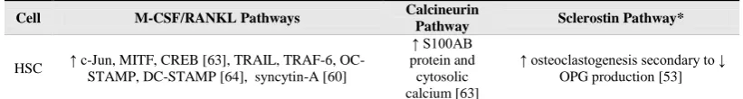
Using NASA’s rotary cell culture system and mouse bone marrow cultures, Sambandam and associates found that simulated microgravity modulated osteoclastogenesis via control of autophagy [59]. Ethiraj et. al. reported similar findings noting that osteoclastogenesis was associated with increased stem cell expression of syncytin-A [60]. In bone, syncytin-A fuses the membranes of mononucleated pre-osteoclasts producing mature multinucleated osteoclasts. Rucci and associates found that modeled microgravity stimulated osteoclastogenesis and bone resorption by increasing RANKL/osteoprotegerin ratios [61] and Saxena et al found that modeled microgravity and hindlimb unloading sensitized osteoclast precursors to RANKL-mediated osteoclastogenesis [62]. Sambandam and associates found that cultured RAW 264.7 osteoclast progenitor cells increased their expression of cytokines, growth factors, proteases and signaling proteins, transcription factors involved in osteoclast differentiation including c-Jun, MITF, and CREB, and cytosolic calcium levels in comparison to controls [63). The same investigators found that simulated microgravity upregulated expression of TRAIL in murine preosteoclast cells in the absence of RANKL stimulation; they also found that simulated microgravity increased the expression of TRAF-6 and fusion genes OC-STAMP and DC-STAMP in preosteoclast cells [64].
Thus, simulated microgravity appears to exert its major effects by enhancing osteoclastogenesis rather than increasing the bone resorptive capacity of mature osteoclasts (see Table 2).
Table 2. Effect of microgravity on osteoclast hematopoietic stem cells precursors.
Cell M-CSF/RANKL Pathways Calcineurin
Pathway Sclerostin Pathway*
HSC ↑ c-Jun, MITF, CREB [63], TRAIL, TRAF-6, OC-STAMP, DC-STAMP [64], syncytin-A [60]
↑ S100AB protein and cytosolic calcium [63]
↑ osteoclastogenesis secondary to ↓ OPG production [53]
*Microgravity-related osteocyte apoptosis increase their secretion of sclerostin [60].
8.0. Discussion
integrity of the sealing zone, so essential for bone resorption, maintained under such adverse conditions? What happens to the complex associations of actin and intermediate filaments, septins and microtubules in osteoclasts subjected to microgravity? Why are M-CSF, RANKL, and calcineurin transcriptional pathways upregulated in hematopoietic stems cells but downregulated in mesenchymal stem cell precursors in zero gravity? And is there any relation between osteoclast survival and space-related changes in osteoclast regulation by immune cells and their cytokines? These intriguing questions should provide an ample basis for future research into the amazingly resilient osteoclast, including the development of agents capable of disabling key elements in its cytoskeleton.
In addition to bone unloading, a number of physiopathological conditions are characterized by excessive osteoclast activity. These include but are not limited to menopause, juvenile Paget’s disease of bone, inflammatory joint diseases, bone cancers such as multiple myeloma, and glucocorticoid therapy [1,2]. Thus, it is not surprising that many of the studies on bone homeostasis have been motivated by the need to find treatments capable of modifying osteoclast activity without inducing osteopetrosis [8].
Biphosphonates have long been used with success to control osteoclast-mediated bone disease; these agents are incorporated into the bone matrix and are ingested by bone resorbing osteoclasts, causing their apoptosis. However, biphosphonates inhibit the stimulatory activity of osteoclasts on osteoblast differentiation and, as a consequence, patients on these drugs suffer from a blockade of de novo bone formation [68]. A recently developed human monoclonal antibody against RANKL, Denosumab, has undesirable side effects and, like biphosphonates, adversely affects osteoblastogenesis [69,70]. An inhibitor of cathepsin K, odanacatib, was shown to prevent pathological bone loss while preserving bone formation but failed in clinical phase III trials due to increased risk of stroke [71]. Scientists have recently developed a humanized monoclonal antibody directed against sclerostin (romosozumab) which is approved for treatment of osteoporosis. Clinical trials have shown that monthly subcutaneous injections of romosozumab is effective in increasing bone formation and density and decreasing bone resorption, results in keeping with the known effects of sclerostin on bone homeostasis [72]. However, there is some concern about potential cardiotoxicity of romosozumab, prompting the need for further observations [73]. Osteoprotegerin-Fc given subcutaneously to mice flown for 12 days in space produced a sustained suppression of bone resorption and, thus, deserves further study [74]. And insulin-like growth factor (IGF)-1, which plays a major role in all phases of bone and cartilage growth, has been shown to increase rodent humerus periosteal bone formation by 37% during a 10 day Space Shuttle flight and [8]. The potential of IGF-1 and other growth factors such as TGF-β and BMP to regulate bone homeostasis in situations of bone unloading merits further investigation.
The United States, Russia, and China are planning to establish colonies on our moon sometime in the 2030s. The force of gravity on the Moon is 1.6 m/s 2 which is 16.7% of Earth’s gravity or ~1 × 10⁻² g, well within the range of simulated microgravity studies already performed. It would seem that once established, colonies on the moon (and eventually on mars) will provide ample opportunity for future studies on the remarkable and resilient osteoclast.
9.0. Conclusion
Astronauts are at risk of losing 1.0 to 1.5% of their bone mass for every month they spend in space despite their adherence to high impact exercise training programs and dietary regimens designed to preserve their musculoskeletal system. This loss is the consequence of microgravity-related impairment of osteocyte and osteoblast function and the consequent upregulation of osteoclast-mediated bone resorption. Further research is needed to better understand how osteoclasts are able to function in zero gravity and to develop more effective interventions to prevent osteoclast-mediated bone disease.
Funding: This research received no external funding.
Conflicts of Interest: The author declares no conflict of interest.
References
1. Okamoto, K; Nakashima, T; Shinohara, M; Negishi-Koga, T; Komatsu, N; Terashima, A; et. al. Osteoimmunology: the conceptual framework unifying the immune and skeletal systems. Physiol. Rev. 2017, 97, 1295-1349.
2. Blangy, A; Bompard, G; Guerit, D; Marie, P; Maurin, J; Maurin, J; et. al. The osteoclast cytoskeleton – current understanding and therapeutic perspectives for osteoporosis. J. Cell Sci. 2020, 133 (13): jcs244798. doi: 1242/jcs.244798.
3. Svitkina, T. The actin cytoskeleton and actin-based motility. Cold Spring Harb. Perspect. Biol. 2018, 10, a018267.
4. LaFlamme, S.E.; Mathew-Steiner, S.; Singh, N.; Colello-Borges, D.; Nieves, B. Integrin and microtubule crosstalk in the regulation of cellular processes. Cell Mol. Life Sci. 2018, 75, 4177–4185.
5. Martínez, P.T.; Navajas, P.L.; Lietha, D. FAK Structure and regulation by membrane interactions and force in focal adhesions. Biomolecules2020, 10, 179.
6. Burke, B. Chain reaction: LINC complexes and nuclear positioning. F1000Research2019, 8, 136.
7. Howard, R.; Scheiner, A.; Cunningham, J.; Gatenby, R. Cytoplasmic convection currents and intracellular temperature gradients. PLoS Comput. Biol. 2019, 15.
8. Smith, J.K. Microgravity, bone homeostasis, and insulin-like growth factor-1. Appl. Sci. 2020, 10, 4433
9. Iwamoto, J.; Takeda, T.; Sato, Y. Interventions to prevent bone loss in astronauts during space flight. Keio J. Med. 2005, 54, 55–59.
10. Cavanagh, P.R.; Licata, A.A.; Rice, A.J. Exercise and pharmacological countermeasures for bone loss during long-duration space flight. Gravit. Space Biol. Bull. 2005, 18, 39–58.
11. Smith, S.M.; McCoy, T.; Gazda, D.; Morgan, J.L.L.; Heer, M.; Zwart, S.R. Space flight calcium: Implications for astronaut health, spaceflight operations, and Earth. Nutrients2012, 4, 2047–2068.
12. Leblanc, A.; Matsumoto, T.; Jones, J.; Shapiro, J.; Lang, T.; Shackelford, L.; Smith, S.M.; Evans, H.; Spector, E.; Ploutz-Snyder, R.; et al. Biphosphonates as a supplement to exercise to protect bone during long-duration spaceflight. Osteoporos. Int. 2013, 24, 2105–2114.
13. NASA—Food For Space Flight. Available online:
https:/www.nasa.gov/specials/apollo50thy/index.html (accessed on 12 June 2020).
14. Goto, Y.; Aoyama, M.; Sekiya, T.; Katika, H.; Waguri-Nagaya, Y.; Miyazawa, K.; et. al. CXCR4+CD45- cells are niche forming for osteoclastogenesis via the SDF-1, CXCL7, and CX3CL1 signaling pathways in bone marrow. Stem Cells. 2016, 34(11), 2733-2741.
15. Ishii, M.; Egen, J.G.; Klauschen, F.; Meier-Schellersheim, M.; Saeki, Y.; Vacher, J.; et. al. Sphingosine-1-phosphate mobilizes osteoclast precursors and regulates bone homeostasis. Nature. 2009, 458(7237), 524-528.
16. Ishii, T.; Shimazu, Y.; Nishiyama, I.; Kikuta, j.; Ishii, M. The role of sphingosine 1-phosphjate in migration of osteoclast precursors; an application of intravital two=photon microscopy. Mol. Cells. 2011, 31(5), 399-403.
17. Tamura, M.; Sato, M. M.; Nashimoto, M. Regulation of CXCL12 expression by canonical Wnt signaling in bone marrow stromal cells. Int. J. Biochem. Cell Biol. 2011, 43(5), 760-767.
19. Cappellen, D.; Luong-Nguyen, N-H.; Bongiovanni, S.; Grenet, O.; Wanke, C.; Šuša, M. Transcriptional program of mouse osteoclast differentiation governed by the macrophage colony-stimulating factor and the ligand for receptor activator of NFkB. J. Biological Chem. 2002, 277(24), 21971-21982.
20. Kim, J.H.; Kin, N. Regulation of NFATc1 in osteoclast differentiation. J. Bone Metab. 2014, 21, 233-241.
21. Zawawi, M.S.F.; Dharmapatni, A.A.S.S.K. ; Cantley, M.D. McHugh, K.P.; Haynes, D.R.; Crotti, T.N. Regulation of ITAM adapter molecules and their receptors by inhibition of calcineurin-NFAT signaling during late stage osteoclast differentiation. Biochem. Biophys. Res. Commun. 2012. 427(2),404-409.
22. Lamothe, B.; Webster, W.K.; Gopinathan, A.; Besse, A.; Campos, A.D.; Darnay, B.G. TRAF-s ubiquitin ligase is essential for RANKL signaling and osteoclast differentiation. Biochem. Biophys. Res. Commun. 2007, 359(4), 1044-1049.
23. Kobayashi, Y.; Udagawa, N.; Takahashi, N. Action of RANKL and OPG for osteoclastogenesis. Crit. Rev. Eukaryot. Gene Expr. 2009, 19(1), 61-72.
24. Negishi-Koga, T.; Takayanagi, H. Ca2+ NFATc1 signaling is an essential axis of osteoclast differentiation. Immunol. Rev. 2009, 231(1), 241-256.23.
25. Takayanagi, H. The role of NFAT in osteoclast formation. Ann. N.Y. Acad. Sci. 2007, 16, 227-237.
26. Asagiri, M.; Takayanagi, H. The molecular understanding of osteoclast differentiation. Bone 2007, 40(2), 251-264.
27. Shinohara, M.; Takayanagi, H. Novel osteoclast signaling mechanism. Curr. Osteoporos. Rep. 2007, 5(2), 67-72.
28. Takayanagi, H. Mechanistic insight into osteoclast differentiation in osteoimmunology. J. Mol. Med. (Berl). 2005, 83(3), 170-179.
29. Lam, J.; Takeshita, S.; Barker, J.E.; Kanagawa, O.; Ross, F.P.; Teitelbaum, S.L. TNF-alpha induces osteoclastogenesis by direct stimulation of macrophages exposed to permissive levels of RANK ligand. J. Clin. Invest. 2000, 106(12), 1481-1488.
30. Kitaura, H.; Sands, M.S.; Aya, K.; Zhou, P.; Hirayama, T. Uthgenannt, B; et. al. Marrow stromal cells and osteoclast precursors differentially contribute to TNF-alpha-induced osteoclastogenesis in vivo. J. Immunol. 2004, 173(8), 4838-4846.
31. Azuma, Y.; Kaji, K.; Katogi, R.; Takeshita, S.; Kudo, A. Tumor necrosis factor-alpha induces differentiation of and bone resorption by osteoclasts. J. Biol. Chem. 2000, 275(7), 4858-4864.
32. Gilbert, L.; He, X.; Farmer, P.; Boden, S.; Kozlowski, M.; Rubin, J.; et. al. Inhibition of osteoblast differentiation by tumor necrosis factor-alpha. Endocrinology 2000, 141(11), 3956-3964.
33. Tanabe, N.; Maeno, M.; Suzuki, N.; Fujisaki, K.; Tanaka, H.; Ogiso, B.; et. al. IL-alpha stimulates the formation of osteoclast-like cells by increasing M-CSF and PGE2 production and decreasing OPG production by osteoblasts. Life Sci. 2005, 77(6), 615-626.
34. Watanabe, Y.; Namba, A.; Aida, Y.; Honda, K.; Tanaka, H.; Suzuki, N.; et. al. IL-1 beta suppresses the formation of osteoclasts by increasing OPG production via an autocrine mechanism involving celecoxib-related prostaglandins in chondrocytes. Mediators Inflamm. 2009; 2009:308596. doi:10.1155/2009/308596.
35. Quinn, J.M.; Itoh, K.; Udagawa, N.; Hausler, K.; Yasuda, H.; Shima, N.; et. al. Transforming growth factor beta affects osteoclast differentiation via direct and indirect actions. J. Bone Miner. Res. 2001, 16(10), 1787-1794.
37. Salamanna, F.; Maglio, M.; Borsari, V.; Giavaresi, G; Aldini, N.N.; Fini, M. Peripheral blood mononuclear cells spontaneous osteoclastogenesis: mechanisms driving the process and clinical relevance in skeletal disease. J. Cell Physiol. 2016, 231(3), 521-530.
38. Guillonneau, C.; Bézie, S.; Anegon, I. Immunoregulatory properties of the cytokine IL-34. Cell Mol. Life Sci. 2017, 74(14), 2569-2586.
39. Mohamed, S.G.K.; Suglyama, E.; Shinoda, K.; Hounoki, H.; Taki, H.; Maruyama, M.; et. al. Interleukin-4 inhibits RANKL-induced expression of NFATc1 and c-Fos: a possible mechanism for downregulation of osteoclastogenesis. Biochem. Biophys. Res. Commun. 2005, 329(3), 839-845.
40. Wei, S.; Wang, M. W-H.; Teitelbaum, S.L.; Ross, F.P. Interleukin-4 reversibly inhibits osteoclastogenesis via inhibition of NF-kappa B and mitogen-activated protein kinase signaling. J. Biol. Chem. 2002, 277(8), 6622-6630.
41. Fujii, T.; Kitaura, H.; Kimura, K.; Hakami, Z.W.; Takano-Yamamoto, T. IL-4 inhibits TNF-α-mediated osteoclast formation by inhibiting RANKL expression in TNF-α-activated stromal cells and direct inhibition of TNF-α-activated osteoclast precursors via a T-cell-independent mechanism in vivo. Bone 2012, 51(4), 771-780.
42. Zhao, B.; Ivashkiv, L.B. Negative regulation of osteoclastogenesis and bone resorption by cytokines and transcriptional repressors. Arthritis Res. Ther. 2011, 13(4): 234. doi: 10.1186/ar3379.
43. Mohamed, S. G-K.; Sugiyama, E.; Shinoda, K.; Taki, H.; Hounoki, H.; Abdel-Aziz, H.O.; et. al. Interleukin-10 inhibits RANKL-mediated expression of NFATc1 in part via suppression of c-Fos and c-Jun in RAW264.7 cells and mouse bone marrow cells. Bone 2007, 41(4), 592-602.
44. Honda, K. Interleukin-6 and soluble interleukin-6 receptor suppress osteoclast differentiation by inducing PGE(2) production in chondrocytes. J. Oral Sci. 2011, 53(1), 87-96.
45. Kitaura, H.; Nagata, N.; Fujimura, Y.; Hotokezaka, H.; Yoshida, N.; Nakayama, K. Effect of IL-12 on TNF-alp[ha-mediated osteoclast formation in bone marrow cells: apoptosis mediated by Fas/Fas ligand interaction. J. Immunol. 2002, 169(9), 4732-4738.
46. Morita, Y.; Kitaura, H.; Yoshimatsu, M.; Fujimura, Y.; Kohara, H.; Eguchi, T.; et. al. IL-18 inhibits TNF-alpha-induced osteoclastogenesis possibly via a T cell-independent mechanism in synergy with IL-12 in vivo. Calcif. Tissue Int. 2010, 86(3):242-248.
47. Horwood, N.J.; Elliott, J.; Martin, T.J.; Gillespie, M.T. IL-12 alone and in synergy with IL-18 inhibits osteoclast formation in vitro. J. Immunol. 2001, 166(8), 4915-4921.
48. Udagawa, N.; Horwood, N.J.; Elliott, J.; Mackay, A.; Owens, J.; Okamura, H.; et. al. Interleukin-18 (interferon-gamma-inducing-factor) is produced by osteoblasts and acts via granulocyte/macrophage colony-stimulating factor and not via interferon-gamma to inhibit osteoclast formation. J. Exp. Med. 1997, 185(6),1005, 1012.
49. Kohara, H.; Kitaura, H.; Fujimura, Y.; Yoshimatsu, M.; Morita, Y.; Eguchi, T.; et. al. IFN-γ directly inhibits TNF-α-induced osteoclastogenesis in vitro and in vivo and induces apoptosis mediated by Fas/Fas ligand interactions. Immunol. Lett. 2011, 137(1-2), 53-61.
50. Hillsley, M.V.; Frangos, J.A. Bone tissue engineering: Role of interstitial fluid flow. Biotechnol. Bioeng. 1994, 43, 573-581.
51. Liu, C.; Zhao, Y.; Cheung, W.-Y.; Gandhi, R.; Wang, L.; Lidan, Y. Effects of cyclic hydraulic pressure on osteocytes. Bone 2010, 46, 1449-1456.
52. Smith, J.K. IL-6 and the dysregulation of immune, bone, muscle, and metabolic homeostasis during spaceflight. Npj Microgravity 2018,4,24
54. Nabavi, N.; Khandani, A.; Camirand, A.; Harrison, R.E. Effects of microgravity on osteoclast bone resorption and osteoblast cytoskeletal organization and adhesion. Bone 2011, 49(5), 965-974.
55. Hughs-Fulford, M. Function of the cytoskeleton in gravisensing during spaceflight. Adv. Space. Res. 2003, 32, 1583-1593.
56. Gerbaix, M.; Gnyubkin, V.; Farlay, D.; Olivier, C.; Ammann, P.; Courbon, G.; et. al. One-month spaceflight compromises the bone microstructure, tissue-level mechanical properties, osteocyte survival and lacunar volume in mature mice skeletons. Sci. Rep. 2017, 7, 2659.
57. Tamma, R.; Colaianni, G.; Camerino, C.; Di Benedetto, A.; Greco, G.; Strippoli, M.; et. al. Microgravity during spaceflight directly affects in vitro osteoclastogenesis and bone resorption. FASEB J. 2009, 23, 2549-2554.
58. Vico, L.; Bourrin, S.; Genty, C.; Palle, S.; Alexandre, C. Histomorphometric analysis of cancellous bone from COSMOS 2044 rats. J. Appl. Physiol. 1993, 75(5), 2203-2208.
59. Sambandam, Y.; Townsend, M.T.; Pierce, J.J.; Lipman, C.M.; Haque, A.; Bateman, T.A.; et. al. Microgravity control of autophagy modulates osteoclastogenesis. Bone 2014, 61, 125-131.
60. Ethiraj, P.; Link, J.R.; Sinkway, J.M.; Brown, C.D.; Parler, W.A.; Reddy, S.V. Microgravity modulation of syncytin-A expression enhance osteoclast formation. J. Cell. Biochem. 2018, 119, 5695-5703.
61. Rucci, N.; Rufo, A.; Alamanou, M.; Teti, A. Modeled microgravity stimulates osteoclastogenesis and bone resorption by increasing osteoblast RANKL/OPG ratio. J. Cell. Biochem. 2007, 100, 464-473.
62. Saxena, R.; Pan, G.; Dohm, E.D.; McDonald, J.M. Modeled microgravity and hindlimb unloading sensitize osteoclast precursors to RANKL-mediated osteoclastogenesis. J. Bone Miner. Metab. 2011, 29, 111-122.
63. Sambandam, Y.; Blanchard, J.J.; Daughtridge, G.; Kolb, R.J.; Shanmugarajan, S.; Pandruvada, S.N.M.; et. al. Microarray profile of gene expression during osteoclast differentiation in modelled microgravity. J. Cell. Biochem. 2010, 111(5), 1179-1187.
64. Sambandam, Y.; Baird, K.L.; Stroebel, M.; Kowai, E.; Balasubramanian, S.; Reddy, S.V. Microgravity induction of TRAIL expression in preosteoclast cells enhances osteoclast differentiation. Sci. Rep. 2016, 6, 25143.
65. Zayzafoon, M.; Meyers, V. E.; McDonald, J. M. Microgravity: the immune response and bone. Immunol. Rev. 2005, 208, 267–280.
66. Burger, E.H.; Klein-Nulend, J. Microgravity and bone mechanosensitivity. Bone 1998, 22, 1275-1305.
67. Lin, C.; Jiang, X.; Dai, Z.; Guo, X.; Weng, T.; Wang, J.; et. al. Sclerostin mediates bone resorption response to mechanical unloading through antagonizing Wnt/beta-catenin signaling. J. Bone Miner. Res. 2009, 24, 1651-1661.
68. LeBlanc, A.; Matsumoto, T.; Jones, J.; Shapiro, J.; Lang, T., Shackelford, L.; et. al. Biphosphonates as a supplement to exercise to protect bone during long-duration spaceflight. Osteoporos. Int. 2013, 24, 2105-2114.
69. Deeks, E.D. Denosumab: a review in postmenopausal osteoporosis, Drugs Aging 2018, 35, 163-173.
70. Vargas-Franco, J.W.; Castaneda, B.; Rédini, F.; Gómez, D.F.; Heymann, D.; Lézot, F. Paradoxical side effects of biphosphonates on the skeleton: what do we know and what can we do? J. Cell. Physiol. 2018, 233, 5696-5715.
71. Stone, J.A.; McCrea, J.B.; Witter, R.; Zajic, S.; Stoch, S.A. Clinical and translational pharmacology of the cathepsin K inhibitor odanacatib studied for osteoporosis. Br. J. Clin. Pharmacol. 2019, 85, 1072-1083.
72. Bandeira, L.; Lewiecki, E.M.; Bilezikian, J.P. Romosozumab for treatment of osteoporosis. Expert Opin. Biol. Ther. 2017, 17, 255-263.